EPHELIDES
Synonym: Freckles
Thomas Wiesner and Raymond L. Barnhill
Key features
Small, uniformly pigmented macules in sun-exposed sites
More common in fair-skinned individuals and more noticeable in summer than in winter
Hyperpigmentation of the basal layer of the epidermis without an increase in melanocyte density
Epidemiology and Pathogenesis
Ephelides are common in fair-skinned individuals, especially those with red hair, and occur more frequently in women. They first appear in early childhood and usually stabilize in number during adulthood, followed by gradual fading. Ephelides result from an increase in sun-induced melanin synthesis and transport of melanosomes from melanocytes to keratinocytes. Frequent sunburns and variants of the melanocortin-1 receptor gene (MC1R) play an important role in the development of ephelides (see Fig. 65.13).
Clinical Features
Ephelides are limited to sun-exposed sites – primarily the face, upper trunk, and dorsal aspects of the arms. Ephelides are well demarcated, uniformly pigmented, oval to irregular-shaped macules that are usually 1 to 3 mm in size. Depending upon the intensity of sun exposure, ephelides vary in color from light to medium brown. In the summer, they are more conspicuous and then fade during the winter. Ephelides are benign with no potential for malignant transformation. However, higher numbers of ephelides correlate with an increased risk of acquired melanocytic nevi and melanoma.
Pathology
There is hyperpigmentation of the basal layer of the epidermis without an increase in the number of melanocytes. Compared to adjacent uninvolved epidermis, melanocytes in ephelides are larger and have more branching of dendrites and stronger DOPA positivity, indicating increased activity.
Differential Diagnosis
Ephelides must be distinguished from simple lentigines, solar lentigines, small café-au-lait macules, and junctional nevi. In addition to being confined to sun-exposed skin, the clinical appearance of ephelides varies depending upon the amount of exposure to UVR (Table 112.1). Café-au-lait macules are usually solitary and larger than ephelides.
Treatment
If ephelides are a cosmetic concern, sun exposure should be minimized and use of sunscreens, hats, and protective clothing recommended. Topical retinoids and hydroquinone may lighten lesions, but it is difficult to achieve an homogeneous clinical effect and potential side effects (e.g. irritation, dyspigmentation) need to be considered. Pigment-specific lasers represent an additional treatment option, but recurrences are common.
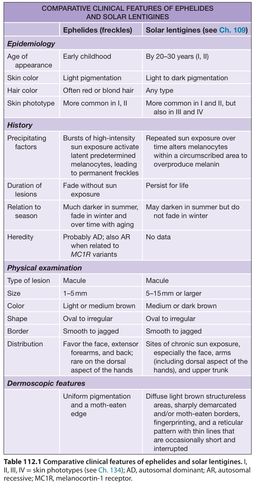
Table 112.1 Comparative clinical features of ephelides and solar lentigines. I, II, III, IV = skin phototypes (see Ch. 134); AD, autosomal dominant; AR, autosomal recessive; MC1R, melanocortin-1 receptor.