MOLECULAR PATHOGENESIS
Cancer usually develops through a stepwise evolutionary process, with tumor progression involving both genetic instability and selective growth of cells with advantageous mutations. Additional contributing factors include genetic predisposition, mutagenic environmental events, and antitumor host response. As discussed in Chapter 107, the hallmarks of cancer can be acquired by the activation of oncogenes or the inactivation of tumor suppressor genes via point mutations, genomic rearrangements (e.g. deletions, inversions, duplications, fusions, and translocations), and epigenetic mechanisms such as microRNA expression and promoter methylation. Genome-wide analysis of genetic aberrations has revolutionized our understanding of the complex interplay of signaling pathways.
In melanoma, initiating oncogenic events frequently affect genes involved in the mitogen-activated protein kinase (MAPK) and phosphoinositide 3-kinase (PI3K) signaling pathways (Fig. 113.1). While these alterations may initially result in senescence, secondary genetic alterations can then result in progression into a malignant tumor. The constitutive activation of MAPK or PI3K signaling that commonly occurs can be due to mutations in several kinase genes or fusions that disrupt the regulatory domain of a kinase gene, as well as copy number alterations. Other mechanisms include: upregulation of telomerase genes which results in unlimited replicative potential; changes in chromatin remodeling that lead to increased gene expression; and disruption of cell cycle regulation via mutations in genes that encode cyclin D1, p16/p14ARF, or p53. Table 113.1 lists some of the key primary and secondary driver mutations identified in melanomas.
Melanomas from different anatomic sites with varying degrees of UV radiation (UVR) damage differ in their characteristic driver mutations (see Table 113.1). Recognition of several distinct mechanisms of melanoma pathogenesis has inspired the current taxonomy of melanocytic neoplasia reflected in the 2018 WHO Classification of Skin Tumours, which integrates site of origin (epithelium- versus non-epithelium-associated) and role of cumulative sun damage (CSD; high-, low-, or non-CSD-related), along with mole phenotype (high vs low nevus count) and frequency of BRAF, NRAS, and other relevant mutations.
Advances in our understanding of the molecular basis of melanoma have paved the way for the era of targeted therapy. For example, discovery of the BRAF V600E mutation, which substitutes a glutamic acid (E) for valine (V) at codon 600, leading to activation of the MAPK pathway, has led to the use of BRAF inhibitors for BRAF-mutant melanomas. These agents are combined with MEK inhibitors, which act downstream of BRAF in the MAPK pathway, for improved efficacy and tolerability. For melanomas lacking the typical point mutations in BRAF, NRAS, or NF1 (so-called “pan-negative” melanomas), identification of MAPK-activating kinase gene fusions in a subset of these melanomas may permit their treatment with other kinase inhibitors. For example, melanomas harboring NTRK fusions can be treated with larotrectinib, a selective TRK inhibitor approved for TRK fusion-positive tumors.
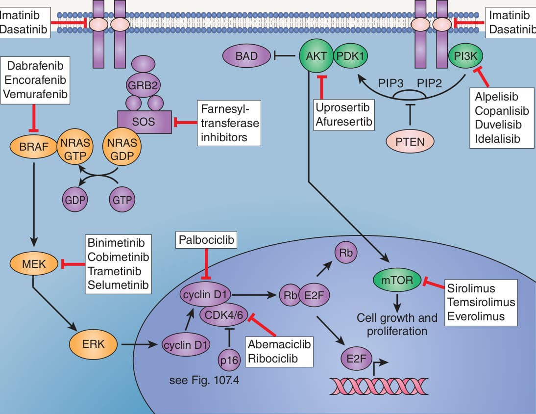
Fig. 113.1 RAS–RAF–MEK–ERK (MAPK) and PI3K–
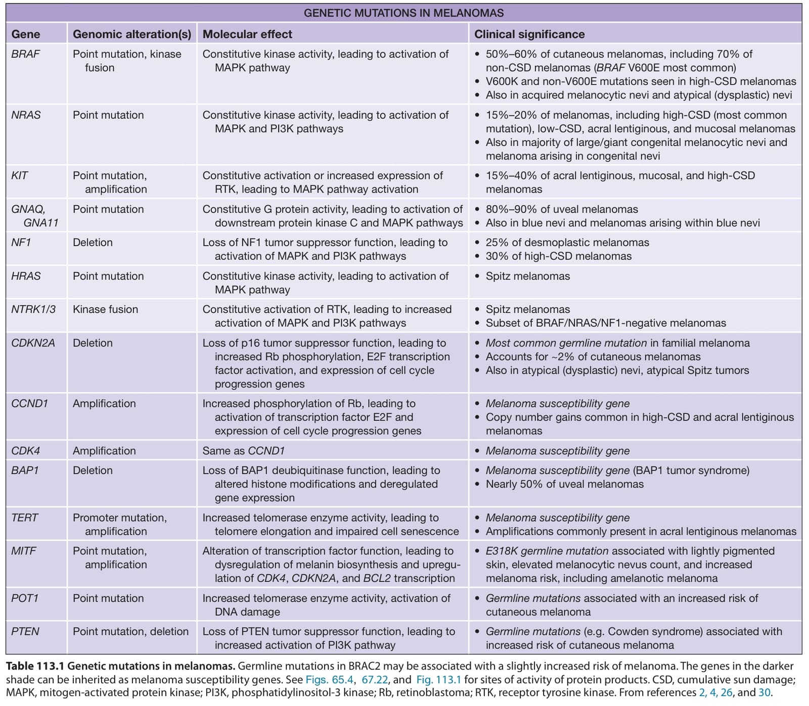
Table 113.1 Genetic mutations in melanomas. Germline mutations in BRAC2 may be associated with a slightly increased risk of melanoma. The genes in the darker shade can be inherited as melanoma susceptibility genes. See Figs. 65.4, 67.22, and Fig. 113.1 for sites of activity of protein products. CSD, cumulative sun damage; MAPK, mitogen-activated protein kinase; PI3K, phosphatidylinositol-3 kinase; Rb, retinoblastoma; RTK, receptor tyrosine kinase. From references 2, 4, 26, and 30.