HOST IMMUNE RESPONSE TO MELANOMA
Melanoma is a highly immunogenic tumor, making it ideal for immunebased therapeutic approaches (Fig. 113.2). Multiple melanoma antigens, recognized by autologous T cells or antibodies, have been described including: (1) mutant tumor antigens (e.g. mutant p16 [CDKN2A]); (2) shared tumor-specific antigens of the cancer/testis family (e.g. MAGE-1, MAGE-3, NY-ESO-1); and (3) cell type-specific differentiation antigens (e.g. tyrosinase, PMEL17/gp100, MART-1/Melan-A). These proteins are processed and presented on the melanoma cell surface as MHC/peptide complexes. CD8+ cytotoxic T cells recognize these antigens via their T cell receptors (TCRs) and, if appropriately activated by costimulatory signals, are able to kill tumor cells. CD4+ T helper cells and antibodies also play a critical role.
Activation of melanoma-specific CD8+ T cells is dependent on the migration of tumor antigen-loaded dendritic cells to a draining lymph node. Here, melanoma antigens are presented to CD8+ T cells in the presence of a costimulatory signal; the latter consists of the binding of B7 on the antigen-presenting cell to CD28 on the T cell (see Ch. 4). Cytotoxic T lymphocyte-associated antigen-4 (CTLA-4) is an inhibitory homolog of CD28 that is upregulated following T cell activation and acts as natural inhibitor of T cell activation by removing the costimulatory signal. Programmed cell death protein 1 (PD-1) is another inhibitory receptor in the CD28 family (see Fig. 128.9). The interaction of PD-1 with its ligands PD-L1 and PD-L2 promotes apoptosis of tumor antigen-specific T cells and reduces apoptosis in regulatory T cells.
Immunotherapy based upon CTLA-4, PD-1, and PD-L1 blockade was first introduced for the treatment of melanoma (see below), beginning with ipilimumab, an anti-CTLA-4 antibody. This was followed by the approval of anti-PD-1 antibodies (nivolumab, pembrolizumab) as first-line monotherapy for metastatic and locally advanced melanoma. Combining anti-CTLA-4 and anti-PD-1 antibodies leads to greater efficacy, but a significant increase in immune-related adverse events (irAEs). Of note, vitiligo is a melanocyte-targeted irAE that occurs in
AKT signaling pathways. The mitogen-activated protein kinase (MAPK) pathway (orange color), also referred to as the MAP kinase pathway, is physiologically activated by growth factor binding to receptor tyrosine kinases. The stimulus is relayed to the nucleus via the GTPase activity of NRAS and the kinase activity of BRAF, MEK, and ERK. Within the nucleus, this results in increased transcription of genes involved in cell growth, proliferation, and migration. The central role of this pathway in melanocytic neoplasia is highlighted by the fact that either NRAS or BRAF is mutated in up to 80% of all cutaneous melanomas and melanocytic nevi. The PI3K–AKT pathway is another important regulator of cell survival, growth, and apoptosis (green color). A key inhibitor of this pathway is PTEN and inactivation of the gene that encodes PTEN via mutations, deletions, or promoter methylation also occurs in cutaneous melanomas. As a result, there is increased activity of the PI3K–AKT signaling pathway. Examples of farnesyltransferase inhibitors are tipifarnib and lonafarnib. BAD, BCL-2 associated agonist of cell death; CDK, cyclin-dependent kinase; E2F, transcription factor that controls transcription of cyclins; ERK, extracellular signal-regulated kinase; GDP, guanosine- 5-diphosphate; GRB2, growth factor receptor-bound protein 2, an adaptor protein that contains one Src homology 2 (SH2) domain and two SH3 domains; GTP, guanosine-5-triphosphate; MEK, MAPK kinase; mTOR, mammalian target of rapamycin (aka sirolimus); p16, protein product of CDKN2A; PDK1, phosphoinositidedependent kinase-1; PI3K, phosphatidylinositol 3-kinase; PIP2, phosphatidylinositol 4,5-bisphosphate; PIP3, phosphatidylinositol 3,4,5-triphosphate; PTEN, phosphatase and tensin homolog; Rb, retinoblastoma protein; SOS, son of sevenless.
2%–25% of patients and its development correlates with improved progression-free survival.
Insights into mechanisms of melanoma-specific host immune responses and immune escape have clearly led to improved immunotherapy in the form of checkpoint inhibitors and hopefully will result in other more effective therapies, including vaccines. However, overcoming the range of immune escape mechanisms still remains a challenge (Table 113.2).
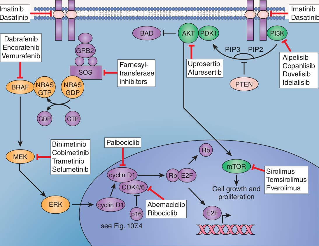
Fig. 113.1 RAS–RAF–MEK–ERK (MAPK) and PI3K–
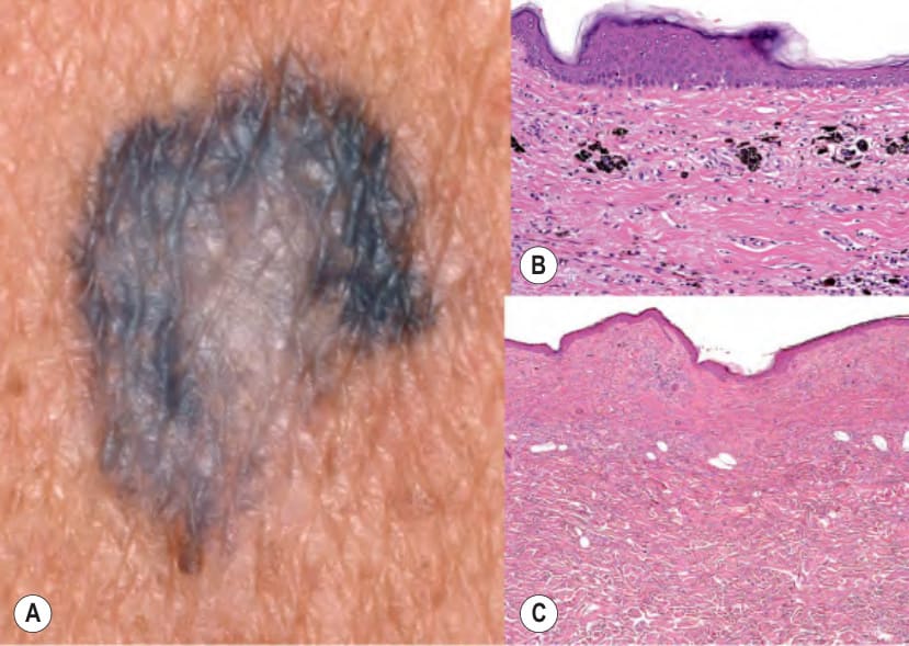
Fig. 113.2 Completely regressed cutaneous melanoma.A, B The gray-black arciform area had only melanophages in the upper dermis; although there were no remaining cells of melanoma, the patient already had nodal metastases. C The central hypopigmented area had fibrosis with sparse inflammation. Complexes of a pre-existing melanocytic nevus were present focally below the zone of regression. Courtesy, Department of Dermatology, Medical University of Graz.
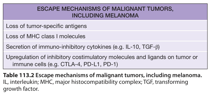
Table 113.2 Escape mechanisms of malignant tumors, including melanoma.