OTHER MELANOMA VARIANTS
There are cutaneous melanomas that may present with unusual manifestations. Some of these are defined by clinical features, others by histologic findings.
Amelanotic Melanomas
Fortunately, the majority of melanomas are clinically pigmented, which aids in visual recognition and diagnosis. Melanomas lacking clinically evident pigment are termed “amelanotic” (Fig. 113.17). All four major histologic subtypes of melanoma discussed above can have amelanotic variants that may confound clinical diagnosis; recognition of dermoscopic features in amelanotic melanoma can be useful (see Figs. 0.38 and 0.39). Amelanotic SSMs, NMs, and LMMs are often biopsied due to clinical suspicion of basal or squamous cell carcinomas. Amelanotic ALMs, including subungual type, are especially challenging and may be mistaken for warts or SCC (see Fig. 113.15B). Amelanotic melanomas do not differ from pigmented melanomas in terms of prognosis or therapy.
Melanoma with Features of a Spitz Nevus (Spitzoid Melanoma and Spitz Melanoma)
Spitzoid melanomas comprise a heterogeneous category of melanomas demonstrating histomorphologic features suggestive of a Spitz nevus, e.g. well-circumscribed margins and epithelioid melanocytes that do not mature with progressively deeper dermal extension. However, unlike benign Spitz nevi, spitzoid melanomas may have features such as a large clinical diameter, deep infiltration into the dermis or subcutis,
marked cytologic atypia of dermal melanocytes, or mitotic figures at the base of the lesion (see Table 112.7).
Within the category of “spitzoid melanoma” is Spitz melanoma, defined by the 2023 WHO classification as the malignant counterpart of the Spitz nevus based upon genetic characteristics. Genomic analyses have revealed that many cases of spitzoid melanomas (e.g. with morphologic features of Spitz nevi) demonstrate genetic features of low-CSD melanoma, including BRAF and NRAS mutations not typically present in Spitz nevi. In contrast, Spitz melanomas demonstrate a distinct spectrum of MAPK-activating alterations more closely associated with Spitz lineage, such as mutations or fusions involving HRAS, ALK, BAP1, ROS1 and NTRK1/3, among others.
A subset of spitzoid lesions demonstrate histopathologic features intermediate between benign Spitz nevi and melanoma and may present a significant diagnostic challenge, particularly since they present more often in pediatric, adolescent, and young adult populations. In the 2023 WHO classification, these lesions are termed Spitz melanocytomas whereas in the past, they were referred to by several terms including atypical Spitz tumors (ASTs), “melanocytic tumors of uncertain malignant potential” (MelTUMPs), or “spitzoid tumors of uncertain malignant potential” (STUMPs). These ambiguous lesions are considered to have biologically indeterminate behavior and are frequently treated with complete excision. Some ASTs may metastasize to regional lymph nodes, further confounding their distinction from melanoma. However, ASTs generally follow a more benign course than melanoma, even with nodal metastases, and the diagnostic and prognostic value of sentinel lymph node biopsy (SLNB) for this class of lesions has been questioned in recent years. In the absence of widely accepted histomorphologic diagnostic criteria, several molecular approaches have been taken in attempts to distinguish these entities from melanoma, e.g. immunohistochemistry, fluorescence in situ hybridization (FISH), comparative genomic hybridization (CGH), gene expression profiling (GEP), next generation sequencing (NGS) analysis (see below).
Desmoplastic Melanoma
Desmoplastic melanoma (DM) is defined histologically by the presence of spindled melanocytes and prominent stromal fibrosis. The typical clinical lesion is a skin-colored, pink, or red plaque or nodule located on sun-exposed skin. The lack of classic clinical findings for melanoma may result in a delay in diagnosis. DM may develop: (1) de novo; (2) in conjunction with an overlying LM, ALM, or other radial growth phase melanoma; or (3) rarely in mucosal sites. Deep tissue samples are necessary to establish the diagnosis (Fig. 113.18), as superficial portions of the tumor show subtle or
non-diagnostic findings which may be mistaken for the fibrosis of a scar or other spindle cell neoplasms. Based upon the degree of desmoplasia and spindled melanocytes within the tumor, “pure” (>90% of tumor consists of a proliferation of spindle cells in a desmoplastic stroma) and “mixed” (<90% of the tumor has a desmoplastic stroma and focally there are nodules of melanoma cells) forms of DM have been described, with differing clinical behavior in the two forms (see below).
Compared to SSM and LMM subtypes, DM is more infiltrative and typically thicker at the time of diagnosis. Incomplete excision and local recurrences commonly occur. A subset of DMs may demonstrate neurotropism (i.e. perineural invasion), with associated higher rates of local recurrence following surgical resection; adjuvant radiation therapy may be considered to improve local control. While deep DMs have significant potential for distant metastasis, conventional T-staging tends to overestimate the likelihood for metastasis. When matched for thickness, several studies have demonstrated improved melanoma-specific survival (MSS) for pure DMs as compared to other melanoma subtypes. Prognosis appears to be worse for the mixed subtype compared to the pure subtype, although data are somewhat conflicting. Regional lymph node metastases are generally uncommon in DM, but mixed DM is significantly more likely than pure DM to have nodal involvement, with positive SLNB rates comparable to conventional melanomas in some studies. Lymphovascular invasion is also more commonly observed in the mixed form.
Clear Cell Sarcoma (“Melanoma of Soft Parts”)
Clear cell sarcoma is an uncommon soft tissue malignancy that demonstrates substantial histopathologic overlap with melanoma, including
the presence of melanosomes by electron microscopy and immunohistochemical expression of S100, HMB-45, Melan-A, and other melanocytic markers. Despite its close histopathologic resemblance to melanoma, leading to the designation “melanoma of soft parts”, the exact cell of origin remains unknown, and its clinical presentation is distinctive from conventional melanoma.
Clear cell sarcoma most often presents on the distal extremities of adolescents and young adults, typically arising within the deep soft tissue in association with tendons and aponeuroses. Tumors are composed of nests and fascicles of oval to spindled cells with vesicular nuclei, basophilic nucleoli, and eosinophilic to clear cytoplasm. Multinucleated giant cells and melanin can frequently be seen. Rare cases of superficial clear cell sarcoma may be histologically challenging to distinguish from primary dermal melanomas or melanoma metastases. However, unlike melanoma, clear cell sarcoma demonstrates characteristic rearrangements of EWSR1; most frequently a reciprocal translocation t(12; 22)(q13; q12) results in a EWS–ATF1 fusion gene. These tumors tend to be aggressive, with a high likelihood of regional and distant metastases.
Melanoma Arising in a Blue Nevus (Blue Nevus-Like Melanoma)
Blue nevus-like melanoma, also known as melanoma arising in blue nevus and formerly as malignant blue nevus, is a rare dermal tumor of melanocytes. It is most commonly located on the head and particularly the scalp (Fig. 113.19). The tumor presents as a blue–black, deeply situated nodule, generally >1 cm in diameter. Histologically, elements of a benign cellular blue nevus are associated with nodular areas of atypical spindle-shaped and bipolar dendritic melanocytes, mitotic figures, necrosis, and melanophages. The clinical course is characterized by a high rate of recurrence and metastasis. The associated cellular blue nevus may have mutations in GNAQ or less often GNA11 (see Table 112.3), with mutations in BAP1, SF3B1, and EIFA1X detectable within the malignant portion.
Primary Dermal Melanoma
Although rare, dermal-based melanomas may mimic cutaneous or lymphatic metastases histologically and appear clinically similar to a cyst or other dermal tumor. These so-called “primary dermal melanomas” are characterized by a lack of overlying epidermal involvement and no regressive features, and they may be misclassified as stage III or IV melanoma at the outset. Although primary dermal melanomas present with an average Breslow depth of 3–9 mm, depending on the study, current evidence suggests a more favorable prognosis when compared to equivalently staged conventional primary melanomas – notably an estimated 5-year survival of 80%–100%. Histologic distinction between primary dermal melanoma and cutaneous metastasis may be challenging, if not impossible, and clinical exclusion of metastatic disease (often via imaging) is essential. Findings of an associated intradermal nevus and focal or weak immunohistochemical staining for proliferation-related markers (cyclin D1, Ki-67, and p53) have been described as potential distinguishing features of primary dermal melanoma.
Ocular Melanoma
Primary ocular melanomas, which are relatively rare (5% of all melanomas), can be divided into conjunctival melanomas and uveal melanomas (iris, choroidal, and ciliary body melanomas). Uveal melanomas frequently (up to 85%) harbor somatic activating mutations in GNA11 or GNAQ. These genes encode members of the q class of G protein α-subunits. Heterotrimeric G proteins are involved in mediating signals between G-protein-coupled receptors (GPCRs) and downstream effectors (see Fig. 65.14). As noted previously, GNAQ (more frequently than GNA11) mutations have been detected in blue nevi but not in cutaneous melanomas.
Although biopsy of the primary tumor is not generally needed for diagnosis, it may be considered for prognostication purposes, with molecular testing via chromosomal analysis or GEP preferred over cytology alone. Chromosomal changes of prognostic significance in ocular melanoma include monosomy 3 and alterations of 8q and 6p. Monosomy 3 reduces the 5-year survival from ~100% to <50%, and a particularly poor outcome has been associated with combined abnormalities in chromosomes 3 and 8. However, chromosomal studies of fine needle biopsies can be limited due to sampling error resulting from tumor heterogeneity and technical failures.
Uveal melanomas can be divided into Class 1 (low metastatic risk) and Class 2 (high metastatic risk) based upon a GEP of 15 genes. The GEP of Class 1 uveal melanomas resembles normal uveal melanocytes with >95% of patients free of metastasis at 4 years. In contrast, the GEP of Class 2 uveal melanomas resembles primitive neural/ectodermal stem cells with <20% of these patients free of metastasis at 4 years. BAP1 mutations are associated with Class 2 uveal melanomas.
The management of uveal melanoma has shifted to eye-sparing approaches, without significantly impacting patient survival. Radiotherapy with proton beams has replaced eye enucleation as the standard treatment. Unfortunately, immune checkpoint inhibitors have not led to responses similar to those seen in cutaneous melanoma. Tebentafusp, a bispecific engager that brings together CD3+ T cells and melanoma cells with the gp100/HLA-A02:01 complex, is approved for unresectable or metastatic uveal melanoma in HLA-A02:01- positive adults. Surveillance imaging with contrast-enhanced magnetic resonance imaging or ultrasound is primarily directed at the liver, the most frequent site of uveal melanoma metastasis.
Mucosal Melanoma
Mucosal melanomas are rare lesions that may occur in the mouth, nasopharynx, larynx, and anogenital mucosae. They tend to occur near the mucocutaneous junctions of squamous and columnar epithelia and account for ~1% of melanomas. As many as 35% of mucosal melanomas are reportedly amelanotic, further complicating clinical diagnosis. Not surprisingly, these tumors tend to be diagnosed at a locally advanced stage with associated poor prognosis. Similar to ALM, mucosal melanomas are a genetically distinct subtype of melanoma. They harbor more and different DNA copy number changes than do cutaneous melanomas and they can harbor activating KIT mutations, which could make the tumor sensitive to KIT-inhibiting drugs (e.g. imatinib; see Fig. 113.1). However, as with ALM, response rates to KIT inhibitors have so far been limited.
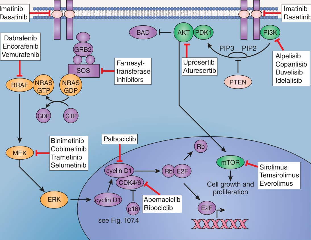
Fig. 113.1 RAS–RAF–MEK–ERK (MAPK) and PI3K–
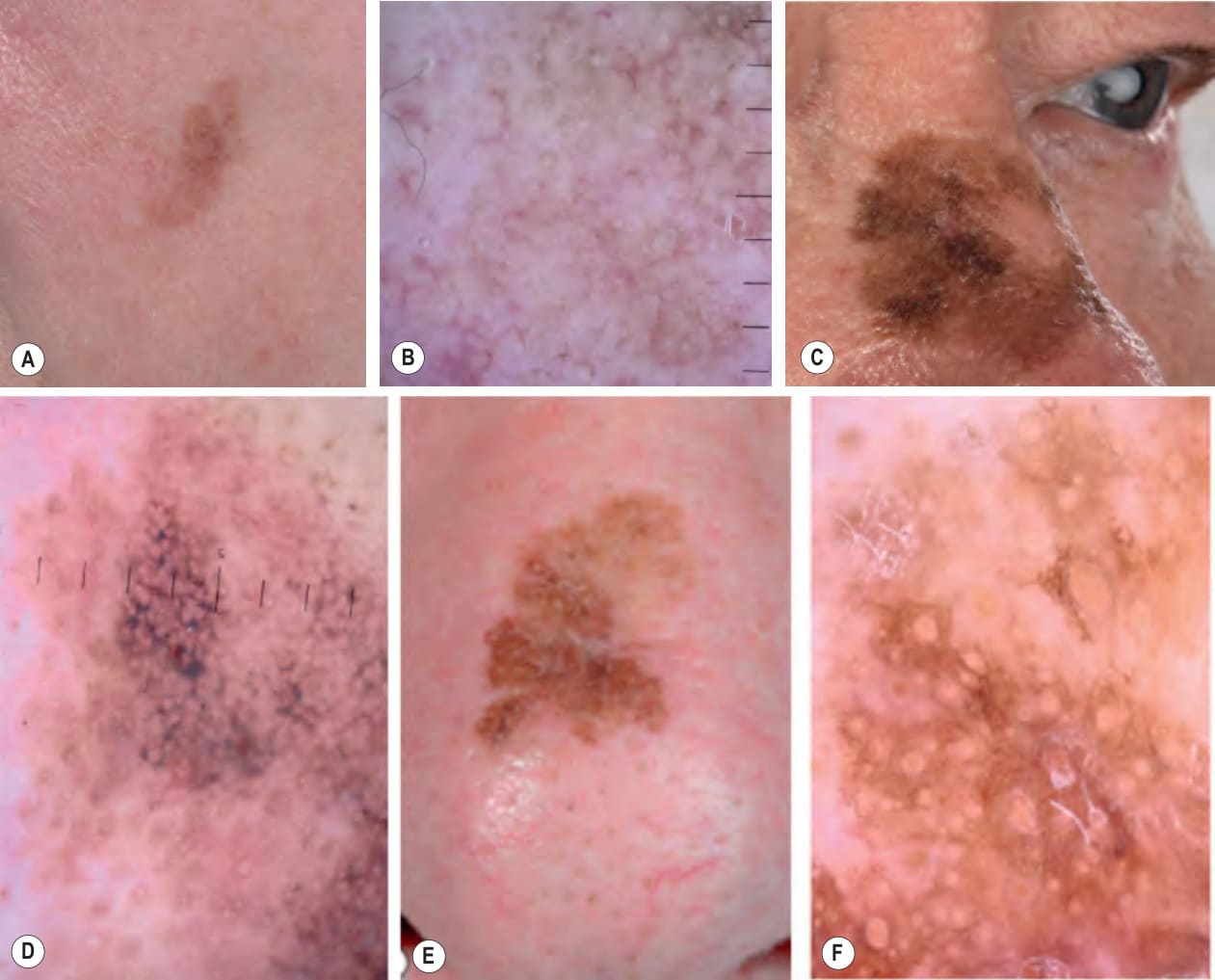
Fig. 113.14 Lentigo maligna (melanoma in situ, lentigo maligna type).A, C An early lentigo maligna presenting as a light brown patch with subtle, asymmetric pigmentation is compared to a more obvious lesion with readily apparent variation in pigmentation. B, D By dermoscopy, follicular openings surrounded by annular pigmentation are seen, referred to as a “circle in a circle”. Findings are more subtle in B and a few rhomboidal structures are seen in D. E A pigmented lesion on the dorsal nose, with irregular borders, light to dark brown color, and marked asymmetry. F Dermoscopy demonstrating annular structures corresponding to follicular openings surrounded by melanoma cells (“circle in a circle”). Courtesy Claus Garbe, MD and Jürgen Bauer, MD.
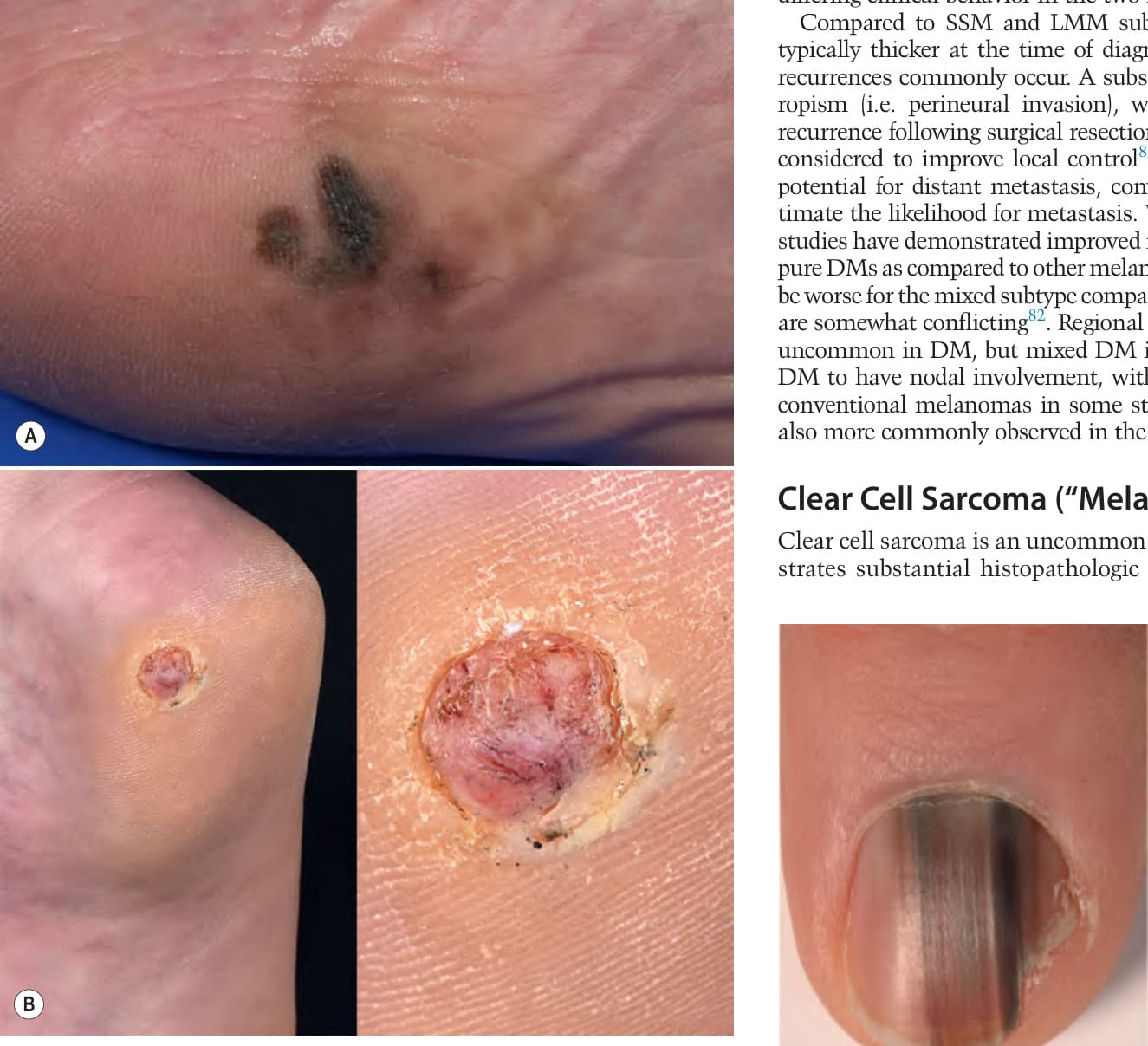
Fig. 113.15 Acral lentiginous melanoma.A Irregularly pigmented lesion on the plantar surface of the foot. Dermoscopic findings include irregular diffuse pigmentation and a parallel ridge pattern (see Fig. 112.14). B Crusted pink nodule on the heel that had been treated for several months as a wart prior to diagnosis. A, Courtesy Claus Garbe, MD and Jürgen Bauer, MD; B, Courtesy Department of Dermatology, Medical University of Graz.
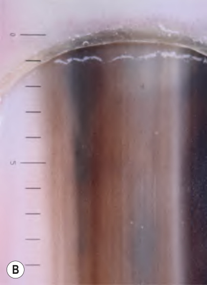
Fig. 113.16 Melanoma of the nail matrix.A, B The longitudinal pigmentation of the nail plate is non-homogeneous, both clinically and dermoscopically. Histologically, the Breslow depth was 0.7 mm. Courtesy Claus Garbe, MD and Jürgen Bauer, MD.
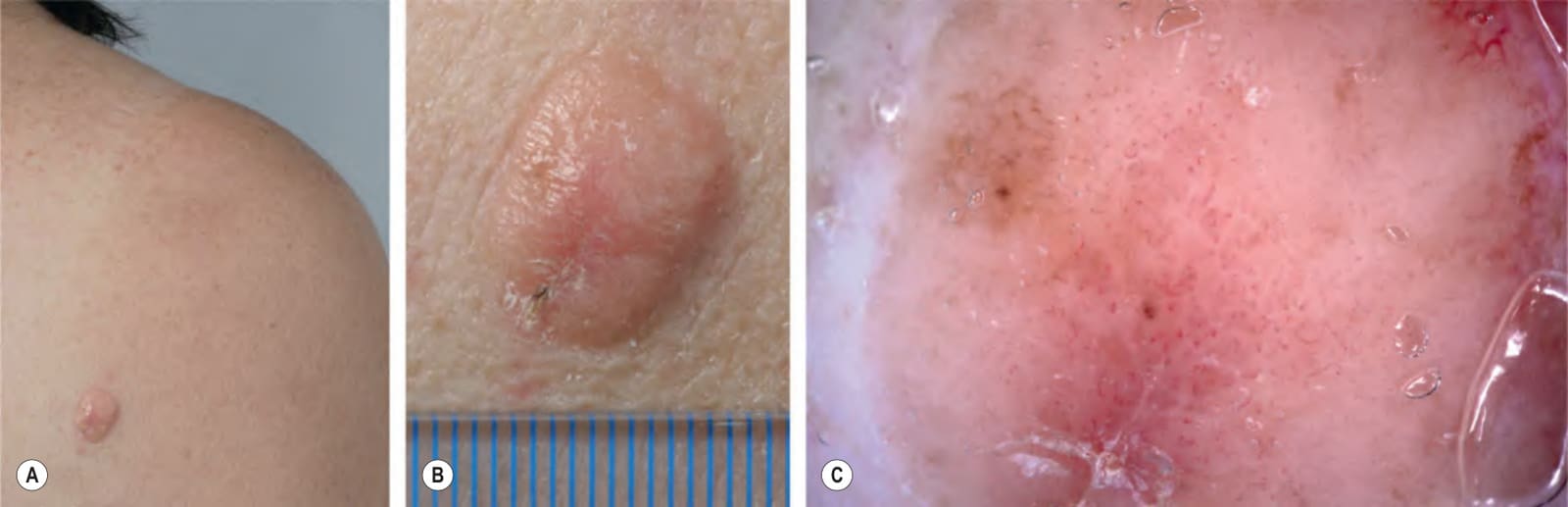
Fig. 113.17 Amelanotic melanoma.A, B Skin-colored to light pink nodule on the right scapula of a female patient; tumor thickness was 4 mm. C By dermoscopy, some pigmented globules allow one to identify the tumor as a melanocytic lesion. Dotted, point, and linear-irregular vessels are suggestive of melanoma. Courtesy Claus Garbe, MD and Jürgen Bauer, MD.
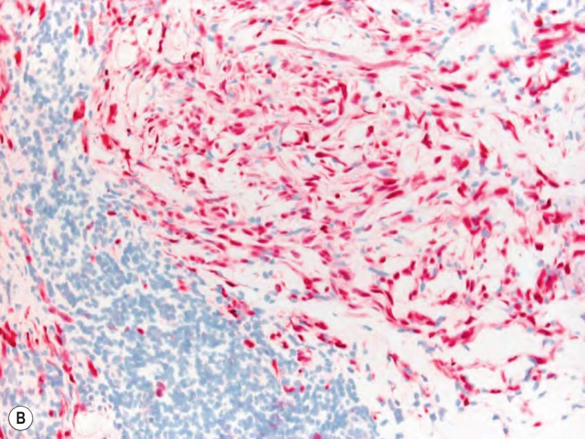
Fig. 113.18 Histopathologic features of a desmoplastic melanoma.A Loosely textured spindle cells (inset) within fibroblastic stroma. In the epidermis and follicular epithelium, there are changes of melanoma in situ with proliferation of atypical melanocytes. Note the lymphoid infiltrates. B S100-positive spindle cells within the dermis. Courtesy Lorenzo Cerroni, MD.
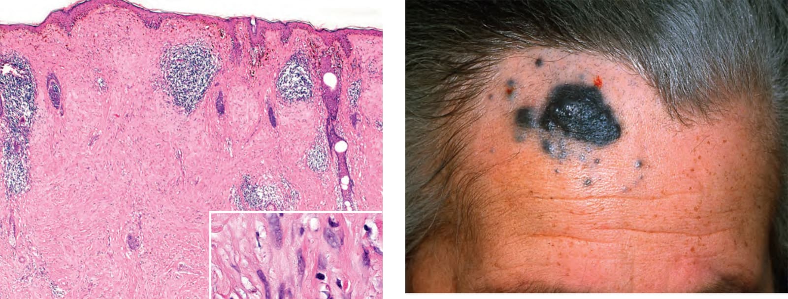
Fig. 113.19 Melanoma arising in a cellular blue nevus (blue nevus-like melanoma). Satellite (lymphatic) metastases are also present. Courtesy Helmut Kerl, MD.