MANAGEMENT
The management of cutaneous melanoma has rapidly changed over the past decade with the advent of molecular tests that aid in diagnosis, in addition to the paradigm-changing use of targeted therapy for BRAF-mutant melanoma and immune checkpoint blockade.
Management of the Primary Melanoma (Stage I and II)
Following histologic diagnosis, the primary cutaneous melanoma site should be re-excised with an appropriate surgical margin determined by the tumor thickness (Table 113.16). The rationale for excisional margins is based on the capacity of melanoma cells to migrate away from the tumor origin, i.e. melanoma may extend wider or deeper than is visibly apparent. The major goal is to prevent local recurrence, including persistent disease-type and satellite (lymphatic) metastasis.
Beginning in 1980, several prospective RCTs led to an evolution of surgical margins, although these studies, with one exception, excluded MIS and invasive melanoma in acral and head/neck sites. In the prior era, invasive melanoma was often excised with a wide margin of 5 cm, independent of tumor thickness. Then a WHO randomized trial demonstrated that 1 cm surgical margins (compared with 3 cm margins) were safe and effective for melanomas <1 mm in thickness and for melanomas up to 2 mm in depth. A subsequent RCT focusing on intermediate-thickness melanoma (1 to 4 mm) found that 2 cm margins
were as effective as 4 cm margins in preventing local metastatic recurrence. Another RCT demonstrated the long-term safety of a 2 cm excision margin for thick melanomas (>2 mm thickness) at a median follow-up of 19.6 years, essentially confirming a maximum of 2 cm surgical margins for T3/T4 melanomas. While current randomized trial evidence is non-existent for MIS (typically excised with 0.5 to 1 cm margins) and insufficient with regard to optimal surgical margins for T1 melanoma, expert international committees have produced fairly consistent guidelines, as summarized in Table 113.16.
Surgical margins in anatomically constrained sites such as the face, ears, digits, and mucous membranes may be narrower to preserve function and/or cosmesis. For MIS of the face, which is typically LM/CSD type, Mohs micrographic surgery (MMS) or staged excision techniques with permanent sections may be employed to provide complete circumferential peripheral and deep histologic margin assessment (CCPDMA), while providing the narrowest possible margins. At the same time, subclinical extension of MIS, LM type, may require margins of 1 cm or greater for complete histologic clearance. Similarly, lentiginous acral and mucosal melanomas are often poorly defined clinically and multifocal in nature. As a result, local recurrences are more frequent in these melanoma subtypes.
Mohs micrographic surgery (MMS)
In the US, the use of MMS for melanoma has nearly tripled over the past two decades. As noted previously, MMS and staged excision using paraffin-embedded permanent section analysis may be considered for the treatment of MIS, LM type, especially in the head and neck region, as well as MIS in acral sites. Utilization of CCPDMA in these surgically challenging sites with significant photodamage has been associated with lower rates of persistent-disease type local recurrence. The use of frozen section immunohistochemistry for MART-1 or other melanocytic markers has further improved intraoperative assessment of tumor clearance, although melanocyte hyperplasia in the setting of chronic actinic damage may complicate histopathologic interpretation of margins. For MIS in non-anatomically constrained sites (e.g. trunk, extremities) and/or with distinct clinical margins (e.g. superficial spreading type), 5 mm surgical margins are typically performed, and MMS/CCPDMA is not generally needed for histologic clearance.
The role of MMS for invasive melanoma remains controversial. Proponents have argued that CCPDMA often results in surgical margins greater than those recommended for conventional excisions, ensuring full tumor resection. Retrospective analyses of the National Cancer Database (NCD) have demonstrated improved overall survival (measured by all-cause mortality) for thin (T1a) invasive melanomas of the head and neck treated with MMS as compared to excision with >1 cm margins, but importantly, MSS is not assessed in the NCD. Other retrospective studies using SEER and NCD data have found similar MSS rates between MMS and standard excision, suggesting at least non-inferiority of MMS in melanoma treatment. Of note, these studies do not provide information about the specific MMS technique employed, the precise surgical margins or other treatment details, local recurrence types (persistent disease vs satellitosis), and MSS rates.
A more recent systematic review and meta-analysis assessed local recurrence following MMS, staged excision, or conventional WLE for nearly 14 000 head and neck melanomas (100 published studies); 51.0% (7138) were treated by WLE, 34.5% (4826) by MMS, and 14.5% (2034) by staged excision. Local recurrence rates were the lowest for MMS (0.61%; 95% CI, 0.1%–1.4%), followed by staged excision (1.8%; 95% CI, 1.0%–2.9%) and WLE (7.8%; 95% CI, 6.4%–9.3%), although definitions of local recurrence and proportions of invasive melanomas varied, with marked heterogeneity among the studies. A subsequent critical review of quality and data reporting in the MMS/staged excision literature noted serious or critical bias in 98% of publications eligible for analysis when modified “risk of bias in non-randomized studies of interventions” (ROBINS-I) criteria were employed. The authors cited the need for longer follow-up periods, clear tumor classifications, and prospective, randomized study designs to improve the quality of future research.
Large, prospective, head-to-head comparisons of MMS and other margin control techniques versus conventional excision are now being proposed. Presently, there is insufficient evidence for national guidelines to recommend the use of MMS to treat invasive melanoma. Current NCCN guidelines do recognize the value of comprehensive histologic margin assessment for MIS, LM/CSD and acral types, along with minimally invasive (T1a) LMM in anatomically constrained sites. It anticipated that greater consensus will emerge from further investigations.
Topical imiquimod
Another area of debate is the treatment of LM with topical imiquimod, a Toll-like receptor (TLR)-7 agonist, rather than with surgical excision. An important distinction is its use in the primary setting (in lieu of surgical resection) versus in the adjuvant setting (after optimal surgery has been performed). Primary therapy is most often utilized in elderly patients with limited function or life expectancy and/or comorbidities that prevent surgical excision, in addition to facial lesions where surgical removal would lead to disfigurement. Adjuvant use of imiquimod may be initiated when the surgical margin is histologically narrow (≤1 mm) or transected, but clinically no residual tumor is evident. Imiquimod may also be used as a neoadjuvant therapy so as to shrink the tumor preoperatively.
Because most published studies consist of case series, there is significant variation in: (1) frequency and duration of applications; (2) pre-treatment and post-treatment disease assessments; (3) use of combination therapies (e.g. cryotherapy, topical 5-fluorouracil); and (4) duration of surveillance. In two studies that described imiquimod as a neoadjuvant therapy prior to staged excision with a 2 mm margin, an 82%–83% histologic clearance rate and 4% recurrence rate were reported; however, an initial excisional biopsy was performed so the application of imiquimod was actually more adjuvant in nature.
In recent analyses, when imiquimod was used as a primary therapy with similar mean follow-up times, clinical clearance rates of 72%–84% were reported. In a single-arm, phase II trial of primary imiquimod for LM diagnosed by partial punch biopsy, histologic clearance rates were somewhat lower at 37% after application 5 days per week for 12 weeks. However, distinguishing residual LM from actinic melanocytic hyperplasia in excisional specimens was a noted challenge in this study, which did not assess long-term response rates. In an open-label, nonrandomized prospective study of 89 patients with MIS, LM type, treated until significant inflammation developed and followed for a median of 4.8 years, 16 (18%) relapsed; a greater number of lesional melanocytes (counted in biopsy specimens) was associated with an increased risk of local recurrence. Two recent systematic reviews suggest the importance of cumulative dose and treatment intensity, with at least 60 applications of imiquimod at >5 times per week associated with the highest likelihood of complete clinical and histologic clearance.
In the adjuvant setting, i.e. following surgery, the clinical clearance rate in one retrospective study was 94% with a mean follow-up of 43 months. Use of imiquimod versus radiation therapy as secondline therapy for LM is currently under investigation in an RCT in Australia, when staged surgical excision with 5 mm margins is not possible, declined by the patient, or fails to clear the tumor histologically (RADICAL Trial, www.clinicaltrials.gov/ct2/show/NCT02394132).
Adjuvant Therapy Considerations for Stage II Melanoma
Based upon results of a phase 3, randomized, double-blind trial, adjuvant pembrolizumab was recently approved for resected stage IIB (T3b/ T4aN0) and IIC (T4bN0) melanoma. The trial involved 976 patients ≥12 years of age who underwent pathologic staging with SLNB to confirm node-negative status. They were treated with adjuvant pembrolizumab (200 mg IV every 3 weeks) for one-year (17 cycles) versus placebo. At a median follow-up of 20.9 months, adjuvant pembrolizumab significantly reduced the risk of recurrence or death (hazard ratio 0.61, 95% CI, 0.45–0.82); the 18-month recurrence-free survival rate was 85.8% for pembrolizumab and 77.0% for placebo. Similar quality-of-life measures were observed in the two study arms, along with manageable immunotoxicities – 16% of patients developed grade 3–4 immunerelated adverse events (irEAs), similar to adjuvant immunotherapy trials in stage III melanoma. However, careful discussion of risk/benefit ratio and referral to medical oncology is recommended to determine the most appropriate stage II melanoma patients for adjuvant therapy.
Local Recurrences
It is critical to understand the biologic differences between local recurrence from inadequate excision of the primary tumor (termed persistent disease-type or “true” local recurrence) from that due to lymphatic metastasis in or around the scar (termed satellitosis). Persistent disease-type local recurrence abuts the surgical excision scar, is histologically defined by in situ and/or radial growth phase, and should be surgically managed according to Breslow thickness of the recurrence (Fig. 113.28A). Local satellite recurrence is an intralymphatic recurrence within or adjacent (≤2 cm) to the excision scar, although AJCC-8 recognizes this distance as arbitrary, since satellite and in-transit (lymphatic) metastases have similar survival outcomes (Fig. 113.29). Importantly, local satellite recurrence reclassifies the melanoma as stage III, prompting staging via imaging, referral for WLE and possible SLNB, and discussion of adjuvant therapy.
Melanomas with a higher frequency of subclinical extension, e.g. acral lentiginous, LM, are at higher risk of persistent disease-type local recurrence (Figs. 113.28A-C & 113.30), whereas melanomas that are thicker, ulcerated, have a higher mitotic rate, and/or lymphovascular invasion are at increased risk of metastatic local recurrence (satellites) despite prior adequate surgical margins. Thick desmoplastic melanomas with “pure” desmoplastic histology, extensive perineural invasion/ neurotropism, and/or head and neck location may exhibit either type of local recurrence (Fig. 113.28D) and thus are sometimes considered for adjuvant radiation therapy following initial resection.
Satellitosis presents as one or more indurated papulonodules within or immediately surrounding the melanoma scar (generally within 2 cm) and is strongly associated with the development of in-transit, regional, and distant metastases. As noted previously, satellite recurrence reclassifies the melanoma as stage III, with consideration of subsequent systemic therapy. There is no evidence that patient survival is adversely affected if local recurrence results from incomplete excision of the primary melanoma (i.e. persistent disease), provided that the recurrent in situ and/or radial growth phase tumor is promptly re-excised and there is no evidence of nodal or distant disease.
Local recurrences of melanoma, particularly the persistent diseasetype, can occur several years (rarely decades) after excision of the primary lesion. When a lesion previously diagnosed histologically as a melanocytic nevus recurs locally more than two years after removal, re-evaluation for possible melanoma should be considered.
Management of Regional Metastatic Melanoma (Stage III)
In ~70% of patients, metastatic spread of melanoma is locoregional, i.e. confined to the site of the primary melanoma, its draining lymph nodes, and the lymphatics between the primary tumor and regional lymph nodes. Locoregional metastasis may manifest as clinically occult lymph node micrometastasis, clinically evident (i.e. palpable) lymph node macrometastasis, or as satellite/in-transit metastasis (discussed above). The vast majority of stage III patients are identified with microscopic nodal disease following SLNB.
Sentinel lymph node biopsy (SLNB) and completion lymph
SLNB was introduced in the 1990s as a method for determining whether the melanoma had microscopically spread to regional nodes and was based upon the presumed migration of melanoma cells from the primary tumor to an initial draining regional lymph node(s). SLNB thereafter replaced elective lymph node dissection (ELND), which was previously considered standard of care for intermediate thickness melanoma until four multicenter, randomized, prospective trials failed to show a survival benefit for patients treated with ELND plus wide excision compared to wide excision alone.
Approximately 15%–20% of patients with clinically node-negative cutaneous melanoma >1 mm in depth will exhibit microscopic lymph node involvement on SLNB. Increased rates of SLNB-positivity occur with greater tumor thickness and other adverse histologic features in the primary melanoma, e.g. ulceration, high mitotic index, lymphovascular invasion. SLNB is optimally performed in the same operative setting as the surgical excision. Preoperative lymphatic mapping is performed to identify the first draining lymph node(s) within the lymph node basin(s) using technetium sulfur colloid and/or blue dye injected into the skin surrounding the melanoma biopsy site. Lymphoscintigraphy demonstrates the lymphatic drainage pattern from a given melanoma site and can be combined with 3D CT imaging. The tumor typically drains to one lymph node basin and 1–2 nodes within that basin, although there can be multiple draining nodal basins in areas of lymphatic overlap such as the mid trunk or head and neck.
The sentinel lymph nodes are intraoperatively defined as: (1) any pathologically enlarged nodes based on palpation; (2) nodes dyed blue (following injection of dye); (3) the node with the highest emission of radiotracer; and (4) any nodes that emit ≥10% of radiotracer compared to the hottest node. Intraoperatively, a small incision is made overlying the
nodal basin and visual inspection, palpation, and a hand-held gamma counter are used to identify and then remove the sentinel nodes in their entirety, followed by histopathology to identify microscopic deposits of tumor via cytomorphologic and immunohistochemical analyses.
Pathologic staging of the sentinel lymph nodes is considered standard for staging and prognostication of primary cutaneous melanomas ≥1 mm in thickness. Numerous publications have identified the status of the sentinel lymph node(s) as the strongest prognostic factor for survival and recurrence. For melanomas ≥1 mm thickness, pathologic nodal staging is recommended in practice guidelines worldwide and by the AJCC and UICC, taking into account patient factors such as age, functional status, and life expectancy (see Table 113.11 & 113.12). For example, if a patient has a medical condition such as dementia or comorbid factors that preclude general anesthesia or raise operative risk, it would be reasonable to forgo SLNB.
The role of SLNB for thinner (T1) melanomas is less clear. It may be considered in AJCC-8 T1b melanoma (<0.8 mm with ulceration or 0.8–1.0 mm with or without ulceration), although rates of SLNB-positivity in this group are still relatively low. T1a melanomas (<0.8 mm without ulceration) rarely exhibit a high mitotic index (>2/mm) or lymphovascular invasion. However, if these histologic factors are evident, SLNB may also be considered, particularly in younger patients who may be more likely to pursue adjuvant therapy and/or surveillance imaging.
Apart from select subgroups of patients with regional nodal disease in the MSLT-1 trial, SLNB has not been demonstrated to improve overall survival and is considered a prognostic, rather than therapeutic, procedure. However, identification of occult stage III disease via SLNB now allows for adjuvant therapies that improve relapse-free survival and may ultimately result in increased MSS. Risks of SLNB include those associated with general anesthesia, surgical site seroma, infection, and rarely, lymphedema in an extremity.
Pursuit of completion lymph node dissection (CLND) following a positive SLNB has declined worldwide following two RCTs (DeCOG and MSLT-2) that failed to show an overall survival benefit in SLNB-positive patients who underwent CLND versus those who were observed but followed with regional nodal ultrasound examination. Where radiologic expertise is available, surveillance nodal ultrasound (or cross-sectional imaging) is generally recommended in lieu of CLND for SLNB-positive patients per current NCCN guidelines, but imaging does not provide pathologic information regarding non-sentinel lymph node status.
Skin satellite and in-transit metastases
As noted previously, satellite and in-transit metastases occur within the lymphatic vessels located between the primary melanoma and the draining lymph node basin. They represent stage III disease and are associated with increased risk for the development of regional nodal and distant metastasis.
In the past, the treatment of choice for satellite/in-transit metastasis was surgical, but intralesional therapy with immune-activating injectables such as talimogene laherparepvec (TVEC) and the TLR9 agonist tilsotolimod and/or systemic therapies can now be used in a neoadjuvant fashion if numerous or extensive lesions are not initially surgically resectable. TVEC is a genetically engineered herpes virus that replicates in cancer cells, causing them to burst and produce GM-CSF, thus stimulating an immune response against the cancer. For locally unresectable disease in a patient who wishes to avoid the side effects of systemic neoadjuvant therapy, TVEC may be an appropriate choice. TVEC was FDA-approved based upon a phase III study that demonstrated a 32% response rate with a complete remission in 17%; other studies have shown a response rate of up to 88%. TVEC has also been investigated in combination with immune checkpoint inhibitors.
For multiple in-transit metastases on an extremity, isolated limb perfusion with melphalan, with or without tumor necrosis factor (TNF), has palliative value. The procedure may be curative, as indicated by reported 5- and 10-year survival rates of 40% and 30%, respectively. Nowadays, however, intralesional and systemic immunotherapies and targeted agents are more commonly employed.
Clinically detectable regional lymph node metastases
If regional lymph node metastases are diagnosed clinically (i.e. palpable) or via imaging, confirmatory core biopsy or fine needle aspiration is recommended. Therapeutic lymph node dissection (TLND) has been considered standard therapy for bulky regional adenopathy and consists of an anatomically complete dissection of the involved nodal basin. The extent of the TLND may be modified according to the anatomic area of lymph node involvement. While TLND has been the standard of care for local basin control of bulky lymphadenopathy, the approach to clinically apparent/macroscopic nodal disease (as well as resectable distant metastases) now includes neoadjuvant immunotherapy and targeted therapy prior to surgical excision.
Adjuvant therapy
The goal of adjuvant therapy is the elimination of clinically inapparent micrometastases, primarily in resected high-risk stage II or III melanoma and occasionally resected stage IV melanoma. Prior to the advent of more effective systemic therapies (see below), there was limited success with chemotherapy and immunostimulants such as bacille Calmette–Guérin (BCG) or Corynebacterium parvum. The latter were tried because of the finding that melanoma cells express tumor antigens recognizable by the immune system. In 1995, the FDA approved the use of high-dose interferon alpha (IFN-α) for adjuvant treatment of stage IIB and III melanoma (per AJCC staging at the time), and in 1998, high-dose interleukin 2 (IL-2) was approved for unresectable melanoma. Flu-like symptoms, including profound malaise, and depression often led to dose reductions of IFN-α while IL-2 was associated with hypotension and capillary leak syndrome. Prior to the 2010s, adjuvant IFN-α (low-and intermediate-dose in Europe and high-dose in the US) was standard treatment for resected stage III melanoma.
Based upon the discovery of genetic mutations in melanomas, including BRAF V600E, there was a paradigm shift in treatment strategies. While the combination of BRAF and MEK inhibitors was initially approved for unresectable stage III and IV melanoma, in 2018, dabrafenib plus trametinib was approved for adjuvant treatment of resected stage III BRAF V600-mutant disease. The second key discovery was the ability of immune checkpoint blockade to selectively stimulate activated T cells (see below). This eventually led to the approval of anti-CTLA-4 (ipilimumab) and anti-PD-1 antibodies (nivolumab, pembrolizumab) as adjuvant therapies for resected stage III melanoma. Based on demonstration of durable relapse-free survival in the seminal prospective randomized trials, adjuvant therapy is considered a standard treatment option for appropriate patients with high-risk resected stage III and stage IV melanoma, although to date overall survival benefit has not been reported.
Management of Distant Metastases (Stage IV)
Over the past decade, effective targeted and immune therapies have revolutionized the treatment of advanced melanoma, leading to marked improvement in relapse-free and overall survival. There is a rapid and high response rate to oral kinase inhibitors (initially targeting BRAF), but development of resistance is an issue. On the other hand, immune checkpoint inhibitors have a slower response rate (apart from dual anti-CTLA-4/PD-1 blockade), but the response is often long-lasting. Prior to the introduction of these therapies, the prognosis of stage IV melanoma was poor, with a 5-year survival rate of <10% and median survival of about 9 months. Within the AJCC substages, differential survival is observed, with soft tissue metastases only (M1a) having a better prognosis than visceral and CNS metastases (M1b, M1c, M1d). For each of these disease sites, i.e. across M categories, an abnormal LDH level is associated with a poorer prognosis, as are a greater number of metastases, multi-organ involvement, and older age at diagnosis (see Table 113.11). In a small proportion of patients with few metastases involving only one or two organs, surgical or other ablative treatment strategies with curative intent may be considered.
Surgery
Despite melanoma’s tendency to disseminate to multiple organs, resection of limited metastases (metastasectomy) may provide palliation and potential long-term survival benefit. Numerous studies have demonstrated clear and durable survival advantages for patients under-going complete resection of metastatic melanoma, particularly isolated pulmonary, nodal or skin metastases, although modern systemic therapeutic approaches are also an option. Factors that positively influence prognosis are isolated non-visceral metastasis and complete resection with free surgical margins. For certain indications, such as isolated lung metastases, median 5-year survival can approach 30% after surgical treatment, with adjuvant systemic therapy further improving relapse-free survival. For brain metastases, surgical resection alone may extend median survival to about 10 months, with a significant improvement in quality of life, but nowadays stereotactic radiosurgery is commonly employed as are systemic therapies (see below). While targeted and immune therapy have become standard of care for resectable and unresectable stage IV melanoma, there are ongoing investigations of combinatorial approaches including neoadjuvant systemic therapy prior to surgery.
Radiation therapy
Although radiation therapy (RT) is rarely indicated for localized cutaneous melanoma, it may be used for primary treatment of unresectable MIS, LM type, and in the adjuvant setting for desmoplastic melanoma at high-risk of recurrence. In the past, adjuvant RT was routinely administered following surgical resection of bulky nodal disease, but then it was found to have no relapse-free or overall survival benefit in a large international RCT. Long-term follow-up did confirm the benefit of RT for improved local control of the lymph node basin, but at the expense of both short- and long-term adverse side effects. For stage III disease, the use of RT has been largely replaced by improved adjuvant systemic therapies.
In stage IV melanoma, RT is used primarily for palliation of symptoms related to distant metastases in patients with incurable disease. Specific indications include pain control as well as spinal cord compression, stabilization of bones affected by metastasis, and brain metastases. The latter are typically treated via stereotactic radiosurgery and fractionated stereotactic radiotherapy; these methods are preferred as both primary and adjuvant treatments due to their superior efficacy and improved side effect profile compared to whole brain radiotherapy. Whole brain radiotherapy is reserved for palliation when stereotactic radiosurgery is not possible or when systemic therapy has failed.
Systemic therapy
Systemic therapy is the mainstay of treatment in patients with distant metastases, with surgery and RT playing a more limited role (see above). Both small-molecule kinase inhibitors that target BRAF and MEK and immune checkpoint inhibitors (ICIs) have revolutionized treatment strategies (Table 113.17). These therapies are far superior and generally less toxic than chemotherapy and systemic IL-2, and they are considered first-line treatments for unresectable stage III and IV melanoma. Clinical trials have or are currently evaluating regimens
in which ICIs and BRAF/MEK inhibitors are administered in combination or sequentially as well as possible advantages of pulse-dosing targeted agents to improve efficacy, combat resistance, and reduce side effects. Of note, severe drug hypersensitivity reactions have been observed when BRAF inhibitors were administered soon after ICIs, with ~20% of patients fulfilling the criteria for DRESS (drug reaction with eosinophilia and systemic symptoms), compared to 5% of patients who received BRAF inhibitors without ICI priming.
Tumor genotyping indicates that ~45% of cutaneous melanomas harbor activating mutations in BRAF and ~15% have activating mutations in NRAS, leading to constitutive activation of the RAS–RAF– MEK–ERK (MAPK) signaling pathway that regulates cell proliferation (see Fig. 113.1). A specific point mutation in BRAF, which leads to the substitution of glutamic acid (E) for valine (V) at codon 600, hence V600E, accounts for 90% of BRAF mutations. Three small molecule inhibitors that selectively target the mutant BRAF kinase have been EMA- and FDA-approved, beginning in 2011. Approval was based upon the results of phase III clinical trials that initially compared dabrafenib and vemurafenib to dacarbazine (DTIC).
The three BRAF inhibitors – vemurafenib, dabrafenib, and encorafenib – exhibit similar rapid therapeutic responses and side effect profiles (see Table 21.16), although photosensitivity to UVA and pyrexia are more common with vemurafenib and dabrafenib, respectively. Unfortunately patients treated with BRAF inhibitor monotherapy develop tumor resistance after an average of six months of treatment. Furthermore, due to paradoxical activation of the MAPK pathway in keratinocytes with wild-type BRAF and oftentimes RAS mutations, cutaneous SCCs and keratoacanthomas develop in up to 25% of patients treated with BRAF inhibitor monotherapy.
Mechanisms of resistance to selective BRAF inhibitors include increased MEK activity, either via mutational activation or overexpression of COT, as well as expression of truncated forms of BRAF (which dimerize and activate the MAPK pathway) and overexpression of platelet-derived growth factor. MEK inhibitors are therefore combined with BRAF inhibitors in an effort to postpone development of resistance (see Table 113.17). Because this combination of kinase inhibitors prolongs the average time to development of treatment resistance to 10 months and improves efficacy, it is considered standard of care for locally advanced or metastatic BRAF-mutated melanoma. Importantly, the cutaneous side effects of the selective BRAF inhibitors, including squamous papillomas, plantar keratoses, and cutaneous SCCs, are significantly reduced with the dual regimen. For patients whose melanomas have activating BRAF (V600) mutations, initial combination targeted therapy may be used when a rapid clinical response is needed in life-threatening situations, but ICIs are normally preferred as first-line therapy, independent of mutational status242a.
In patients with NRAS mutations, BRAF inhibitors are ineffective, but administration of MEK inhibitor monotherapy can result in objective tumor responses in ~25% of patients. A subgroup of melanoma patients, especially those with acral lentiginous or mucosal melanoma, have amplifications and/or mutations in KIT which lead to enhanced activity of the KIT receptor and potential targeted therapy (see above and Fig. 113.1).
Immunotherapy is based on the concept that the immune system is able to fight cancer, and melanoma is one of the prototypic and most highly immunogenic malignancies. Therefore, a therapeutic effect might be achieved by infusing effectors of the immune system such as antibodies, cytokines, or killer cells. Not only has the advent of ICIs transformed the treatment of melanoma, it has positively impacted numerous other cancers. Additional anti-melanoma immunotherapies include injection of oncolytic agents (see above), cancer vaccination, and adoptive cell therapy. In the latter, non-myeloablative lymphodepleting chemotherapy is followed by infusion of expanded autologous tumor-infiltrating lymphocytes.
Immune checkpoint blockade Currently, ICIs are considered first-line therapy in patients with unresectable stage III or IV disease, either with or without an activating BRAF mutation. While the anti-CTLA-4 antibody ipilimumab was the first to be FDA- and EMA-approved for advanced melanoma (see Table 113.17), it has largely been supplanted by anti-PD-1 antibodies (nivolumab, pembrolizumab). As single agents, the latter have superior efficacy and less toxicity when compared to ipilimumab alone. Combined anti-CLTA- 4/-PD-1 blockade is also considered first-line and is associated with a significantly greater response rate but higher incidence of side effects.
These antibodies antagonize inhibitory receptors on T cells, thus leading to stimulation of existing immune responses against melanoma antigens via activation of cytotoxic CD8+ T cells (see Fig. 128.9). This strategy is in contrast to simply enhancing existing immunostimulatory mechanisms as was done for decades in melanoma vaccine trials.
Cytotoxic T lymphocyte-associated antigen-4 (CTLA-4) blockade. Full T cell activation requires stimulation through the T cell receptor as well as a costimulatory signal provided by the binding of B7 on an antigen-presenting cell to CD28 on the T cell. CTLA-4 is a homolog of CD28 and is an inhibitory T cell receptor that is upregulated following T cell activation. The normal function of CTLA-4 is to compete with CD28 to bind B7 in order to downregulate T cell activation, acting as a natural “brake” by removing the costimulatory signal. The CTLA-4–B7 interaction can be blocked with an anti-CTLA-4 monoclonal antibody, which has a higher affinity for CTLA-4 than B7 (see Fig. 128.9). Thus, the inhibitory signal is prevented and the “brake” on T cell activation released.
Two fully human anti-CTLA-4 monoclonal antibodies (mAbs) have been investigated, tremelimumab and ipilimumab, with the latter receiving FDA and EMA approval for the treatment of metastatic melanoma. As a single agent, objective response rates of 11% were achieved and a significant portion of these responses were durable. At three years, the survival rate for ipilimumab plus dacarbazine was 21% versus 12% for dacarbazine alone. Although responses to anti-CTLA-4 mAbs may persist, they may take as long as 12 weeks or even longer to develop.
Side effects from CTLA-4 blockade are autoimmune-related and include colitis, hepatitis, thyroiditis, hypophysitis, myocarditis, and a wide range of skin reactions from dermatitis to leukoderma. Of note, development of vitiligo-like leukoderma is correlated with an improved clinical response. Additional cutaneous and systemic side effects are outlined in Table 21.18. Systemic corticosteroids or steroid-sparing agents are often employed to treat these autoimmunerelated phenomena. Although the combination of ipilimumab plus the anti-PD-1 antibody nivolumab can lead to an enhanced objective response (61%) when compared to ipilimumab alone (11%), it results in a significant increase in grade 3 and grade 4 side effects. Newer regimens have employed alternative dosing of combination ipilimumab and nivolumab to try to reduce toxicity while maintaining efficacy.
Blockade of programmed cell death 1 (PD-1) and its ligand (PD-L1). PD-1 is an inhibitory immune receptor of the CD28 family that plays an important role in the immune escape of tumors. The interaction of PD-1 (CD279) with its ligand PD-L1 (B7-H1; CD274) inhibits T cell proliferation and effector functions and induces apoptosis of tumorspecific T cells. Blockade of either PD-1 or PD-L1 can restore the immune functions of dysfunctional T cells, including enhancement of cytokine production and cytolysis. Since PD-L1 is widely expressed on tumor cells, this blockage focuses on T cell–cancer cell interactions, i.e. the T cell’s effector phase (see Fig. 128.9).
Two anti-PD-1 antibodies, nivolumab and pembrolizumab, have been approved as first-line monotherapy for metastatic melanoma (see Table 113.17), with markedly less toxicity compared to ipilimumab. As with ipilimumab, anti-PD-1 therapy works equally well in BRAF-wild type and BRAF-mutated melanomas. Compared to an overall one-year survival rate of 42% in dacarbazine-treated patients, the one-year survival rate for patients who received nivolumab was 73%, and the objective response rate was 40% (compared to 14% for dacarbazine). As monotherapies, nivolumab and pembrolizumab are more effective than ipilimumab, e.g. nivolumab led to a median progression-free survival (PFS) of 6.9 months versus 2.9 months for ipilimumab while pembrolizumab resulted in an objective response of 33% versus 12% for ipilimumab. A more provocative finding was the enhanced clinical response and reduced incidence of colitis seen in patients receiving anti-PD-1 checkpoint inhibitors who had a diverse microbiome, and the use of oral antibiotics within one month of starting ICIs could impair therapeutic effectiveness. This is presumably because intestinal bacteria play a role in the development of a T cell immune response.
The irAEs seen with anti-PD-1 antibodies overlap with those of anti-CTLA-4 antibodies and include dermatitis, pneumonitis, and nephritis (see Table. 21.18). However, a higher proportion of patients receiving CTLA-4 inhibitors experience grade 3 or higher irAEs compared to those treated with PD-1 inhibitors. As with ipilimumab, patients who had an objective response were more likely to develop vitiligo-like leukoderma. Utilizing antibodies that target PD-L1 rather than PD-1 represents another strategy and these antibodies have been approved for the treatment of other carcinomas that express PD-L1 (see Table 113.17). Of note, in patients with PD-L1-negative tumors, the combination of nivolumab plus ipilimumab prolonged median PFS to a greater extent than did nivolumab alone (11.2 months vs 5.3 months) whereas the two treatment regimens led to a similar PFS in patients with PD-L1-positive tumors. However, further data did not support PD-L1 expression as a biomarker for response to ICIs, and melanoma guidelines do not recommend utilizing PD-L1 expression in clinical decisionmaking. Lastly, given the durable survival benefit with single agent immunotherapy in pivotal anti-CTLA-4 and -PD-1 antibody trials, ICIs are recommended as frontline therapy over targeted agents in patients with metastatic or unresectable disease242a,253,254.
Blockade of other immune checkpoints. There are multiple ligandreceptor interactions between T cells and dendritic cells that represent potential therapeutic targets. Recently, the combination of relatlimab, an inhibitor of LAG3 on T cells, plus nivolumab was approved for the treatment of unresectable or metastatic melanoma. Fewer grade 3–4 irAEs were observed compared to dual checkpoint blockade with nivolumab plus ipilimumab. Progression-free survival with the combination was 10.1 months compared to 4.6 months for nivolumab alone. Additional potential treatments include sotigalimab, which interrupts interactions between CD40L on T cells and CD40 on dendritic cells. Given the rapidly evolving immunotherapeutic options in melanoma, attention to guidelines-based care is critical.
Cancer vaccines Cancer vaccines have long been pursued in the hope of enhancing immune recognition and effector antitumor immune responses via improved antigen presentation and elicitation of effector memory T cell responses that are durable. Increased knowledge of the relevant antigenic epitopes capable of eliciting antitumor immunity has prompted a variety of vaccine approaches. Although vaccines are well tolerated, they rarely have been monitored with methods that are now recognized to be critical for detecting whether or not the vaccine induced an immune response. Not surprisingly, the large phase III trials of older crude autologous tumor cell vaccines did not demonstrate clinical evidence of antitumor activity, either in advanced disease or in the adjuvant setting.
Peptide vaccines have been studied in large, multicenter ECOG trials in the US and led to the induction of immune responses in more than a third of vaccinated patients. However, none showed an improvement in overall survival, with the exception of a combination of gp100 peptide vaccine plus IL-2, which improved progression-free survival compared with IL-2 alone. More recently, mRNA vaccines are being investigated for induction of antitumor immunity in a variety of cancers and hold promise for melanoma.
Over the past four decades, cytotoxic chemotherapy has demonstrated a low but reproducible level of activity against metastatic melanoma. Drugs most commonly employed include dacarbazine (DTIC)/ temozolomide, carboplatin/cisplatin, vindesine/vinblastine, BCNU/ fotemustine, and paclitaxel/docetaxel. Unfortunately, despite the identification of multiple chemotherapeutic agents with activity against melanoma both in vitro and in phase I/II clinical trials, to date no significant impact on survival exists for any chemotherapeutic regimen. In those countries where targeted therapy and ICIs are readily available, chemotherapy is now administered as salvage therapy. However, in lower-income countries, chemotherapy may be a first-line treatment due to the high cost of newer agents.
The most widely used single chemotherapeutic agent remains dacarbazine (DTIC). It has shown the greatest effectiveness as a single agent in most trials – an ~10%–15% response rate can be achieved with a median response duration of 5 to 6 months. The oral alkylating agent temozolomide (a prodrug of MTIC, the active metabolite of DTIC) has some efficacy in treating CNS metastases and is equal in efficacy to DTIC. Fotemustine is a member of the nitrosourea family and has some activity against melanoma, including brain metastases.
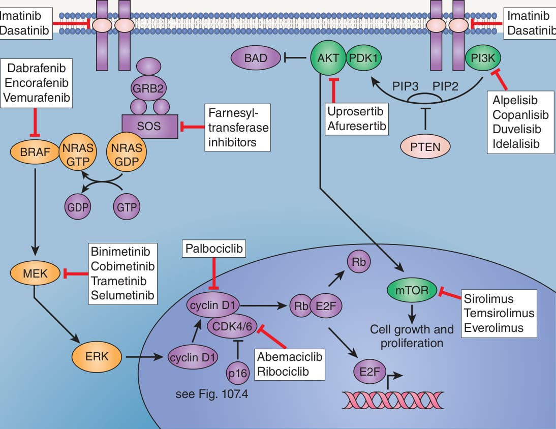
Fig. 113.1 RAS–RAF–MEK–ERK (MAPK) and PI3K–
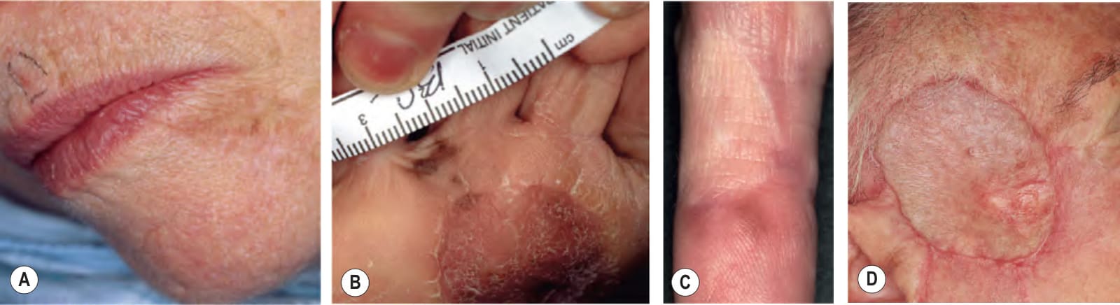
Fig. 113.28 Local recurrences of cutaneous melanoma.A Persistent disease-type local recurrence of lentigo maligna (melanoma in situ) along the upper lateral margin of the excision of the chin; inked lesion on the central upper lip is an actinic keratosis. B Recurrent in situ acral lentiginous melanoma at the 10 o’clock position on the split-thickness skin graft. C Recurrent acral lentiginous melanoma on the second finger. The lentiginous types of melanoma have a propensity to recur locally due to persistence of subclinical melanoma (mainly in situ) as well as the presence of “field melanocytes” that harbor the same genetic alterations as the primary tumor but are morphologically indistinguishable from normal melanocytes, thus leading to “false negative” histologic margins. Staining with PRAME may highlight positive nevoid melanocytes at the site of the tumor, possibly reducing the risk of recurrence, but data are still very limited. D Amelanotic nodule representing local recurrence of a desmoplastic melanoma under the skin graft. A,B,D, Courtesy Jean L. Bolognia, MD; C, Courtesy Department of Dermatology, Medical University of Graz.
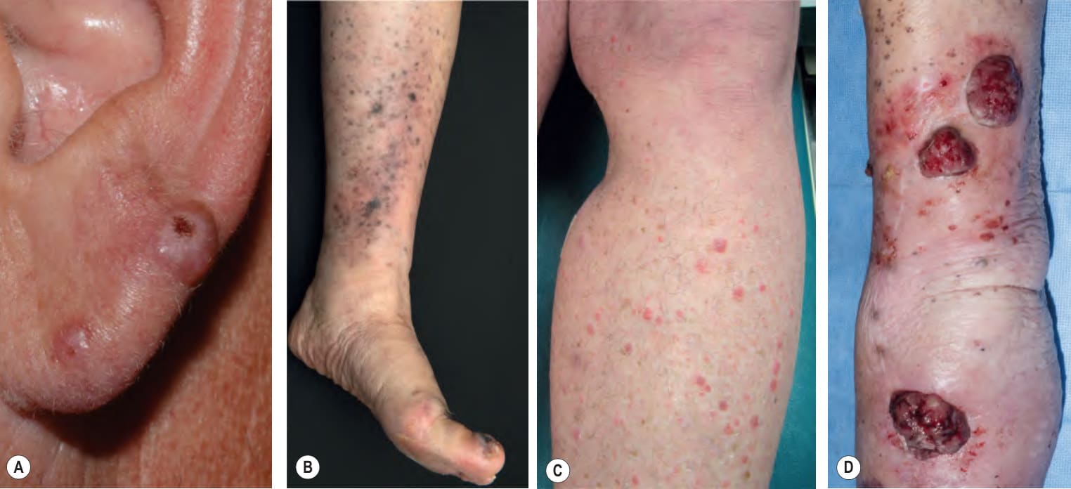
Fig. 113.29 Satellite and in-transit (lymphatic) metastases of cutaneous melanoma.A Amelanotic melanoma of the external ear with satellitosis. B Ulcerated melanoma of the hallux with multiple in-transit metastases on the shin and calf, consisting of numerous brown to gray-black patches, papules, and small plaques. C In-transit metastases consisting of multiple firm pink papules on the leg. D In-transit metastases consisting of multiple friable tumors on the arm as well as smaller blue–gray papules. A, Courtesy Sonya K. Burton, MD; C, Courtesy, Department of Dermatology, Medical University of Graz; D, Courtesy Edward Cowen, MD.
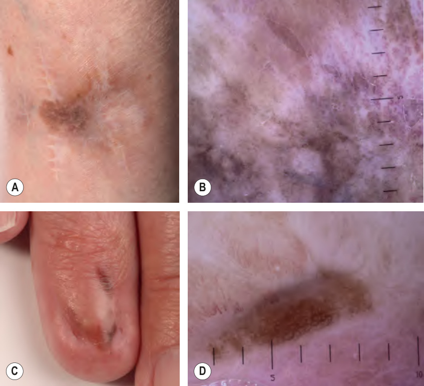
Fig. 113.30 Local recurrences of cutaneous melanoma.A Recurrent lentigo maligna on the lower leg; note the depressed scar due to repeated excisions. B By dermoscopy, an asymmetric pattern with multiple colors, ranging from light brown to dark blue–gray. C Recurrent acral lentiginous melanoma of the nail unit. The lentiginous types of melanoma have a propensity to recur locally due to persistence of subclinical melanoma (mainly in situ and/or in radial growth phase). D By dermoscopy, an asymmetric pattern with an atypical network inferiorly and multiple colors including blue–gray and gray–white. Courtesy Claus Garbe, MD and Jürgen Bauer, MD.
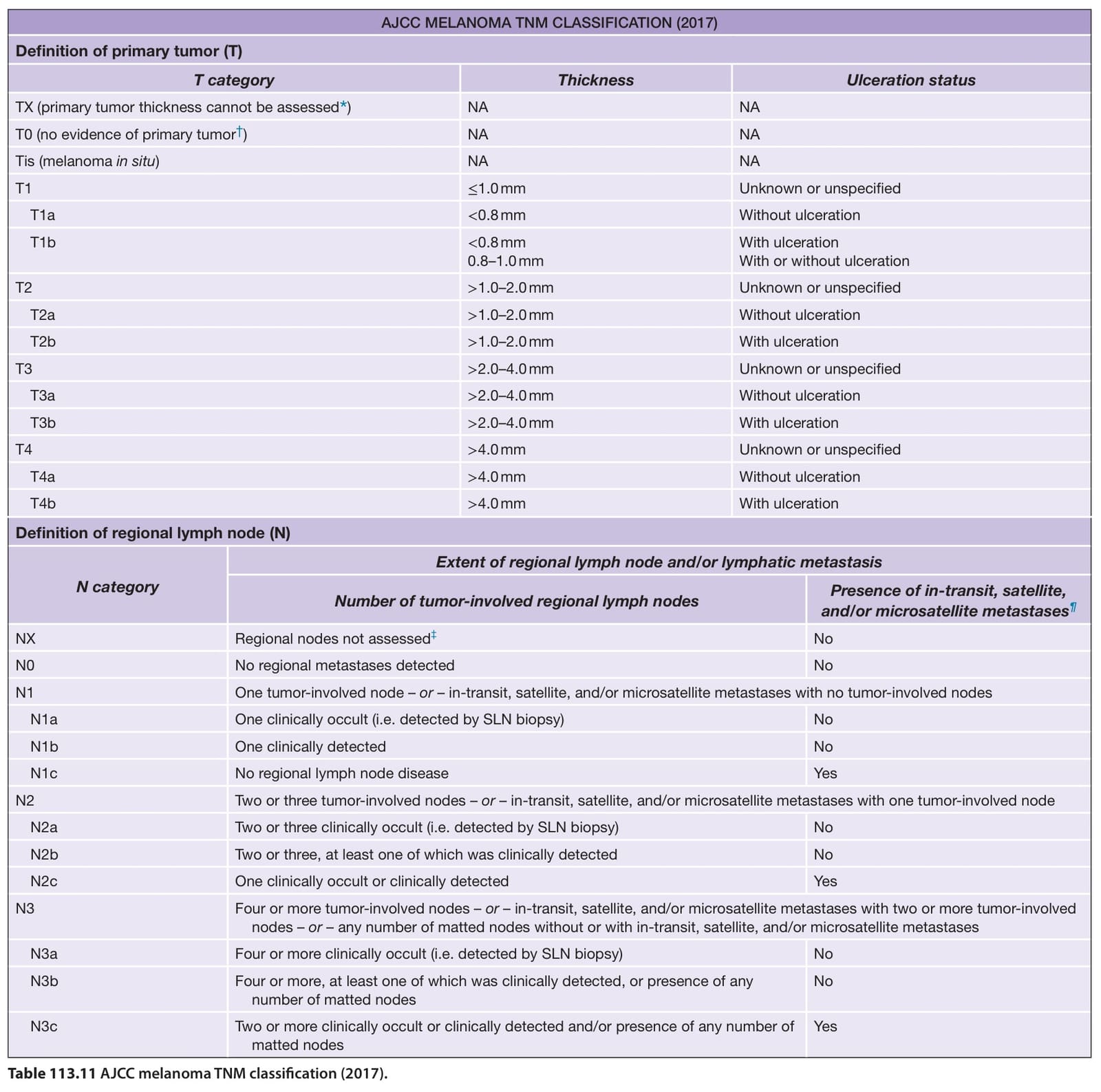
Table 113.11 AJCC melanoma TNM classification (2017).
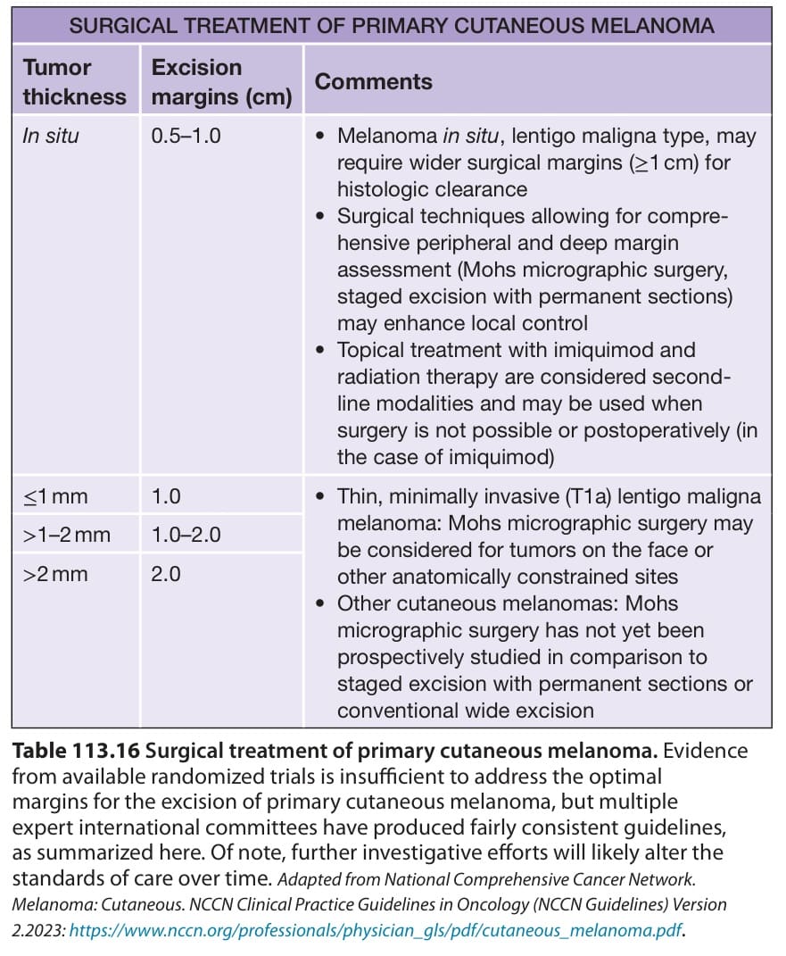
Table 113.16 Surgical treatment of primary cutaneous melanoma. Evidence from available randomized trials is insufficient to address the optimal margins for the excision of primary cutaneous melanoma, but multiple expert international committees have produced fairly consistent guidelines, as summarized here. Of note, further investigative efforts will likely alter the standards of care over time. Adapted from National Comprehensive Cancer Network. Melanoma: Cutaneous. NCCN Clinical Practice Guidelines in Oncology (NCCN Guidelines) Version 2.2023: https://www.nccn.org/professionals/physician_gls/pdf/cutaneous_melanoma.pdf.
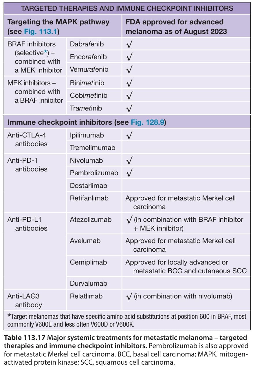
Table 113.17 Major systemic treatments for metastatic melanoma – targeted therapies and immune checkpoint inhibitors. Pembrolizumab is also approved for metastatic Merkel cell carcinoma. BCC, basal cell carcinoma; MAPK, mitogenactivated protein kinase; SCC, squamous cell carcinoma.