INTRODUCTION
In this chapter we use the term neoplasm synonymously with tumor in a general, albeit imperfect, sense, consistent with its common usage: an abnormal proliferation (“new growth”) of cells that appears to be relatively autonomous (i.e. not reactive). Of note, common infantile hemangiomas are discussed separately (see Ch. 103) as are telangiectatic lesions (see Ch. 106) and vascular malformations (see Ch. 104); however, in some patients, vascular malformations may be associated with a number of the cellular vascular proliferations discussed herein. The chapter also includes a review of vascular lesions that were previously designated as vascular tumors (angiomas) but have since been reclassified as distinctive vascular malformations, e.g. glomuvenous malformation (previously “glomangioma”), verrucous venulocapillary malformation (previously verrucous “hemangioma”). In addition, several entities with established or suspected features of reactive processes that mimic neoplasms are discussed as is Kaposi sarcoma, a virus-associated process of as-yet-uncertain position on the hyperplasia–neoplasia spectrum, but with an associated mortality rate. Two benign vascular proliferations of more seemingly straightforward infectious etiology (bacillary angiomatosis and verruga peruana) are covered elsewhere (see Ch. 74). With these caveats in mind, a broad working classification of vascular neoplasms and neoplastic-like conditions is presented in Table 114.1. Selected vascular neoplasms and neoplasticlike proliferations with their associated syndromes and characteristic laboratory findings are outlined in Fig. 114.1.
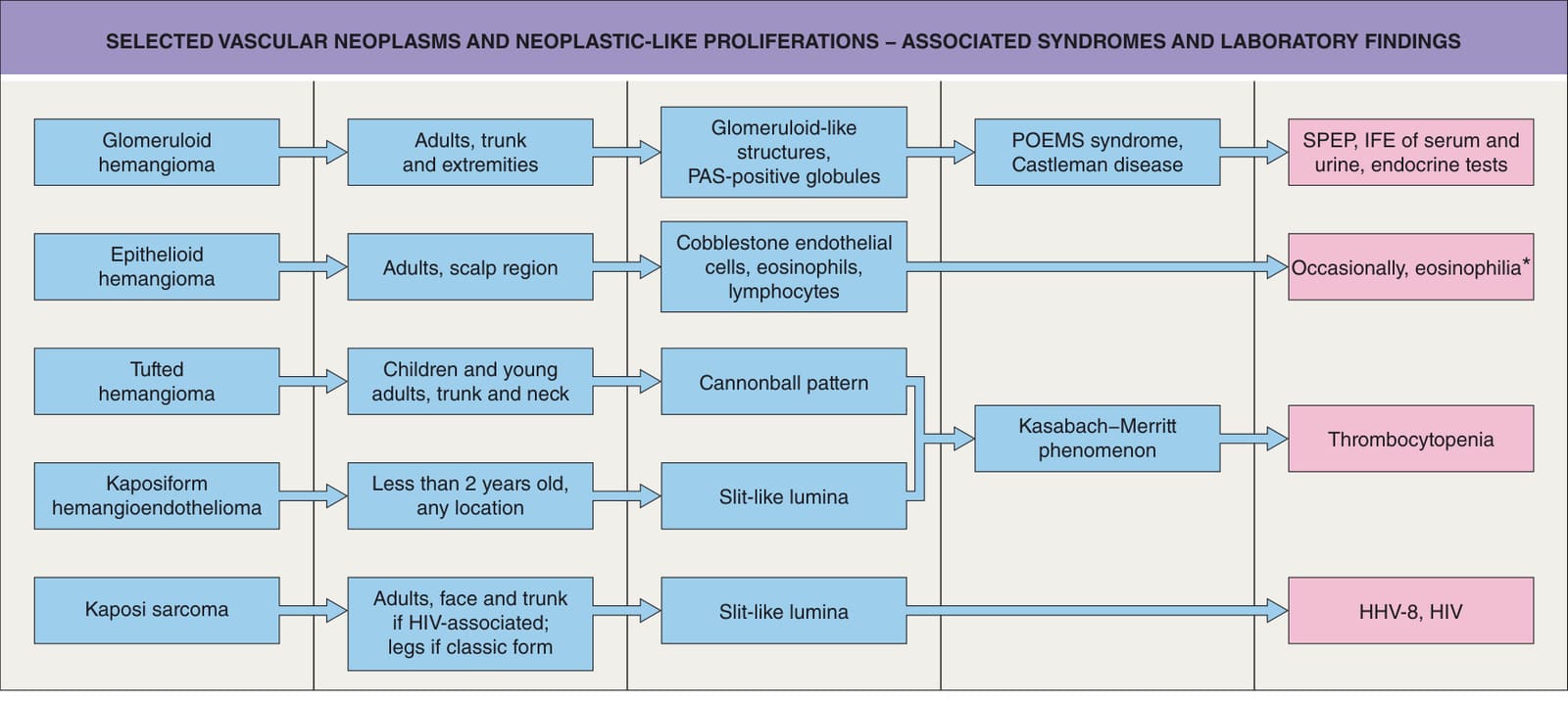
*Fig. 114.1 Selected vascular neoplasms and neoplastic-like proliferations – associated syndromes and laboratory findings. If eosinophilia present, need to consider Kimura disease. IFE, immunofixation electrophoresis; SPEP, serum protein electrophoresis; HHV-8, human herpesvirus 8; HIV, human immunodeficiency virus; PAS, periodic acid Schiff stain.
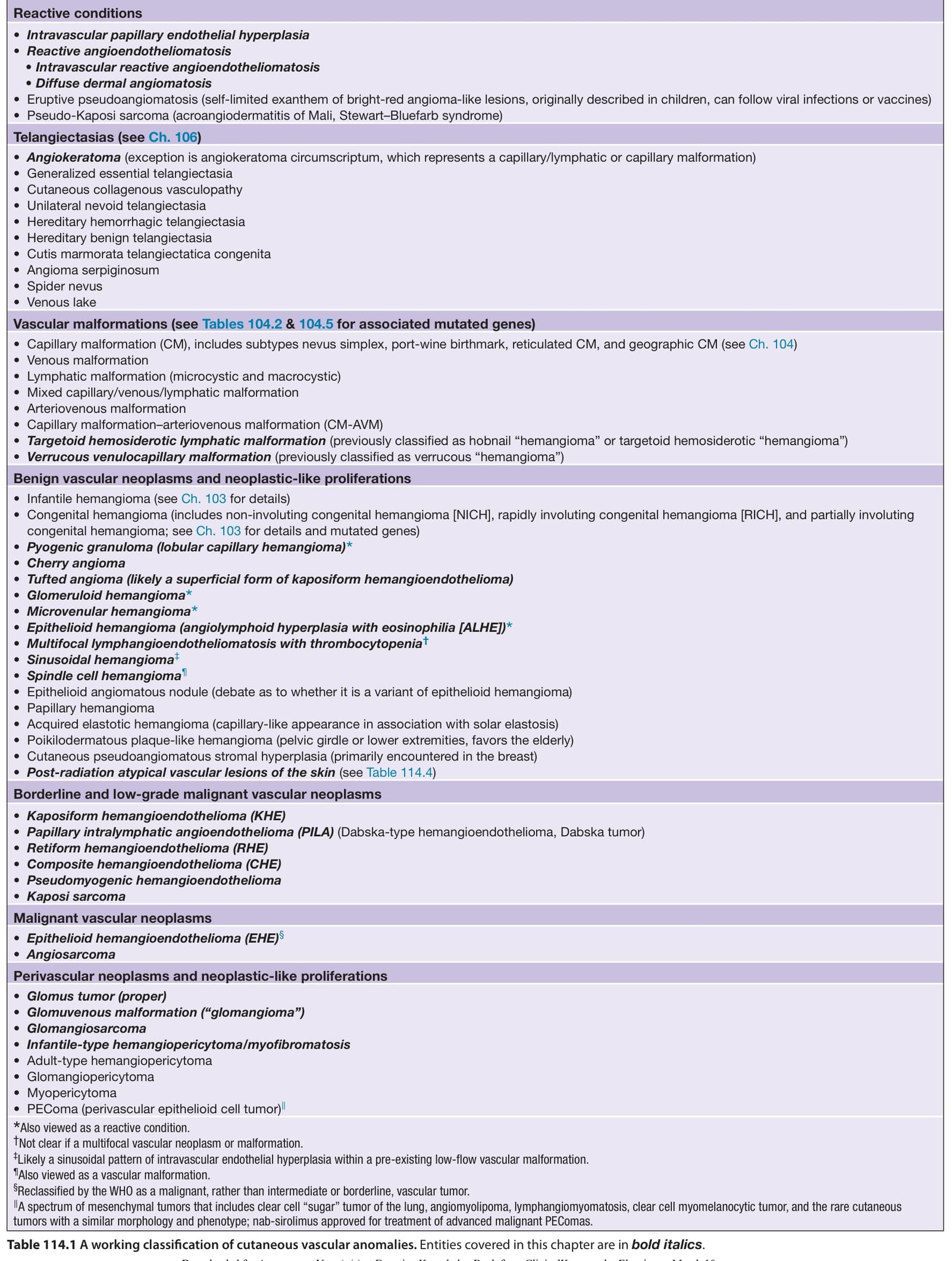
Table 114.1 A working classification of cutaneous vascular anomalies. Entities covered in this chapter are in bold italics.