Angiokeratomas
Key features
Small, dark, vascular, and variably keratotic lesions that result from dilation of superficial vessels
Angiokeratoma circumscriptum is a capillary–lymphatic or capillary malformation
Angiokeratoma corporis diffusum results from several lysosomal storage disorders and is associated with systemic manifestations
Introduction
Angiokeratomas are well-circumscribed vascular lesions consisting of superficial vascular ectasia and hyperkeratosis. Five variants have been recognized. With the exception of angiokeratoma circumscriptum (which represents a capillary–lymphatic or capillary malformation), angiokeratomas result from ectatic dilation of pre-existing vessels in the papillary dermis.
Clinical features
Most commonly, they present as a small, warty, black papule on the lower extremities, but may occur anywhere on the body (Fig. 114.4). The lesions are thought to result from injury to or chronic irritation of the wall of a venule in the papillary dermis. Solitary lesions may be confused with melanoma due to their dark color. Dermoscopy will readily distinguish between these two entities (see Ch. 0).
Synonym: Angiokeratoma of Fordyce
These angiokeratomas may arise in the second or third decade but are most commonly seen in older age groups. In women, angiokeratomas occur most commonly on the labia majora and rarely the clitoris while in men, they develop primarily on the scrotum and rarely the penis. The lesions are red–purple to black in color, may be single or multiple, and arise along superficial vessels (Fig. 114.5). In some patients, they may be associated with thrombophlebitis, varicoceles, and inguinal hernias. Vulvar lesions may be associated with vulvar varicosities, hemorrhoids, or increased venous pressure during pregnancy.
This disorder is characterized by the development of multiple, often clustered angiokeratomas, usually in a bathing trunk distribution. Lesions vary in number (from only a few to numerous) and usually begin to appear during late childhood or adolescence. X-linked recessive Fabry disease is the best-known entity with this clinical presentation and results from a deficiency of the lysosomal enzyme α-galactosidase A (see Ch. 63). This leads to the accumulation of the neutral glycolipid ceramide trihexidose within lysosomes of multiple cell types. Other enzyme deficiencies associated with angiokeratoma corporis diffusum are outlined in Table 63.7.
Lesions usually develop between the ages of 10 and 15 years and are most commonly situated on the dorsal and lateral aspects of the fingers and toes. They may also occur on the dorsa of the hands and feet and rarely on the elbows and knees. Angiokeratoma of Mibelli may be associated with chilblains and acrocyanosis. In rare instances, ulceration of the fingertips can occur. There is a familial predisposition and the disorder may be transmitted in an autosomal dominant fashion with variable penetrance.
This entity usually develops during infancy or childhood as either a plaque of multiple discrete papules (Fig. 114.6) or hyperkeratotic papules and nodules that often become confluent. They occur on the trunk, arms, or legs and are unilateral in most patients. There is a female predominance.
Pathology
Marked dilatation of the papillary dermal vessels is seen in association with an acanthotic, variably hyperkeratotic epidermis. Elongated rete ridges may partially or completely enclose vascular channels, and a collarette may be present at the margin of the lesions. In Fabry disease, vacuoles can be detected within endothelial cells and pericytes. The amount of glycolipid is small and may be difficult to detect in routinely prepared sections. However, the deposits stain positively with PAS and anti-GB3 antibody. They can also be demonstrated by electron microscopy.
Differential diagnosis
Clinically, angiokeratomas should be distinguished from other vascular lesions as well as acral pseudolymphomatous angiokeratomas (see Ch. 121). Darkly colored or thrombosed angiokeratomas may resemble cutaneous melanoma.
Treatment
Patients may request removal for cosmetic reasons. This may be achieved by shave excision, diathermy or laser therapy, the choice of which would largely depend on the size of the lesion.
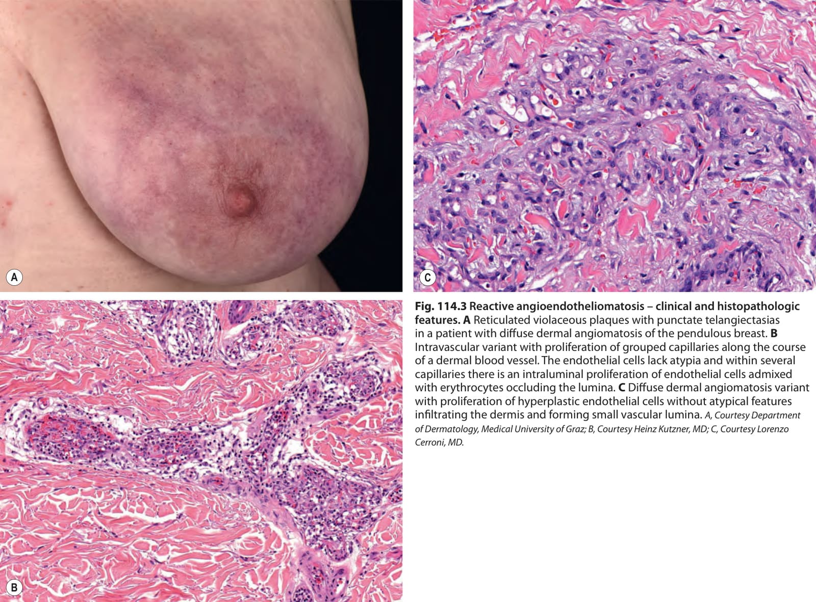
Fig. 114.3 Reactive angioendotheliomatosis – clinical and histopathologic features.A Reticulated violaceous plaques with punctate telangiectasias in a patient with diffuse dermal angiomatosis of the pendulous breast. B Intravascular variant with proliferation of grouped capillaries along the course of a dermal blood vessel. The endothelial cells lack atypia and within several capillaries there is an intraluminal proliferation of endothelial cells admixed with erythrocytes occluding the lumina. C Diffuse dermal angiomatosis variant with proliferation of hyperplastic endothelial cells without atypical features infiltrating the dermis and forming small vascular lumina. A, Courtesy Department of Dermatology, Medical University of Graz; B, Courtesy Heinz Kutzner, MD; C, Courtesy Lorenzo Cerroni, MD.
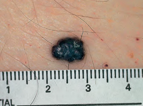
Fig. 114.4 Solitary angiokeratoma. Because of their dark color, these lesions may resemble cutaneous melanoma. Dermoscopy will readily distinguish between these two entities. Courtesy Jean L. Bolognia, MD.
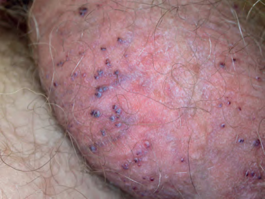
Fig. 114.5 Scrotal angiokeratomas. These lesions typically arise along super-ficial vessels.
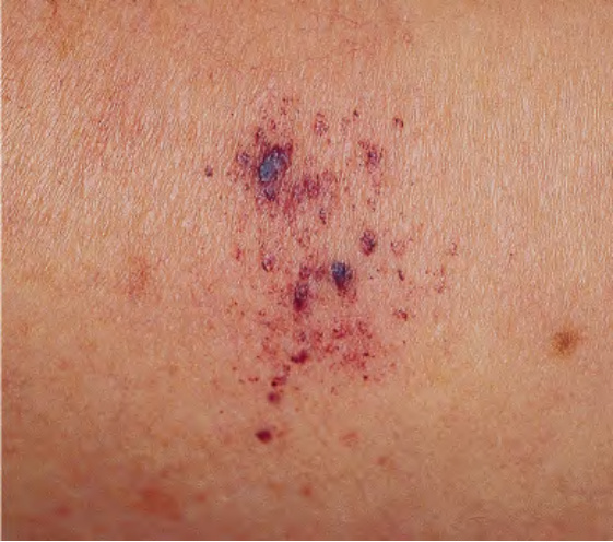
Fig. 114.6 Angiokeratoma circumscriptum. These grouped red–violet papules had been present since childhood. Courtesy Jean L. Bolognia, MD.