INTRODUCTION
Smooth muscle, adipose, and cartilage neoplasms arising in the skin and subcutis comprise a wide spectrum of benign and malignant neoplasms that show myocyte, adipocyte, and chondrocyte differentiation, respectively. Tumors of fat, especially lipomas, occur most commonly. These neoplasms often present as solitary lesions with significant overlap in clinical appearance. In addition, the clinical differential diagnosis can be rather broad (Fig. 117.1). As with other cutaneous tumors, multiple lesions raise the possibility of an inherited disease (e.g. hereditary leiomyomatosis and renal cell cancer).
In this group of neoplasms, the histopathologic diagnosis is established primarily on the basis of characteristic morphologic features, including architecture and cytomorphology, as well as identification of the predominant direction and level of differentiation (Fig. 117.2). Determination of whether a tumor is benign or malignant also requires assessment of features such as circumscription and if atypia and increased mitotic activity are present. Great care is necessary when applying traditional morphologic criteria for malignancy to cutaneous mesenchymal neoplasms because some clinically less aggressive tumors may exhibit prominent atypical features. In other words, aggressive behavior is not always predictable based solely on morphologic findings. For several of these neoplasms, immunohistochemical studies provide helpful diagnostic information (Table 117.1). Cytogenetic analysis and molecular studies are increasingly being used for diagnosis and classification of many soft tissue tumors as there can be relatively histiotypespecific molecular features (see below).
A number of tumor-related and patient-related factors may influence the choice of treatment, including tumor type and size, anatomic location, patient’s life expectancy, presence of comorbidities, and cosmetic concerns. For the majority of solitary, benign, or relatively non-aggressive malignant neoplasms, excisional surgery with clear margins is the treatment of choice. However, removal of a benign tumor is often done at the patient’s request rather than for medical reasons. Other modalities, e.g. cryotherapy, laser ablation (multiple leiomyomas), liposuction (large lipomas), may be preferable in certain patients. For more aggressive malignant tumors, the therapeutic goal is complete eradication, via either wide local excision or Mohs micrographic surgery, while trying to preserve normal function and avoiding mutilating surgery. As expected, the most significant predictor of recurrences is surgical margin status. In some dermal sarcomas, extension into the subcutis represents a negative prognostic factor.
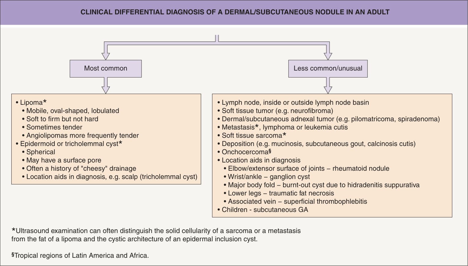
Fig. 117.1 Clinical differential diagnosis of a dermal/subcutaneous nodule in an adult. GA, granuloma annulare.
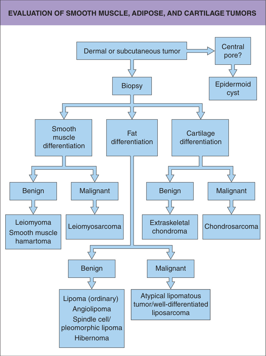
Fig. 117.2 Evaluation of smooth muscle, adipose, and cartilage tumors.
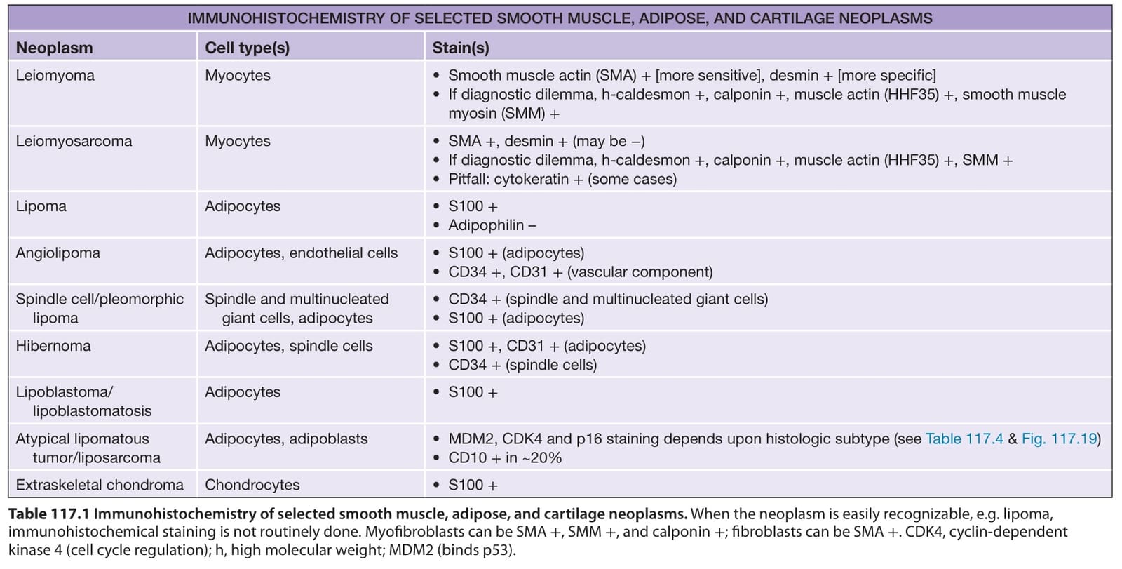
Table 117.1 Immunohistochemistry of selected smooth muscle, adipose, and cartilage neoplasms. When the neoplasm is easily recognizable, e.g. lipoma, immunohistochemical staining is not routinely done. Myofibroblasts can be SMA +, SMM +, and calponin +; fibroblasts can be SMA +. CDK4, cyclin-dependent kinase 4 (cell cycle regulation); h, high molecular weight; MDM2 (binds p53).