Lipoma
Key features
Common benign tumor of mature fat
Asymptomatic, soft to rubbery, subcutaneous nodule arising at any site but rare on the hands and feet
Multiple lipomas are seen in familial multiple lipomatosis,
Madelung disease, adiposis dolorosa, Gardner syndrome, PTEN hamartoma tumor syndrome, and Proteus syndrome
Introduction
(Ordinary) lipomas are benign tumors composed of mature adipocytes. They are among the most common neoplasms in humans, and they represent the most common mesenchymal neoplasm. Lipomas are often solitary, but 5%–10% of patients have multiple lipomas, usually in the setting of a lipomatosis or less often a multisystem syndrome.
Epidemiology
Lipomas develop at any age, but they usually become clinically apparent between the fourth and sixth decades of life. Most studies report the incidence of lipomas to be slightly higher in men than in women.
Pathogenesis
Little is known about the pathogenesis of lipomas. Of note, the incidence of lipomas is increased in overweight individuals, diabetics, and patients with elevated serum cholesterol. In familial multiple lipomatosis, there is clearly a genetic component. In approximately two-thirds of patients, clonal chromosomal aberrations are present (Table 117.4).
Clinical features
Classically, lipomas present as solitary, painless, soft (to rubbery), mobile, subcutaneous nodules, with a normal overlying epidermis. They are often oval in shape and may feel multilobular. Most lipomas measure a few centimeters in diameter, but occasionally reach over 10 cm in size. Although lipomas may develop within any fatty tissue, sites of predilection are the neck, proximal extremities, forearms, and buttocks; lipomas also occur on the trunk, but are very rarely seen on the hands and feet. They present primarily in adults over the age of 30 years.
Lipomas usually exhibit an initial slow growth phase, then remain stable in size with no tendency to involute. The rate of growth may accelerate during periods of weight gain, but weight loss, even when extreme, does not seem to affect their size. Weight loss, however, may result in lipomas becoming more clinically apparent.
Multiple lipomas may be observed in patients with a lipomatosis or in the setting of a multisystem syndrome, such as Proteus syndrome.
Infiltrating or diffuse lipomatosis is characterized by non-encapsulated mature fat infiltrating subcutaneous tissue, muscle, skin, and, at times, even fascia and bone. This entity usually occurs before 30 years of age and is rarely congenital. Diffuse lipomatosis has been described
in association with tuberous sclerosis (see Ch. 61) and paralytic poliomyelitis. The lower extremities are most commonly affected, but involvement of the head, neck, face, and upper extremities have all been reported. Pelvic involvement may lead to urinary tract, intestinal, and even vena caval obstruction.
Familial multiple lipomatosis is also known as familial multiple lipomas, multiple circumscribed lipomas, hereditary multiple lipomas, and discrete lipomatosis. This disorder is characterized by multiple lipomas occurring in several members of one kindred. The tumors are discrete, mobile, and surrounded by a capsule, as opposed to the diffuse and infiltrative tumor growth observed in benign symmetric lipomatosis (Madelung disease). Favored sites include, but are not limited to, forearms and thighs (Fig. 117.9). The neck and shoulders are generally spared.
Proteus syndrome is a complex sporadic disorder due to mosaicism for an activating mutation in the AKT1 oncogene. It is characterized by asymmetric, disproportionate (i.e. progressive and distorting) overgrowth of multiple tissues and multiple hamartomatous malformations (see Ch. 62). The tissue overgrowth tends to plateau after adolescence.
Lipomatosis arising in the setting of Proteus syndrome is associated with overgrowth of multiple tissues in a mosaic pattern, and this can lead to partial gigantism of the head or limbs, vascular malformations, and skeletal deformities as well as areas of lipohypoplasia (see Table 104.5). Connective tissue nevi (particularly cerebriform lesions on the palms and soles) and epidermal nevi are also commonly observed in these patients.
The PIK3CA (phosphatidylinositol-4,5-bisphosphate 3-kinase catalytic subunit alpha)-related overgrowth spectrum includes: (1) hemihyperplasia–multiple lipomatosis which is characterized by multiple lipomas in association with asymmetric (but non-progressive and non-distorting) overgrowth, mild macrodactyly, capillary malformations, and thickened plantar skin with prominent creases; and (2) CLOVES syndrome which is characterized by congenital lipomatous overgrowth (typically not progressive), vascular malformations, epidermal nevi, and skeletal/spinal abnormalities (see Table 104.5). Other individuals with Proteus-like features have a germline mutation in PTEN plus a postzygotic “second hit” in affected tissues and this results in segmental overgrowth, lipomatosis, arteriovenous malformations, and epidermal nevi (SOLAMEN).
Benign symmetric lipomatosis is also known as Madelung disease in the American literature, benign symmetric lipomatosis of Launois– Bensaude in the European literature, and multiple symmetric lipomatosis in OMIM. It consists of extensive symmetric fat deposits in the head, neck, and shoulder girdle area (Fig. 117.10). When the mediastinum is involved, complications related to a space-occupying mass may ensue. Most patients are men, and a high percentage suffer from alcoholism; this may account for several disorders reported in association with benign symmetric lipomatosis, such as peripheral neuropathy, squamous cell carcinomas of the head and neck, and macrocytic anemia.
Large symmetric fatty deposits in the supraclavicular region, lateral chest, and abdomen.
Pathogenic variants in MT-TK, which encodes mitochondrial tRNA lysine, have been described in a subset of patients with benign symmetric lipomatosis. When patients have additional findings such as partial lipoatrophy, myopathy, and hypertriglyceridemia, the possibility of mutations in LIPE (lipase E, hormone sensitive type; see Ch. 101) or MFN2 (mitofusin 2, a mitochondrial protein) can be considered. In one family, heterozygosity for a pathogenic variant in CAPSL was observed, with its protein product, calcyphosine-like, hypothesized to play a role in adipogenesis.
Multiple painful lipomas are present primarily on the arms, trunk, and in the periarticular soft tissue in adiposis dolorosa (Dercum disease). This disease most commonly affects premenopausal women, and it is associated with weakness and psychiatric symptoms such as depression. Pain is intermittent, but it may be debilitating, necessitating sophisticated pain management. Pain is caused by lipomas compressing adjacent nerves.
In addition, lipomas may be part of Gardner syndrome. This syndrome consists of polyposis of the colon (eventuating in colonic carcinoma unless there is surgical intervention), odontomas, multiple epidermoid cysts (often with foci of matrical differentiation), osteomas, leiomyomas, and desmoid fibromatosis (see Ch. 63). Retinal pigment epithelial hamartomas (RPEH) represent an early characteristic finding in Gardner syndrome. Mutations in APC are responsible for this syndrome as well as familial adenomatous polyposis.
Multiple lipomas are also seen in Bannayan–Riley–Ruvalcaba syndrome and Cowden syndrome, autosomal dominantly inherited hamartoma syndromes due to mutations in the tumor suppressor gene PTEN. As outlined in Table 63.3, these two disorders have overlapping features and exist along a disease spectrum. Nowadays they are referred to as the PTEN hamartoma tumor syndrome.
Pathology
Lipomas are composed of a uniform population of mature fat cells with small, uniform, and eccentric nuclei (Fig. 117.11). The fat cells are arranged in lobules with capillaries scattered throughout the lesion. Mitotic figures are absent. Microscopically, lipomas may be indistinguishable from mature adipose tissue. Some lipomas contain lipocytes with intranuclear vacuoles that need to be distinguished from lipoblasts. Fibrolipoma and myxolipoma represent lipomas with an admixture of significant amounts of fibrous tissue and myxoid stroma, respectively. Occasionally, foci of cartilaginous tissue and osseous metaplasia may be present. Secondary changes, due primarily to trauma, include focal fat necrosis and/or aggregates of foamy macrophages.
Differential diagnosis
An epidermoid cyst without a pore is the most common entity in the clinical differential diagnosis, including for subfrontalis lipomas of the
forehead. An ultrasound examination can be done to distinguish between these two entities (see Fig. 117.1). As lipomas are histologically indistinguishable from normal fat, a definitive diagnosis may not be possible if the specimen is too small to demonstrate the overall architecture.
Treatment
Small solitary lipomas are usually cured by simple excision. Local recurrence after excision is more common with larger-sized lipomas. Larger lesions, or systemic lipomatoses, may be amenable to liposuction (see Ch. 156).
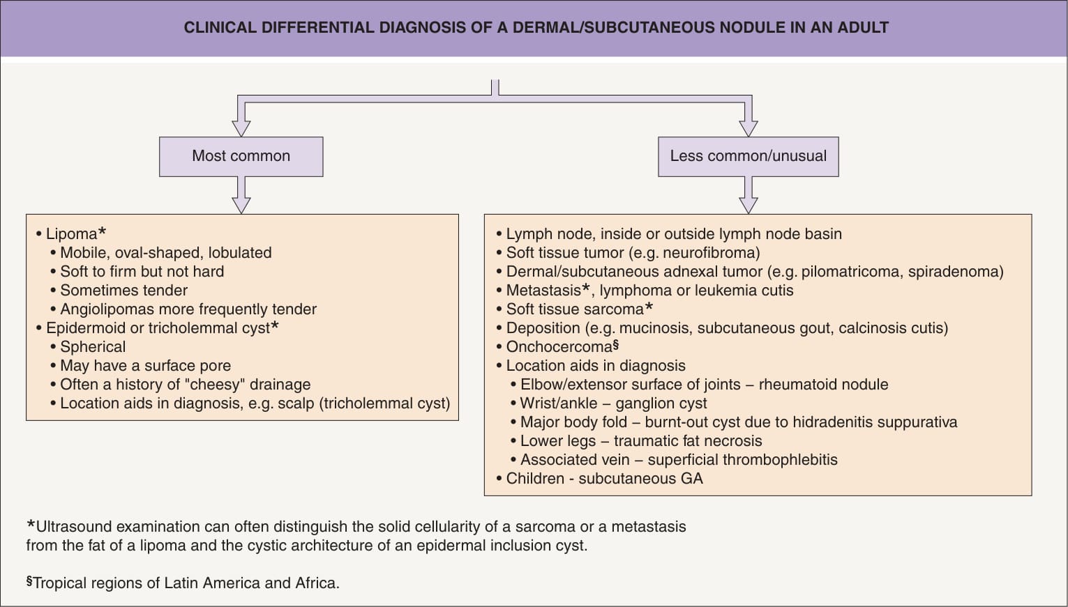
Fig. 117.1 Clinical differential diagnosis of a dermal/subcutaneous nodule in an adult. GA, granuloma annulare.
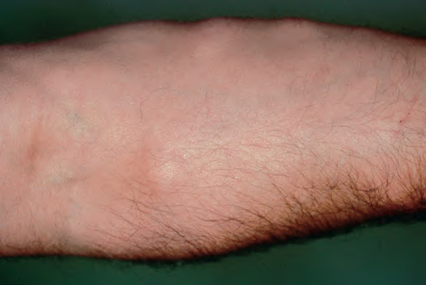
Fig. 117.9 Familial multiple lipomatosis. Several discrete lipomas are present on the forearm.

Fig. 117.10 Benign symmetric lipomatosis (Madelung disease).
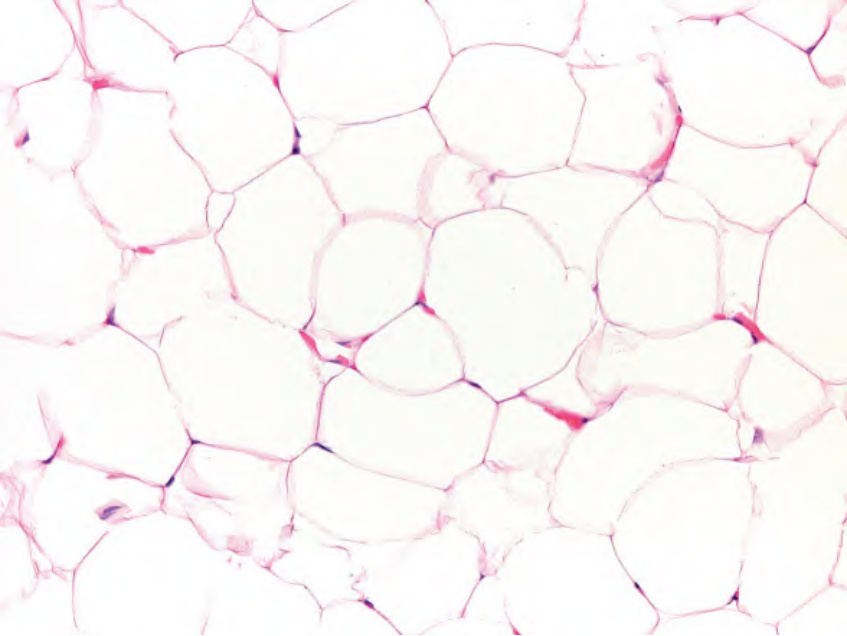
Fig. 117.11 Lipoma (ordinary). A uniform population of mature adipocytes with small and eccentric nuclei is seen.
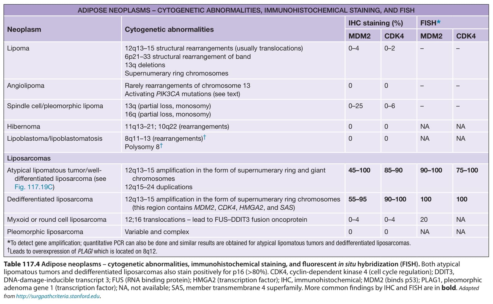
Table 117.4 Adipose neoplasms – cytogenetic abnormalities, immunohistochemical staining, and fluorescent in situ hybridization (FISH). Both atypical lipomatous tumors and dedifferentiated liposarcomas also stain positively for p16 (>80%). CDK4, cyclin-dependent kinase 4 (cell cycle regulation); DDIT3, DNA-damage-inducible transcript 3; FUS (RNA binding protein); HMGA2 (transcription factor); IHC, immunohistochemical; MDM2 (binds p53); PLAG1, pleomorphic adenoma gene 1 (transcription factor); NA, not available; SAS, member transmembrane 4 superfamily. More common findings by IHC and FISH are in bold. Adapted from http://surgpathcriteria.stanford.edu.