Angiolipoma
Key features
Soft subcutaneous nodule, usually less than 2 cm in diameter
Typically located on the forearms of young adults
More often painful than are ordinary lipomas
Well-circumscribed lesion composed of mature fat cells inter- mingled with small, focally thrombosed vessels
Introduction
Angiolipomas are benign subcutaneous tumors composed of mature fat and a vascular component.
Epidemiology
Patients with angiolipomas are typically young adults in their late teens or early twenties; ~5% of cases are familial.
Pathogenesis
The pathogenesis of angiolipomas is not known. A small number of cases are familial, thus suggesting a genetic component. Unlike lipomas, hibernomas, and many other fatty tumors, angiolipomas usually have a normal karyotype. Rarely, rearrangements of chromosome 13 have been observed. In one study of 18 angiolipomas, activating mutations in PIK3CA were identified in the majority (78%) of the tumors, with each having the same mutation [c.1634A>C (p.E545A)] within exon
9. These findings suggest that activation of the PI3K/AKT pathway may play a key role in the formation of angiolipomas.
Clinical features
Angiolipomas usually measure <2 cm in diameter and are clinically similar to ordinary lipomas. However, they may be painful. In approximately two-thirds of patients, the tumors are found on the forearms. Less common sites are the trunk and upper arms. An estimated two-thirds of patients have multiple tumors.
Pathology
Mature adipose tissue is admixed with a variable number of small vessels (Fig. 117.12). Occasional blood vessels are occluded by fibrin thrombi. A cellular variant is characterized by dense cellular angiomatous tissue with a prominent number of spindle cells. Mast cells are more numerous than in ordinary lipomas, and older lesions may contain areas of fibrosis.
Differential diagnosis
Clinically, angiolipomas cannot be reliably distinguished from other benign fatty tumors, although the presence of pain may be a clue. The histopathologic differential diagnosis includes an ordinary lipoma, but no lower threshold exists for the minimal number of vessels required to warrant the diagnosis of angiolipoma. The identification of thrombosed vessels may be helpful in this situation, as they are usually not seen in ordinary lipomas. Highly vascular or cellular angiolipomas may be difficult to distinguish from Kaposi sarcoma, spindle cell lipoma, and spindle cell hemangioendothelioma.
Treatment
Surgical excision is curative and, to date, no malignant transformation has been reported. Angiolipomas tend to be more circumscribed than ordinary lipomas and are therefore more readily excised in toto.
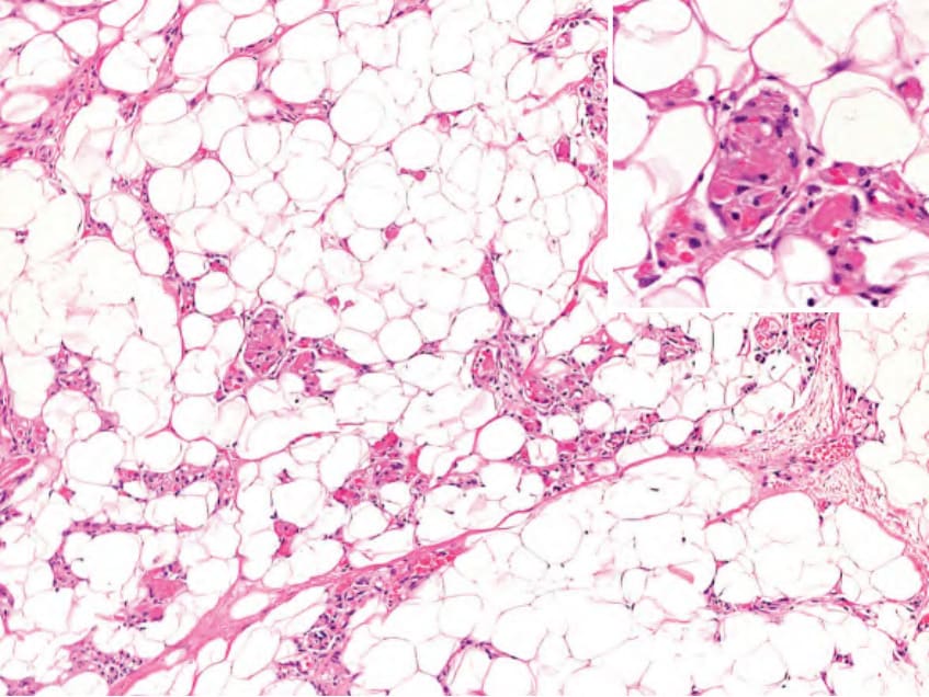
Fig. 117.12 Angiolipoma. Mature fat is admixed with a variable number of small blood vessels. Occasionally, the vessels are occluded by fibrin thrombi (inset).