Hibernoma
Synonyms: Lipoma of immature adipose tissue Lipoma of embryonic fat Fetal lipoma
Key features
Rare benign fatty tumor derived from brown fat
Except for a larger average size, clinically indistinguishable from a lipoma
The most common locations are the interscapular area, thighs, neck, and chest
Introduction
A hibernoma is a rare benign tumor that is derived from brown fat. Brown fat is prominent in hibernating animals, but it is also found in humans. Brown fat first appears in the human fetus and persists into childhood. In children, brown fat is most prominent in the interscapular area, neck, mediastinum, anterior abdominal wall, and around some of the intraperitoneal and retroperitoneal organs. Brown fat gradually disappears with increasing age, and, in adults, it only persists in the neck and around the kidneys, adrenal glands, and the aorta. The main function of brown fat is heat production.
History
This neoplasm was first described in 1906 by Merkel as a pseudo-lipoma of the breast. The term hibernoma was first introduced by Gery in 1914.
Epidemiology
Hibernomas occur primarily in adults. Individuals who develop these tumors are younger than patients with ordinary lipomas and are typically in their thirties. In a series of 170 hibernomas, patient ages ranged from 2 to 75 years, with a mean age of 38 years. The series included nine children.
Pathogenesis
Little is known about the pathogenesis of hibernoma. Characteristic clonal chromosomal abnormalities consist of structural rearrangements of 11q13–21.
Clinical features
Hibernomas are slow-growing tumors located in the subcutis or, occasionally, within skeletal muscle. They are clinically indistinguishable from ordinary lipomas, and the most common locations are the upper trunk (interscapular area, shoulder, chest), neck, extremities, and abdominal cavity/retroperitoneum. Hibernomas can measure up to 25 cm in diameter (average size, 10 cm).
Pathology
On cut sections, hibernomas have a characteristic tan to deep red– brown color. Histologic examination shows pronounced lobulation. Highly vascular interlobular septa surround individual lobules. Common to hibernomas are the characteristic brown fat cells with a small central nucleus and multivacuolated to granular eosinophilic cytoplasm (Fig. 117.15). The cellular membrane is distinct. These typical hibernoma cells are admixed with variable numbers of mature fat cells and pale multivacuolated cells. Rarely, myxoid, lipoma-like, or spindle cell variants may be observed. Adipocytes stain positively for CD31 and this can aid in establishing the diagnosis (see Table 117.1).
Differential diagnosis
Except for a larger average size, hibernomas are clinically indistinguishable from lipomas. However, their histopathologic features are characteristic.
Treatment
Surgical excision is curative. In a clinicopathologic study of 66 cases, none recurred after complete excision.
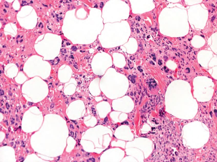
Fig. 117.14 Pleomorphic lipoma. Numerous floret-like multinucleate cells are present in this tumor. Sometimes only a few floret-like multinucleate cells are seen and the majority of cells are mature adipocytes.
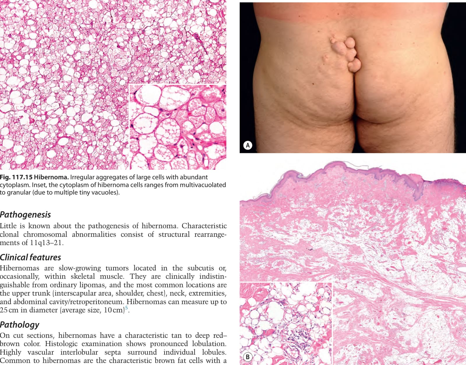
Fig. 117.15 Hibernoma. Irregular aggregates of large cells with abundant cytoplasm. Inset, the cytoplasm of hibernoma cells ranges from multivacuolated to granular (due to multiple tiny vacuoles).
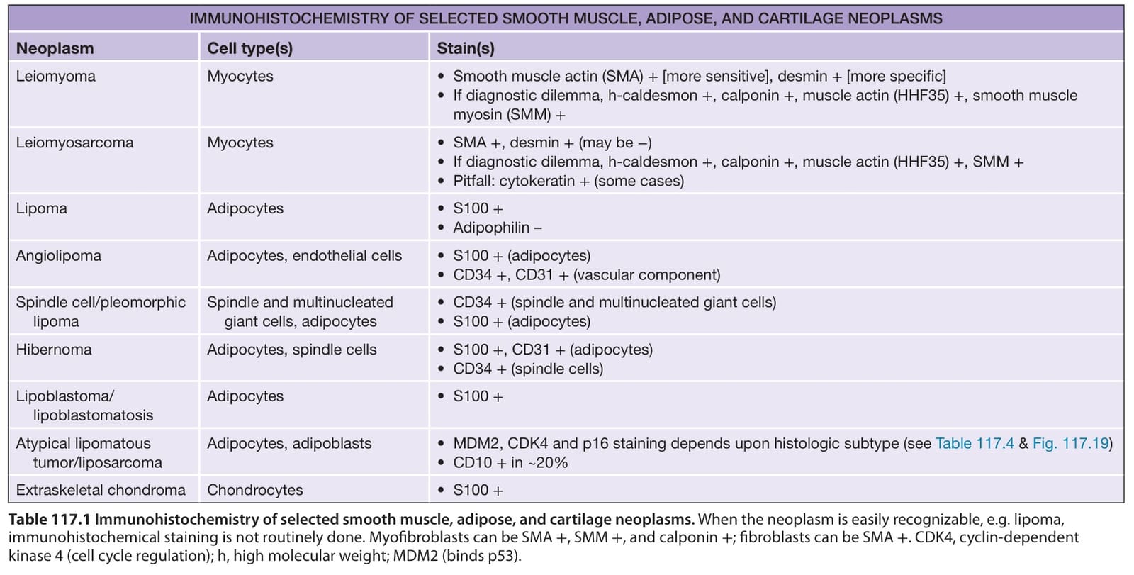
Table 117.1 Immunohistochemistry of selected smooth muscle, adipose, and cartilage neoplasms. When the neoplasm is easily recognizable, e.g. lipoma, immunohistochemical staining is not routinely done. Myofibroblasts can be SMA +, SMM +, and calponin +; fibroblasts can be SMA +. CDK4, cyclin-dependent kinase 4 (cell cycle regulation); h, high molecular weight; MDM2 (binds p53).