CLINICAL FEATURES
Signs and Symptoms
Many children and adults with cutaneous mastocytosis have few, if any, symptoms. When symptoms do occur, they are due to the diverse physiologic effects of secreted mast cell mediators, such as histamine, eicosanoids, and cytokines (Fig. 118.3). These complaints and findings may range from pruritus and flushing to abdominal pain and diarrhea to palpitations, dizziness, and syncope. Of interest is the relative absence of pulmonary symptoms in mastocytosis. Complaints of fever, night sweats, malaise, weight loss, bone pain, epigastric distress, and problems with mentation (cognitive disorganization) often signal the presence of systemic disease. There are even rare reports of deaths in children and adults in association with extensive mast cell mediator release.
Symptoms of mastocytosis can be exacerbated by exercise, heat, friction, or other trauma to skin lesions. In addition, alcohol, narcotics, salicylates and other nonsteroidal anti-inflammatory drugs (NSAIDs), polymyxin B, and anticholinergic medications have been implicated in precipitating symptoms of mastocytosis. Some systemic anesthetic agents may potentially induce anaphylaxis (see below).
Cutaneous Lesions
Childhood-onset mastocytosis, which almost always affects the skin, has three major clinical presentations: (1) solitary or few (≤3) lesions, referred to as “mastocytomas”: 15%–50% of patients; (2) multiple
(<10 to >100) lesions, referred to as “maculopapular” mastocytosis: 45%–75% of patients; and (3) diffuse cutaneous involvement: <5%–10% of patients. A mastocytoma appears as a tan or yellow–tan to brown nodule or plaque, which may be only slightly elevated and typically has a leathery texture (Fig. 118.4). These lesions can be congenital or become evident during infancy, and they favor the distal extremities. Bullae may arise within a mastocytoma, leading to misdiagnoses such as bullous impetigo.
Children with multiple lesions of mastocytosis most often present with large, polymorphic yellow–tan to pink–brown or brown plaques and nodules, which are frequently admixed with smaller lesions (Fig. 118.5D–F). This cutaneous variant is associated with an earlier onset during infancy and is more likely to spontaneously resolve. Less often, only small, monomorphic tan to reddish-brown macules or papules are evident (Fig. 118.5A,B), similar to the typical urticaria pigmentosa presentation in adults (see below). Multiple lesions favor the trunk and classically spare the central face, palms, and soles.
Diffuse cutaneous mastocytosis presents within the first few months of life as thickened skin with a leathery texture and variable hyperpigmentation in a generalized distribution (Fig. 118.6); however, systemic disease is typically absent and spontaneous resolution usually occurs. This term should be reserved for diffuse skin involvement rather than the confluence of individual lesions. Less common presentations of mastocytosis in children include ill-defined tan or telangiectatic patches;
small, firm brown nodules; and yellowish patches with superimposed doughy papules (“xanthelasmoid” mastocytosis).
Blistering following urtication is common in infants and young children with mastocytosis, especially those with large, thick lesions or the diffuse form. The latter patients may present with extensive bullae and erosions (see Fig. 118.6; see Ch. 34). The tendency to blister usually remits by 3 to 5 years of age, and it is believed to result from the release of mast cell serine proteases.
The most common cutaneous lesions of adult-onset mastocytosis are small (<1 cm), monomorphic, reddish-brown macules and papules (Fig. 118.7). This pattern is now referred to as maculopapular but the term urticaria pigmentosa is still commonly employed. In patients with skin type I, the macules and papules may appear pink to red in color. Lesions are most numerous on the trunk and proximal extremities and appear less frequently on the face, distal extremities, or palms and soles. Individual lesions can resolve, but the total number usually increases over time. Close inspection reveals variable hyperpigmentation and fine telangiectasias. These lesions are most often associated with indolent systemic mastocytosis (see below).
Less commonly, yellow–tan to red–brown or violaceous nodules or papulonodules develop, representing a hallmark of indolent disease (see below). Severe infiltrative mastocytosis may progress to diffuse thickening of the skin, especially in flexural sites; facial involvement may acquire a leonine appearance. A rare form of cutaneous mastocytosis
is telangiectasia macularis eruptiva perstans (TMEP), which is characterized by macules and patches composed of telangiectasias without significant hyperpigmentation (Fig. 118.8). Mastocytomas are extremely rare in adults.
Formation of an urticaria-like wheal upon firmly stroking or rubbing of skin lesions (Darier sign) results from release of mast cell mediators and is indicative of the diagnosis (Fig. 118.9). Darier sign is readily demonstrated in pediatric patients, especially with nodular lesions, and if vigorously induced may precipitate systemic symptoms. This sign tends to be more subtle in typical adult mastocytosis lesions and TMEP. These differences reflect lesional mast cell density, which is higher in mastocytomas and childhood maculopapular mastocytosis than in adultonset cutaneous mastocytosis. Dermographism may also be observed in the uninvolved skin of pediatric and adult mastocytosis patients.
Numerous reddishbrown macules and papules.
Systemic Manifestations
Skeletal lesions frequently occur in adult patients with mastocytosis but are rare in children (see Fig. 118.1). Bone involvement can be associated with skeletal pain. In one study of 142 adults with mastocytosis, 40% had skeletal involvement, with a similar frequency in those with childhood-onset and adult-onset disease. Bony lesions may appear as radio-opacities, radiolucencies, or a mixture of the two. The skull, spine, and pelvis are most commonly involved. In an earlier study of 58 adult systemic mastocytosis patients, 57% had diffuse bone involvement, whereas only 2% had focal lesions. Demineralization was the most common change in patients with diffuse skeletal disease, followed by osteosclerosis and mixed lesions of osteosclerosis and osteoporosis. Osteoporosis is more prevalent in men with mastocytosis and may be the presenting manifestation of the disease.
Undecalcified iliac crest biopsies from adult mastocytosis patients have demonstrated increased numbers of mast cells and evidence of enhanced cortical and trabecular bone turnover. These observations led to the hypothesis that mast cells and their mediators are directly responsible for the associated skeletal changes. For example, mast cellderived heparin and SCF have been implicated in promoting osteoporosis via stimulation of osteoclastic activity. Osteoclasts have been shown to express KIT receptors on their surface and to be activated by SCF. Mast cell histamine, however, is also capable of promoting bone sclerosis through activation of fibroblasts, and mast cell-derived IL-6 appears to induce both bone resorptive and fibrotic activities.
The bone marrow is involved in nearly all adult patients with systemic mastocytosis. In general, a bone marrow biopsy appears to be more reliable than an aspirate for identifying abnormal mast cell morphology and distribution. Multifocal mast cell aggregates are typically located in perivascular and paratrabecular locations. Bone marrow involvement is relatively uncommon in children with mastocytosis, and bone marrow biopsy is not routinely recommended.
Splenomegaly is relatively common in patients with advanced systemic mastocytosis but rare in those with indolent systemic disease. Increased numbers of mast cells and eosinophils are frequently observed in the spleen, as are various degrees of fibrosis and hematopoiesis. In general, lymph node enlargement is uncommon in mastocytosis patients but it does occur in those with more advanced systemic disease. Among 58 systemic mastocytosis patients, 26% had peripheral lymphadenopathy, whereas 19% had central nodal disease. Histologically, early involvement of lymph nodes often consists of only clusters of mast cells, while in more advanced disease, mast cell infiltrates involve the paracortex and are often accompanied by eosinophils.
Gastrointestinal (GI) symptoms such as abdominal pain, diarrhea, nausea and vomiting resulting from mast cell mediator release may occur spontaneously or be precipitated by alcohol, aspirin, NSAIDs, or certain foods. Approximately 25% of patients with isolated cutaneous disease and 70% of those with systemic mastocytosis report GI symptoms. Diarrhea in patients with mastocytosis is usually episodic; it can result from malabsorption, increased motility, and/ or acid hypersecretion, with the former due to mast cell infiltration and the latter two symptoms likely related to the release of mast cell histamine and prostaglandins. Patients with systemic mastocytosis are at increased risk of gastritis and peptic ulcers, which occasionally lead to
GI hemorrhage. A number of radiographic changes in the GI tract have been described, including urticaria-like lesions and thickened gastric, duodenal and jejunal folds, as well as mucosal nodules and/or peptic ulcers. Biopsies of mucosal nodules have demonstrated numerous mast cells with varying numbers of eosinophils. Gastrointestinal stromal tumors (GISTs) frequently have underlying somatic activating KIT mutations and are occasionally seen together with cutaneous mastocytosis in families with germline activating KIT mutations (see Fig. 118.2). Hepatomegaly most commonly occurs in advanced systemic mastocytosis and is not always associated with abnormal liver function tests.
A mixed organic brain syndrome with a constellation of symptoms – including irritability, fatigue, headache, poor attention span and motivation, limited short-term memory, inability to work effectively, and difficulty in interacting with other people – has been described in patients with mastocytosis. It has been hypothesized that these symptoms may be secondary to mast cell mediators. Electroencephalographic studies in these patients range from normal to changes consistent with a toxic or metabolic process.
Classification of Mastocytosis
The World Health Organization (WHO) classification scheme for mastocytosis was proposed in 2001 and then modified in 2008 and 2016 (Table 118.1). Cutaneous mastocytosis represents the largest group and includes nearly all affected children and some adults. Childhood-onset mastocytosis resolves by adolescence in ~50%–70% of patients, with persistence into adulthood in the remainder. The WHO has also established criteria for the diagnosis of systemic mastocytosis (SM; Table 118.2). From a dermatologic perspective, appreciation of indolent and smoldering SM is important as these patients frequently have cutaneous lesions.
Although patients with indolent systemic mastocytosis (ISM) have detectable mast cell infiltrates in multiple organs (e.g. bone marrow, liver, spleen, lymph nodes), organ impairment is generally absent and there are few, if any, systemic symptoms. Patients with ISM frequently have slightly increased numbers of spindle-shaped, hypogranulated bone marrow mast cells (BMMCs) that express CD25 ± CD2, which are not expressed in normal BMMCs. CD25 appears to be the most reliable marker for BMMCs with gain-of-function KIT mutations in exon 17 (see Fig. 118.2). Progression of ISM to more severe forms of SM appears to be unusual. In a study of 145 adults with ISM, the cumulative probabilities of disease progression to more advanced SM at 10 and 25 years were 1.7% and 8.4%, respectively. Compared to individuals with ISM, patients with the smoldering SM (SSM) have increased disease burden manifesting as “B” findings – e.g. hepatosplenomegaly, lymphadenopathy, serum tryptase levels >200 ng/ ml (see Table 118.2) – and are more likely to be anemic and an older age. The overall prognosis for SSM patients is less favorable than ISM due to an increased risk for developing more advanced systemic disease.
SM with an associated hematologic neoplasm (SM-AHN) does not usually present with cutaneous lesions, but affected individuals may
have “B” and “C” findings (see Table 118.2). In some patients, the hematologic neoplasm precedes the development of SM. The overall prognosis of SM-AHN patients depends upon the severity of the hematologic disease.
Patients with aggressive SM (ASM) have “C” findings with organ dysfunction secondary to mast cell infiltrates, notably in the liver (e.g. ascites, portal hypertension), bone (pain, pathologic fractures), gastrointestinal tract (malabsorption), and spleen (hypersplenism) (see Table 118.2). They also may develop cytopenias, leukocytoses (eosinophilia, basophilia, monocytosis), and/or thrombocytosis as a reflection of abnormal myelopoiesis. Serum tryptase levels are often elevated, reflecting a significantly increased total body mast cell burden. The overall prognosis appears to depend upon the response to treatment (Fig. 118.10).
Mast cell leukemia (MCL) is rare and is divided into acute and chronic forms. Diagnosis requires ≥10% mast cells among nucleated peripheral blood cells and ≥20% atypical mast cells in the bone marrow. However, because mature mast cells are rarely observed in the blood
of normal individuals, their presence should raise the suspicion of an “aleukemic” form of MCL in which patients have fewer circulating mast cells. Most MCL patients do not have cutaneous lesions, but those with an acute onset frequently exhibit “B” and “C” findings. Multi-organ failure, including of the bone marrow, often occurs over weeks to a few months in the acute form, with an expected survival of a year or less from the time of diagnosis. Chronic MCL, however, has a more indolent course and survival is similar to SSM.
The presence of mutations in KIT and other genes may also have clinical implications in patients with mastocytosis. Advanced forms of mastocytosis are associated with gain-of-function KIT mutations in not only BMMC progenitors but also bone marrow mesenchymal stem cells, leading to involvement of lymphocytic or myeloid cell lineages. In a study of 39 SM patients with a KIT D816V mutation, additional mutations in genes such as TET2, SRSF2, ASXL1, RUNX1, and CBL were found in 25% of those with ISM/SSM and 89% of those with more advanced forms of SM. These additional mutations were associated with decreased overall survival. In contrast, patients with familial disease often lack KIT mutations in codons 815 and 816; their overall prognosis appears to be good but is less well defined.
An approach to the initial evaluation of a patient with cutaneous mastocytosis is presented in Fig. 118.11. A thorough history is crucial, with inquiries into the duration and progression as well as factors that provoke symptoms. For childhood-onset mastocytosis, the diagnosis is usually established clinically by recognition of characteristic skin lesions with a positive Darier sign (see Fig. 118.9). If the diagnosis is in doubt, confirmation can be achieved via a skin biopsy. No further testing is indicated for most children. However, additional evaluation should be performed in children who develop severe mast cell mediator-related symptoms or have evidence of hepatosplenomegaly, with the latter representing a marker of systemic involvement (see Fig. 118.11).
Dermatologists have an opportunity to make the initial diagnosis of mastocytosis in adults who have cutaneous involvement, which can be confirmed by a skin biopsy demonstrating increased numbers of mast cells and/or the presence of an activating KIT mutation. Most of these patients have ISM, with fewer individuals having isolated cutaneous disease. For adults with skin lesions of mastocytosis, a baseline CBC with differential, serum tryptase level, liver function tests (LFTs), and DEXA scan of the lumbar spine and femur should be performed (see Fig. 118.11).
Recent guidelines from the American Initiative in Mast Cell Diseases (AIM) and European Competence Network on Mastocytosis (ECNM) recommend complete staging including a bone marrow biopsy in all adult mastocytosis patients to assist with prognostication. However, other clinicians (including the author) do not believe that a bone marrow biopsy is required in adults with cutaneous lesions who have a normal or slightly elevated serum tryptase level as well as a normal CBC and normal LFTs. Of note, there is agreement that a bone marrow biopsy is indicated in those who have B or C findings (see Table 118.2). Detection of the KIT D816V mutation in the peripheral blood can help to determine the likelihood of SM and follow disease progression over time. A provisional diagnosis of “mastocytosis in the skin” is given to adults with skin lesions of mastocytosis in whom a bone marrow biopsy has not been performed, and a scoring system has been developed to assess the risk of SM in these individuals (see Fig. 118.11).
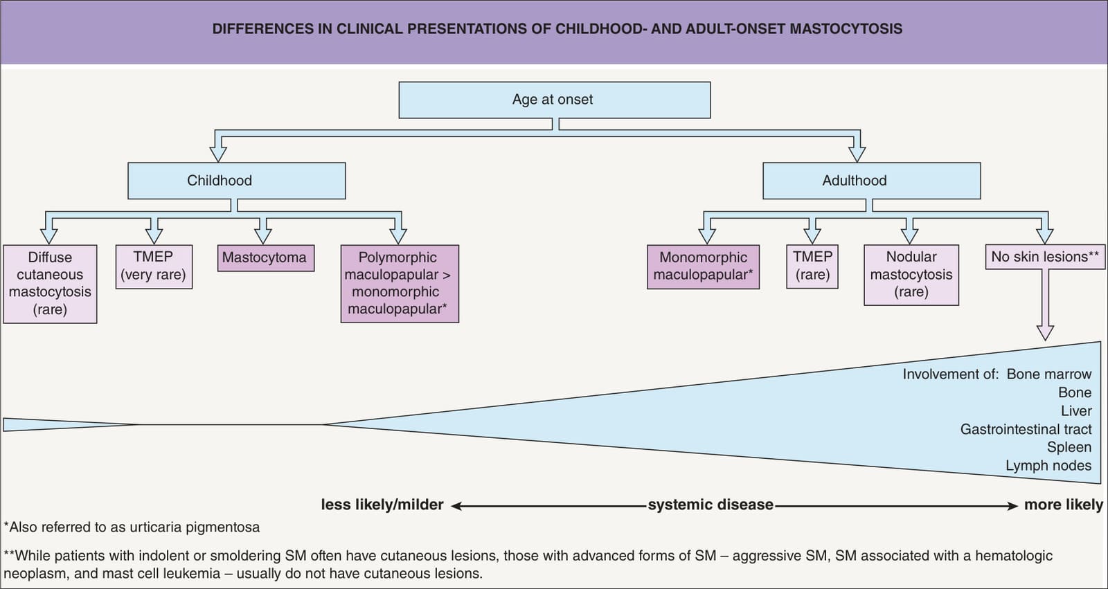
Fig. 118.1 Clinical presentations of childhood and adult-onset mastocytosis. The darker boxes represent the most common forms. Based upon the WHO classification scheme (see Tables 118.1 & 118.2), most children and a small proportion of adults have isolated cutaneous mastocytosis. Most adults with skin lesions have indolent systemic mastocytosis (SM), while those with advanced forms of SM usually do not have cutaneous involvement. In general, children have a benign course with a tendency for spontaneous remission. SM, systemic mastocytosis; TMEP, telangiectasis macularis eruptive perstans.
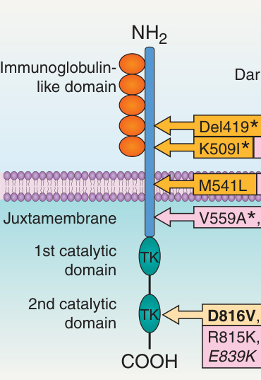
Fig. 118.2 KIT receptor and location of amino acid changes (due to KIT mutations) in pediatric and adult patients with mastocytosis. For example, D816V means that a mutation in codon 816 resulted from valine (V) being replaced by aspartic acid (D). The plasma membrane-bound KIT receptor is composed of three distinct regions: (1) the extracellular region (five immunoglobulin-like repeats [orange]); (2) the transmembrane region; and (3) the intracellular region (two tyrosine kinase domains [green]). The most common site for activating mutations is exon 17, which contains codon 816. The result is constitutive ligand-independent activation of the receptor. TK, tyrosine kinase domain (one binds ATP and the other has phosphotransferase activity).
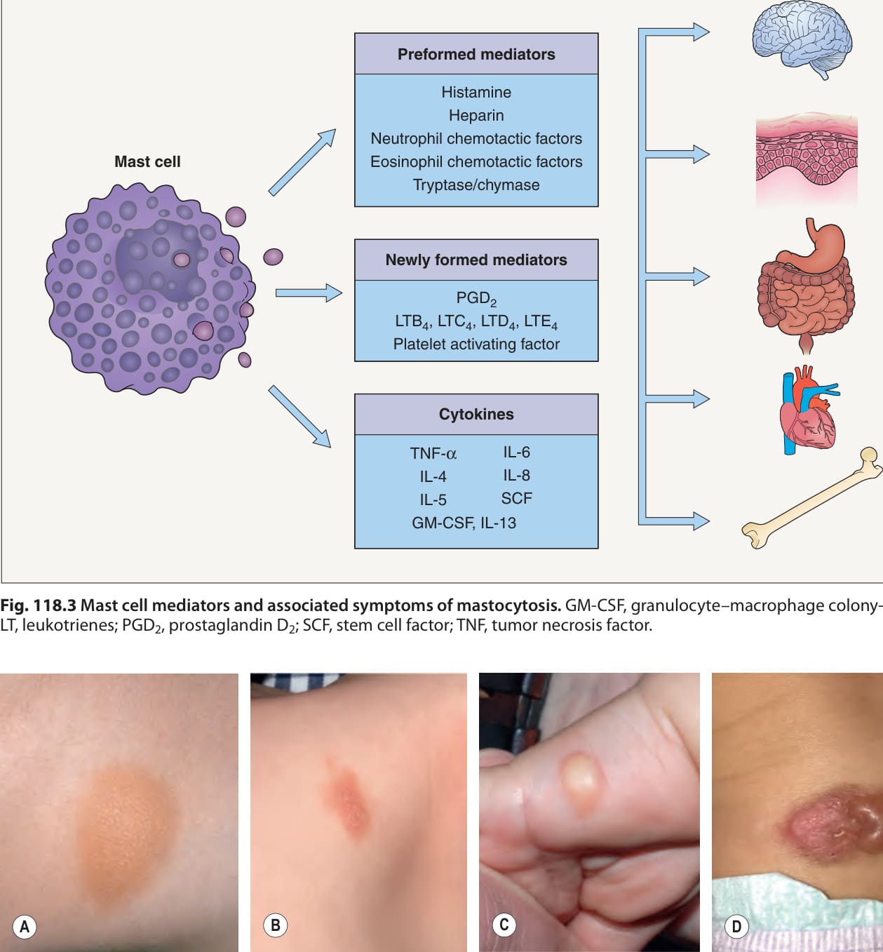
Fig. 118.3 Mast cell mediators and associated symptoms of mastocytosis. GM-CSF, granulocyte–macrophage colony-stimulating factor; IL, interleukins; LT, leukotrienes; PGD, prostaglandin D; SCF, stem cell factor; TNF, tumor necrosis factor.
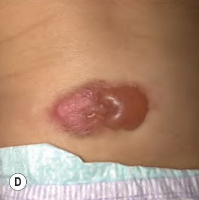
Fig. 118.4 Mastocytomas in young children. Tan (A) and pink–tan (B) plaques with a leathery surface. C Swelling and a subtle rim of erythema resulting from urtication of a yellowish nodule on the wrist. D Bullae formation within a pink–brown plaque on the back. A, Courtesy Antonio Torrelo, MD; B–D Courtesy Julie V. Schaffer, MD.
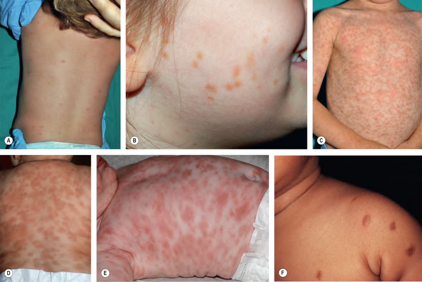
Fig. 118.5 Maculopapular cutaneous mastocytosis in children. Patients can present with variable numbers and sizes of papules, which may be scattered (A), clustered (B), or nearly confluent (C), as well as predominantly plaques (D, E) or papulonodules (F). As in solitary lesions, the degree of associated hyperpigmentation can vary. Note the Darier sign (C, D). A, C, Courtesy Antonio Torrelo, MD; B, D, E Courtesy Julie V. Schaffer, MD.
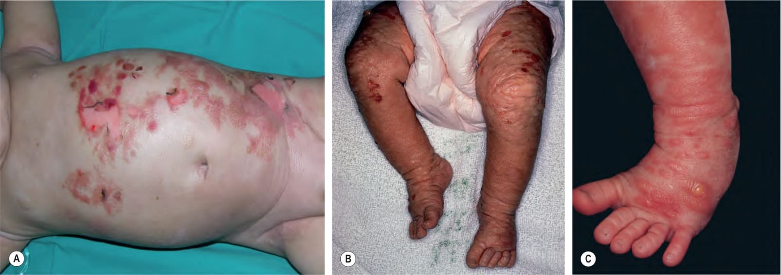
Fig. 118.6 Diffuse cutaneous mastocytosis and blistering in infants. Diffusely infiltrated skin with multiple erosions and a leathery texture that may be subtle (A) or striking (B). This appearance has been compared to pigskin. C Spontaneous blister superimposed on a plaque. A, Courtesy Antonio Torrelo, MD.
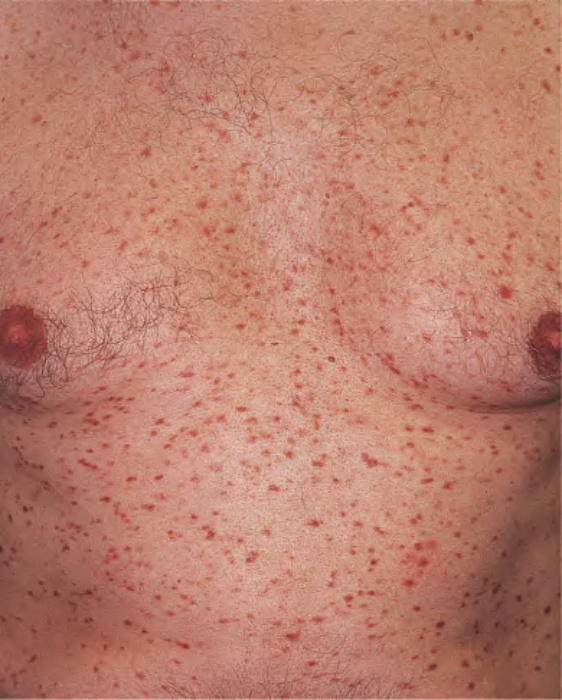
Fig. 118.7 Adult maculopapular mastocytosis (“urticaria pigmentosa”).
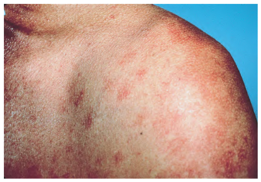
Fig. 118.8 Telangiectasia macularis eruptiva perstans. Multiple lesions composed of telangiectasias are present.
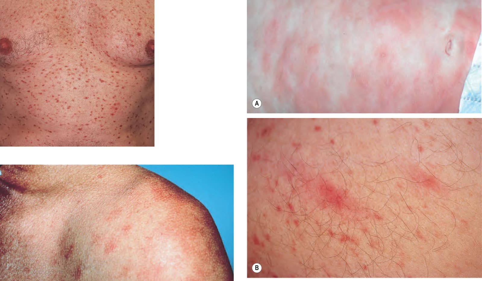
Fig. 118.9 Darier sign in mastocytosis.A An infant with extensive cutaneous involvement. B An adult with macular and papular lesions. A, Courtesy Julie V. Schaffer, MD; B, Courtesy Thomas Horn, MD.
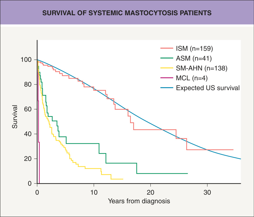
Fig. 118.10 Survival of patients with systemic mastocytosis (SM). The life expectancy of patients with indolent SM (ISM) was not significantly different from the age- and sex-matched US population from the appropriate time period, and leukemic transformation rarely occurred. Based upon these data, the authors recommended that, in general, therapy for ISM should be symptomdirected. ASM, aggressive SM; SM-AHN, SM with associated hematologic neoplasm; MCL, mast cell leukemia. From Lim K-H, Tefferi A, Lasho TL, et al. Systemic mastocytosis in 342 consecutive adults: survival studies and prognostic factors. Blood 2009;113:5727–36.
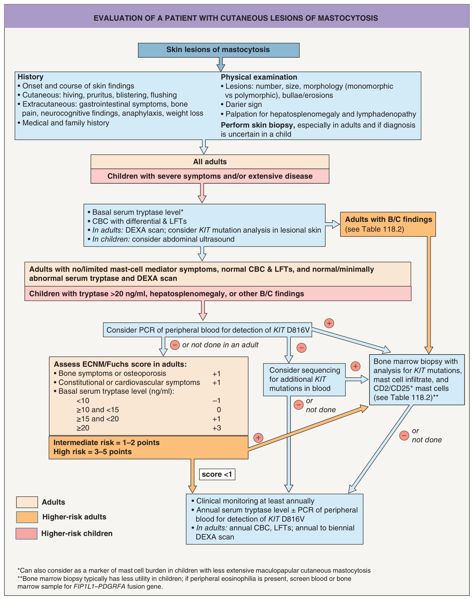
Fig. 118.11 Evaluation of a patient with cutaneous lesions of mastocytosis.
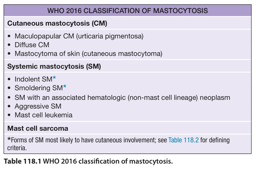
Table 118.1 WHO 2016 classification of mastocytosis.
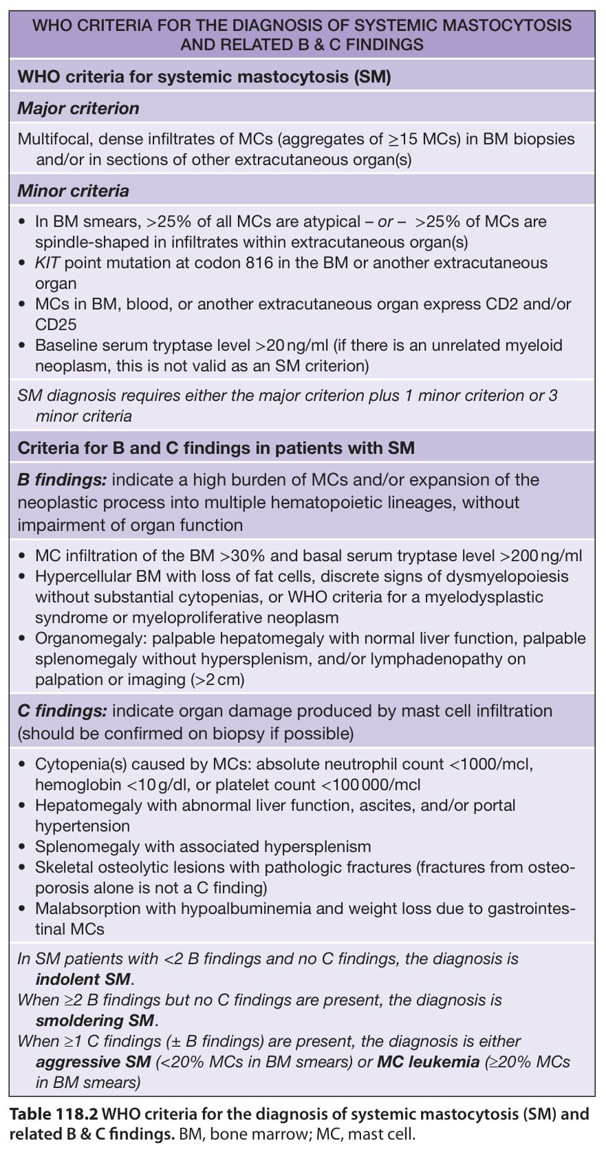
Table 118.2 WHO criteria for the diagnosis of systemic mastocytosis (SM) and related B & C findings. BM, bone marrow; MC, mast cell.