PATHOLOGY
Direct Studies
The diagnosis of mastocytosis is established by the presence of characteristic mast cells in one or more organs. For patients with cutaneous lesions, an increased number of mast cells can be demonstrated in a biopsy of lesional skin (Fig. 118.12). Eosinophils may be found within the dermis; hyperpigmentation of the basal layer of the epidermis with a few melanophages in the upper dermis is a common finding. Special stains, such as toluidine blue, Leder, and Giemsa, or monoclonal antibodies that recognize CD117 (KIT) or tryptase are helpful in identifying tissue mast cells (Fig. 118.13).
Mast cell densities in childhood maculopapular mastocytosis and mastocytomas are 40-fold and 150-fold higher than in normal skin, respectively, and are therefore relatively easy to identify. In comparison, the density is ~30% lower in adults. For nodular, papular, and macular lesions (including TMEP) of mastocytosis, mast cells have been quantified using a morphometric technique and shown to have densities of 63.2% (± 8.2% SEM), 16.1% (± 4.8% SEM), and 3.5% (± 1.8% SEM), respectively, compared to 0.4% (± 0.1% SEM) in normal skin. In childhood mastocytosis, mast cells in lesional skin typically have a round or cuboidal shape, whereas they tend to appear fusiform or dendritic in adults. KIT mutations can often be detected in lesional skin and may help to confirm the diagnosis.
Biopsies of normal-appearing skin from patients with mastocytosis have normal concentrations of mast cells and thus are not useful for diagnosis. A biopsy of the GI tract or bone marrow may be indicated in patients with mast cell mediator-related symptoms who lack skin lesions. Increased mast cell numbers in association with variable numbers of eosinophils can be observed in GI biopsies. The bone marrow of patients with systemic disease frequently shows multifocal, spindle-shaped mast cells that express CD25 ± CD2. The detection of >25% atypical mast cells in extracutaneous tissues and the expression of CD25 and/or CD2 represent minor WHO diagnostic criteria for SM (see Table 118.2). Although coexpression of CD30 in BMMCs was initially reported to predict more advanced mastocytosis, a study of 142 patients demonstrated similar CD30 positivity across all systemic mastocytosis subgroups. Because mast cells may appear hypogranular in tissues of patients with systemic disease, the combination of monoclonal antibodies against CD117 (KIT) and tryptase may be useful for their identification.
Indirect Studies
Detection of circulating mast cell mediators and/or their metabolites can offer indirect evidence of mastocytosis. Two forms (α and β) of mast cell tryptase have been identified. Serum α-tryptase levels are elevated in patients with SM, regardless of whether or not they are experiencing acute symptoms, and therefore may be useful in assessing total body mast cell burden. However, β-tryptase is often detected both in mastocytosis patients and in patients without mastocytosis who are experiencing anaphylactic symptoms. Total (α and β) serum tryptase levels correlate with the extent of mast cell disease. In one study, half of those patients with total serum tryptase levels between 20 and 75 ng/ml had evidence of SM, whereas all patients with levels >75 ng/ml had systemic involvement. Of note, a total serum tryptase level >20 ng/ml represents one of the minor criteria for SM (see Table 118.2).
Urinary excretion of histamine, MeImAA (1,4-methylimidazole acetic acid [histamine’s major metabolite that is more persistently elevated]), and PGDM (major prostaglandin D metabolite) are historical markers of mastocytosis disease that have low sensitivity and specificity. Plasma levels of IL-6 are also elevated in patients with mastocytosis, correlating with severity of bone marrow pathology, organomegaly, and extent of skin involvement.
Diagnosis of mastocytosis in the skin requires one major criterion – clinically typical skin lesions associated with the Darier sign – plus one minor criterion. The minor criteria are: (1) increased number of mast cells in lesional skin; and (2) detection of an activating KIT mutation in lesional skin. Additional investigations can be performed as directed by extracutaneous findings.
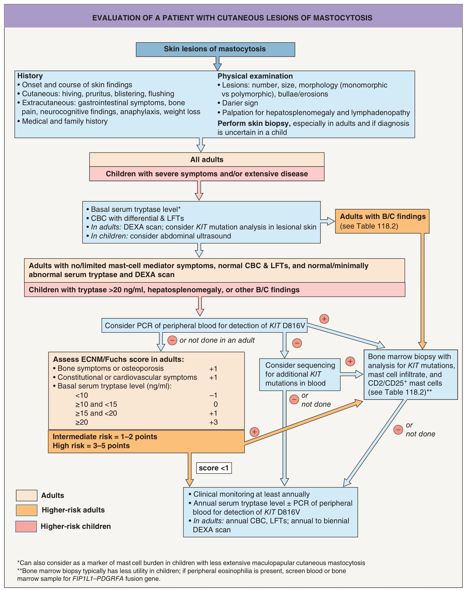
Fig. 118.11 Evaluation of a patient with cutaneous lesions of mastocytosis.
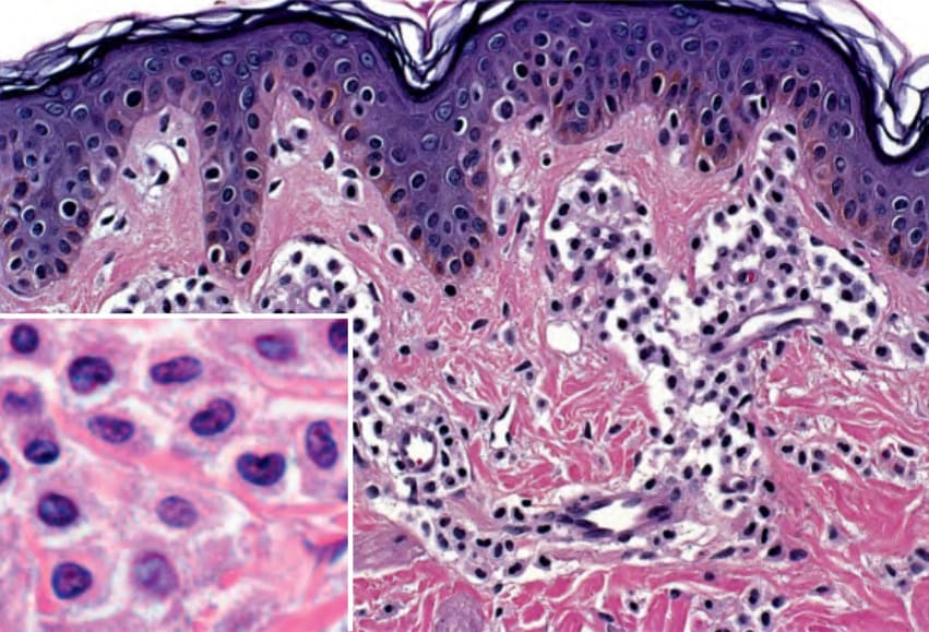
Fig. 118.12 Cutaneous mastocytosis – histologic features. Mast cells within the dermis have a rounded or cuboidal appearance. Granules are seen within the amphophilic cytoplasm (inset). Courtesy Lorenzo Cerroni, MD.
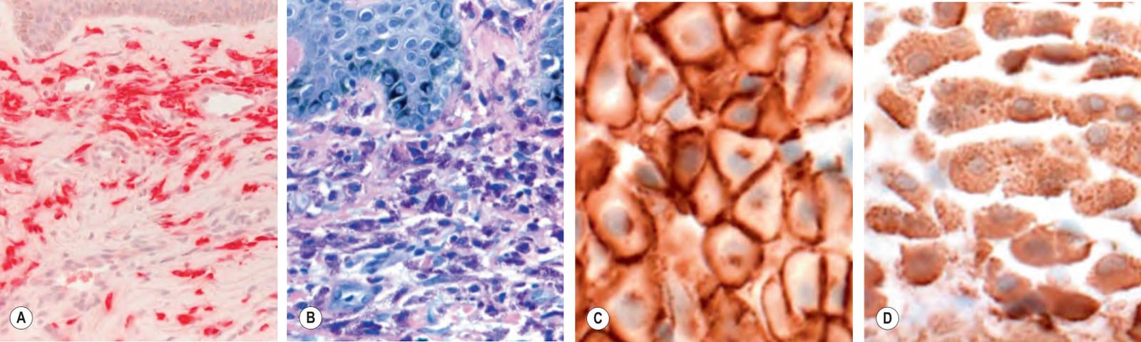
Fig. 118.13 Special stains to detect dermal mast cells.A The Leder method utilizes naphthol AS-D chloroacetate esterase and the mast cell granules appear red. B With the Giemsa stain, the mast cell granules stain metachromatically purple. Immunohistochemical staining with monoclonal antibodies that recognize CD117/KIT receptor (C) or tryptase (D) can also be employed. A, B, Courtesy Lorenzo Cerroni, MD; C, D, Courtesy Antonio Subtil, MD.
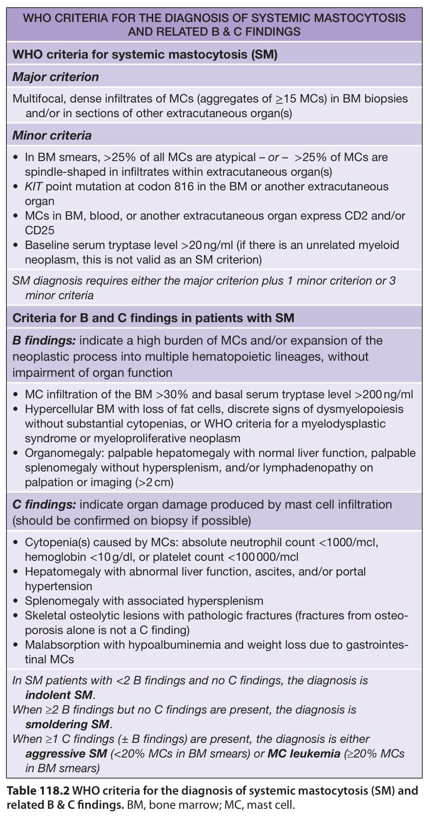
Table 118.2 WHO criteria for the diagnosis of systemic mastocytosis (SM) and related B & C findings. BM, bone marrow; MC, mast cell.