DIFFERENTIAL DIAGNOSIS
The lesions of childhood and adult mastocytosis are so characteristic that they are only occasionally confused with other skin disorders. Mastocytomas may be misdiagnosed as café-au-lait macules, arthropod bites, Spitz or congenital melanocytic nevi, pseudolymphomas, or juvenile xanthogranulomas. Childhood maculopapular lesions that spontaneously urticate may be mistaken for urticaria but are distinguished by the associated hyperpigmentation. The differential diagnosis for blisters due to mastocytosis in children may include bullous arthropod bites, bullous impetigo, herpes simplex viral infection, linear IgA bullous dermatosis and, less often, other autoimmune bullous dermatoses. Infants with diffuse cutaneous mastocytosis can develop widespread blisters, which may be mistaken for epidermolysis bullosa or toxic epidermal necrolysis. Demonstration of increased mast cells in either the blister fluid or a skin biopsy helps to establish the diagnosis of mastocytosis. Nodular scabies in children has occasionally been misdiagnosed as mastocytosis.
The smaller maculopapular lesions typical of adult mastocytosis may at first glance appear as lentigines or melanocytic nevi, but mastocytosis lesions usually have associated erythema. The differential diagnosis of TMEP includes other telangiectatic conditions, but some hyperpigmented maculopapular lesions are often evident. Lastly, if mast cells are not readily detected due to the presence of other leukocytes within the dermis, the patient may be misdiagnosed as having a histiocytic disorder or vasculitis.
Mast cell activation syndrome (MCAS) is characterized by symptoms of mast cell mediator release, but unlike mastocytosis there is no evidence of mast cell proliferation or persistent skin lesions. Serum tryptase levels may be slightly elevated in MCAS, and treatment with antihistamines, leukotriene inhibitors, and/or omalizumab is often of benefit. Of note, the term mast cell activation disorders is sometimes used to encompass the various forms of mastocytosis and MCAS.
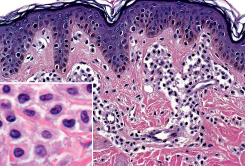
Fig. 118.12 Cutaneous mastocytosis – histologic features. Mast cells within the dermis have a rounded or cuboidal appearance. Granules are seen within the amphophilic cytoplasm (inset). Courtesy Lorenzo Cerroni, MD.