TREATMENT
Treatment of patients with mastocytosis is directed primarily at alleviating symptoms, since there is no cure for this disorder. Many patients with cutaneous mastocytosis and ISM have few, if any, symptoms and therefore require little or no therapy. Mastocytosis patients should be provided with information on environmental triggers (e.g. friction, heat) and potential mast cell degranulating agents (Table 118.3). Systemic anesthetic agents that have been directly or indirectly implicated in precipitating anaphylactoid reactions in mastocytosis patients are also listed in Table 118.3. In contrast, local injections of lidocaine can be used safely in these patients. It has been recommended that mastocytosis patients undergoing general anesthesia be monitored postoperatively for at least 24 hours, since delayed anaphylaxis can occur.
Antihistamines are often helpful in controlling the symptoms associated with mastocytosis. The second-generation antihistamines cetirizine, loratadine, and fexofenadine are preferred because they have longer half-lives and more specific antagonism of the H1 receptor (see Table 18.5). Use of multiple agents and higher than standard doses are often required for symptom control. For example, the author recommends fexofenadine 360 mg in the morning and up to 40 mg of cetirizine at night for control of histamine-related symptoms in adults. In some instances, the addition of an H2 antagonist (e.g. cimetidine, famotidine, nizatidine) may prove beneficial, especially in patients with gastric acid hypersecretion.
Oral cromolyn sodium (disodium cromoglycate; 400–800 mg/day), a mast cell stabilizer with poor oral absorption, may alleviate GI manifestations and (to a lesser degree) cutaneous and CNS symptoms associated with mastocytosis. Use of topical cromolyn for cutaneous mastocytosis has also been described but has limited efficacy.
Narrowband ultraviolet B or psoralen plus UVA (PUVA) phototherapy administered up to four times a week may help to control pruritus and cutaneous whealing in patients with mastocytosis. While this treatment can reduce skin mast cell histamine content, it does not eliminate the mast cell infiltrates.
Potent topical corticosteroids, especially under occlusion for 6 weeks or more, may eliminate pruritus and cutaneous whealing as well as reduce the number of lesional skin mast cells, but this can also lead to skin atrophy. Intralesional injections of triamcinolone acetonide have also been successful in clearing cutaneous mast cell infiltrates. In SM patients, relief of cutaneous and GI symptoms with prednisone alone or in combination with cyclosporine has been described. However, if possible, systemic corticosteroids should be avoided due to the risk of worsening associated osteoporosis. Mastocytosis patients with decreased bone density may benefit from treatment with bisphosphonates or denosumab, a RANK ligand inhibitor.
Some patients with severe mastocytosis experience recurrent, lifethreatening episodes of hypotension following mast cell mediator release, and therefore should be provided with a premeasured epinephrine (adrenaline) preparation (e.g. EpiPen®, Auvi-Q®) for
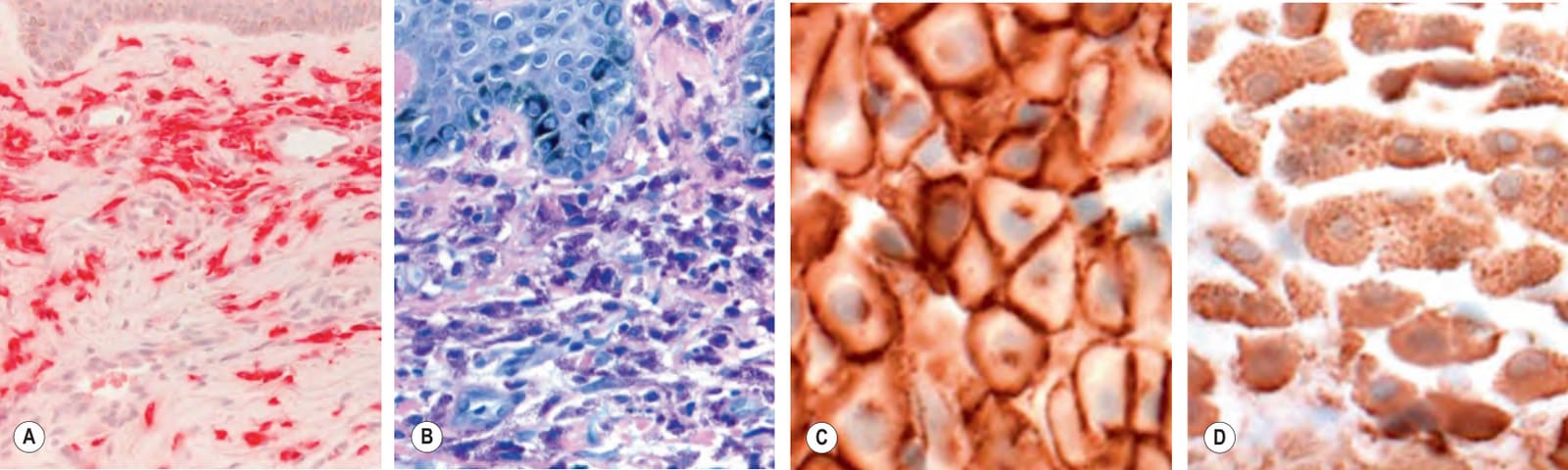
Fig. 118.13 Special stains to detect dermal mast cells.A The Leder method utilizes naphthol AS-D chloroacetate esterase and the mast cell granules appear red. B With the Giemsa stain, the mast cell granules stain metachromatically purple. Immunohistochemical staining with monoclonal antibodies that recognize CD117/KIT receptor (C) or tryptase (D) can also be employed. A, B, Courtesy Lorenzo Cerroni, MD; C, D, Courtesy Antonio Subtil, MD.
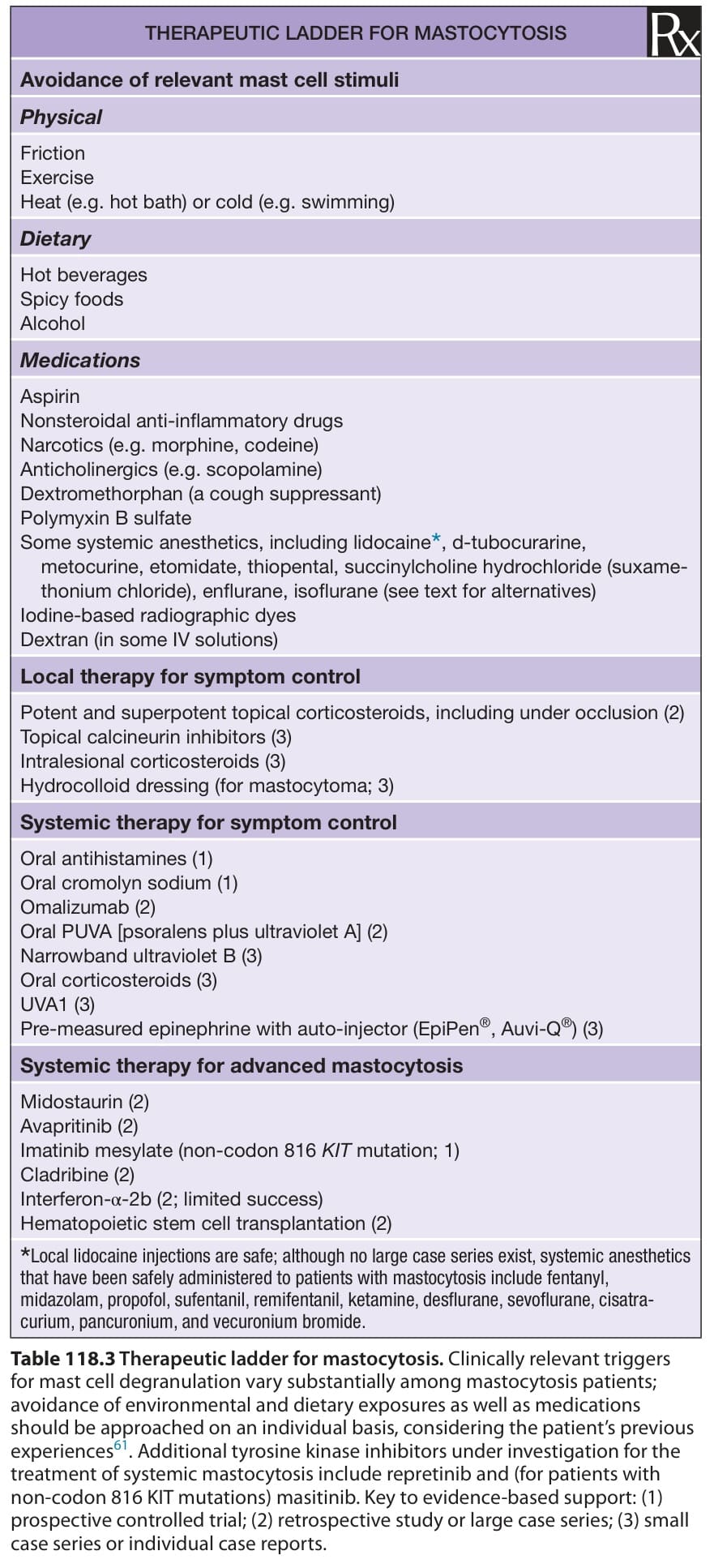
Table 118.3 Therapeutic ladder for mastocytosis. Clinically relevant triggers for mast cell degranulation vary substantially among mastocytosis patients; avoidance of environmental and dietary exposures as well as medications should be approached on an individual basis, considering the patient’s previous experiences. Additional tyrosine kinase inhibitors under investigation for the treatment of systemic mastocytosis include repretinib and (for patients with non-codon 816 KIT mutations) masitinib. Key to evidence-based support: (1) prospective controlled trial; (2) retrospective study or large case series; (3) small case series or individual case reports.
emergency use. In some instances, these patients may experience additional similar attacks within hours of the initial event; prednisone 20–40 mg/day for 2–4 days may suppress these recurrent reactions.
Omalizumab, a humanized murine monoclonal antibody to IgE approved for the treatment of asthma and chronic spontaneous urticaria, has been reported to control symptoms in patients with mastocytosis who are resistant to antihistamine therapy. In a series of 40 adults with cutaneous mastocytosis or ISM, omalizumab (150–300 mg every 2 weeks) induced a >50% reduction in mast cell mediator-related symptoms in 22 patients (55%), with a median time to response of 2 months. In this and most other reports, omalizumab reduced symptoms, but not serum tryptase levels or skin lesions, in mastocytosis patients. However, a 50%–90% reduction in maculopapular lesions was reported in two mastocytosis patients treated with omalizumab (300 mg monthly) for 5–7 months. At present, omalizumab’s mechanism of action in mastocytosis remains unclear.
Multitargeted tyrosine kinase inhibitors are capable of limiting proliferation and promoting apoptosis of D816V-expressing mast cells. Among such agents, midostaurin and avapritinib are FDA-approved for the treatment of aggressive SM (including ASM, SM-AHN, and MCL), with overall response rates of ≥60% and ≥75%, respectively, and a median survival of ~30–40 months for midostaurin in this patient group. Imatinib mesylate is a tyrosine kinase inhibitor that blocks KIT and PDGF receptors, as well as the BCR-ABL oncoprotein associated with chronic myelogenous leukemia. However, the active site for imatinib within the KIT receptor is proximal to the location affected by the common codon 816 mutations (see Fig. 118.2). The resulting amino acid substitutions impede the binding of imatinib to KIT and thereby negate its potential therapeutic effect. On the other hand, imatinib may alleviate the signs and symptoms of mastocytosis in patients with other KIT mutations (e.g. del419, K509I, F522C, V560G; see Fig. 118.2) as well as in those with wild type KIT or a myeloproliferative disorder associated with the FIP1L1–PDGFRA fusion gene. Other genes mutated in SM and their respective pathways may serve as potential therapeutic targets in the future.
Among chemotherapeutic agents, intravenous cladribine has the most evidence of efficacy in reducing skin lesions and BMMCs in patients with advanced SM, including those with the D816 KIT mutation. However, a recent registry-based analysis found significantly longer overall and disease-free survival of patients with advanced SM treated with midostaurin compared to those who received cladribine. For patients with SM-AHN, chemotherapy should be directed at the associated hematologic disorder.
Local radiation therapy with ~20–40 Gy over a 7- to 14-day period may benefit patients with pain due to bone involvement. Splenectomy can be helpful in mastocytosis patients with hypersplenism who experience significant cytopenias and may improve survival in patients with aggressive disease. Interferon α-2b has been used with limited success in patients with aggressive forms of mastocytosis, and non-myeloablative allogeneic hematopoietic stem cell transplantation has been performed with variable benefit in patients with life-threatening SM.
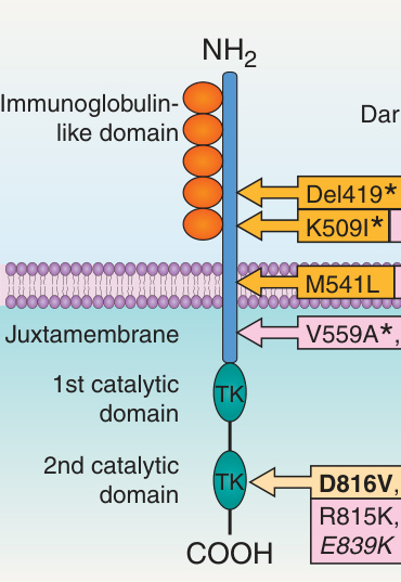
Fig. 118.2 KIT receptor and location of amino acid changes (due to KIT mutations) in pediatric and adult patients with mastocytosis. For example, D816V means that a mutation in codon 816 resulted from valine (V) being replaced by aspartic acid (D). The plasma membrane-bound KIT receptor is composed of three distinct regions: (1) the extracellular region (five immunoglobulin-like repeats [orange]); (2) the transmembrane region; and (3) the intracellular region (two tyrosine kinase domains [green]). The most common site for activating mutations is exon 17, which contains codon 816. The result is constitutive ligand-independent activation of the receptor. TK, tyrosine kinase domain (one binds ATP and the other has phosphotransferase activity).