PATHOLOGY
In addition to routine histology and immunohistochemical phenotyping, which when combined are sufficient for the diagnosis of most cases of pCBCL, molecular investigations are becoming increasingly important for precise stratification of patients with aggressive B cell lymphomas.
Primary Cutaneous Follicle Center Lymphoma
In PCFCL, nodular or diffuse infiltrates are seen within the entire dermis, often extending into the subcutaneous fat (Fig. 119.6A).
The epidermis is usually spared. A clear-cut follicular pattern with formation of neoplastic germinal centers was observed in only a minority of cases (25%) in the series from Graz (Fig. 119.7). However, a higher percentage of cases may show at least some follicular architecture. In those with a follicular pattern, the neoplastic follicles show morphologic features of malignancy, including the presence of a reduced or absent mantle zone, the lack of tingible body macrophages, and a monomorphism of the follicles – so-called “dark” and “clear” areas are no longer recognizable (see Fig. 119.7 [inset]).
In both the follicular and diffuse variants, centrocytes (small to large, cleaved follicle center cells) predominate within the neoplastic infiltrate (Fig. 119.6B) and are admixed with a variable number of centroblasts (large, non-cleaved follicle center cells with prominent nucleoli), immunoblasts, small lymphocytes, histiocytes, and, in some cases, eosinophils and plasma cells. In contrast to nodal FCLs, grading is not used for the classification of PCFCL. On a cytomorphologic basis, some cases of PCFCL with a diffuse pattern of growth and a predominance of large centrocytes have the appearance of a large cell lymphoma, but the prognosis of these cases is similar to that of lesions without the large cell morphology. Patients with a monotonous proliferation of centroblasts and/or immunoblasts should be classified as having diffuse large B cell lymphoma. An accompanying infiltrate of small T lymphocytes and histiocytes/macrophages is usually present, and, in some instances, these cells can be predominant. In rare cases, a population of monoclonal
plasma cells belonging to the neoplastic clone can be observed, representing a pitfall in the differential diagnosis with PCMZLD21a,21b.
In addition to cases characterized by numerous large cells, morphologic variants of PCFCL include those with a predominant spindle cell morphology, which can simulate sarcomas or other spindle cell tumors histopathologically, thus representing a diagnostic pitfall. Lesions in which a few B cell blasts are admixed with numerous T lymphocytes have been classified as “T cell/histiocyte-rich B cell lymphomas”, and in the skin probably represent another rare morphologic variant of PCFCL. Of note, when there is nodal involvement with this latter morphologic variant, it is classified as a large B cell lymphoma.
The tumor cells express B cell-associated antigens (CD20, CD79a, PAX-5); they are CD5− and CD43− (see Table 0.13 for the specificities of CD markers). When follicles are present, they are characterized by an irregular network of CD21+ follicular dendritic cells. Bcl-6 (a marker of germinal center cells and other lymphoid cells) is positive in virtually all cases, irrespective of the pattern of growth. In most cases with a follicular growth pattern, and in a minority of those with a diffuse growth pattern, neoplastic cells also stain positively for CD10. The presence of small clusters of Bcl-6+ cells outside the follicles is considered strongly suggestive of a diagnosis of PCFCL. Of note, Bcl-6 is also expressed by a subset of T lymphocytes (follicular T helper lymphocytes), but these cells are never arranged within clusters in cases of PCFCL.
In the vast majority of cases, staining for the protein product of BCL-2 (Bcl-2) yields negative results within neoplastic follicles, representing a major difference from follicular lymphomas arising within lymph nodes. Positivity for Bcl-2 by >75% neoplastic lymphocytes has been related to a higher rate of cutaneous recurrence23a. A useful, though somewhat counterintuitive, distinguishing immunohistochemical feature is the lower degree of proliferative activity within malignant follicles as detected by the anti-Ki-67 antibody (expressed by proliferating cells), in contrast to the strong Ki-67-positivity in reactive follicles. In cases with large cell morphology (predominance of large cleaved cells), immunohistochemical staining for MUM-1 is either negative or is positive in a minority of neoplastic cells (in contrast to strong expression of MUM-1 by DLBCLLT; see below). The inter-chromosomal 14;18 translocation, typically found in nodal follicular lymphomas, is present in a minority of PCFCLs. Therefore, the presence of the 14;18 translocation and/or expression of Bcl-2 should raise suspicion that the patient has a systemic lymphoma involving the skin. Analysis of the genes that encode the joining segment (JH) and other regions of the immunoglobulin heavy chain (IGH) reveals the presence of a monoclonal rearrangement in the majority of patients (60%–70%). When gene expression in PCFCLs was analyzed via cDNA microarrays, a germinal center cell signature pattern was observed.
Analysis of morphologic, immunohistochemical, and molecular data suggests that follicular lymphomas originating in the lymph nodes and the skin, although characterized by a similar morphologic pattern, have different pathogenic mechanisms.
Nodular lymphoid infiltrate within the entire dermis. Monotypic expression of kappa immunoglobulin light chain (left inset) with negativity for lambda light chain (right inset).
Primary Cutaneous Marginal Zone B Cell Lymphoproliferative Disorder
Histology of PCMZLD shows a patchy, nodular, or diffuse infiltrate involving the dermis and subcutaneous fat. The epidermis is spared. A characteristic pattern can be observed at scanning magnification (Fig. 119.8): nodular infiltrates (sometimes containing reactive germinal centers) are surrounded by a pale-staining population of small- to medium-sized cells with indented nuclei, inconspicuous nucleoli, and abundant pale cytoplasm – variously described as marginal zone cells, centrocyte-like cells, or monocytoid B cells (Fig. 119.9A). In addition, plasma cells (at the margins of the infiltrate), lymphoplasmacytoid cells, small lymphocytes, and occasional large blasts are observed. Eosinophils are also a common finding. In some patients, there may be a granulomatous reaction with epithelioid and giant cells. Cases with a predominance of lymphoplasmacytoid lymphocytes were once classified as cutaneous immunocytomas, but are now considered variants of PCMZLD. PAS-positive intranuclear inclusions (Dutcher bodies) are sometimes observed and represent a valuable clue to the diagnosis (Fig. 119.9B). Occasionally, the predominant cell type is a plasma cell (Fig. 119.9C), and although such cases used to be classified as primary cutaneous plasmacytomas, they are now also considered to be variants of PCMZLD. Finally, rare cases are characterized by the predominance of large blastoid cells resembling plasmablasts (Fig. 119.9D). Solitary cutaneous nodular amyloidosis (cutaneous amyloidoma) harboring a small monoclonal population of plasma cells may represent a peculiar, rare variant of PCMZLD.
The centrocyte-like cells stain positively for CD20, CD79a, Fc receptor-like 4 (FCRL4/IRTA1), and Bcl-2; they are negative for CD5, CD10, and Bcl-6. In the overwhelming majority of cases, intracytoplasmic monotypic expression of immunoglobulin light chains (either κ or λ, but not both) can be observed (see Fig. 119.8; Fig. 119.10). The monoclonal population of B lymphocytes is often characteristically arranged at the periphery of the cellular aggregates. Staining for proliferation markers also shows increased positivity at the periphery of the nodules (as well as in reactive germinal centers). IgG4 expression is observed in less than half of cases with plasmacytic differentiation but is not associated with systemic IgG4-related disease. Monoclonal rearrangement of IGH can be detected in the majority of cases (60%–80%).
PCMZLDs express class-switched immunoglobulins, e.g. the same clone can switch from IgM to IgG or IgA. Although the B lymphocyte changes antibody production from one class to another, the cell retains affinity for the same antigen. PCMZLD are also characterized
by aberrant somatic hypermutations allowing binding to different antigens. A minority of PCMZLD express IgM (non-switched cases), but this is associated with a higher frequency of extracutaneous dissemination.
A particular translocation, t(14;18)(q32;21), that involves IGH and MALT1 has been detected in a subset of PCMZLD (~30%) as well as MALT lymphomas arising in organs other than the skin. In gene expression studies utilizing cDNA microarrays, PCMZLDs had a plasma cell signature pattern. Other genetic aberrations include trisomy 3 (with upregulation of FOXP1) and rarely an 11;18 or a 3;14 translocation. However, in more than 50% of patients, no molecular abnormalities have been found.
Primary Cutaneous Diffuse Large B Cell Lymphoma, Leg Type
In DLBCLLT, a dense diffuse infiltrate is seen within the dermis and subcutis. The infiltrate usually involves the entire papillary dermis extending to the dermal–epidermal junction. Involvement of the epidermis by clusters of large atypical cells, simulating the Pautrier microabscesses found in cutaneous T cell lymphoma, can be observed in some cases (B cell epidermotropism), representing a potential diagnostic pitfall. The neoplastic infiltrate consists predominantly of immunoblasts (large round cells with abundant cytoplasm and prominent nucleoli) and centroblasts (Fig. 119.11). Of note, cases of pCBCLs with a predominance of large cleaved cells (i.e. large centrocytes) are classified among the PCFCLs (see above). Reactive small lymphocytes are usually few in number, and mitoses are frequent. The common finding of immunoglobulin gene hypermutations is evidence that DLBCLLT is a large cell lymphoma, representing post germinal center lymphocytes that originated from lymphocytes of the germinal center.
Neoplastic cells are positive for B cell markers (CD20, CD79a, PAX-5, IgM), but there can be (partial) loss of antigen expression. Bcl-2, MUM-1 (see above), FOX-P1, and MYC are expressed by neoplastic cells in most patients. These markers are useful in the differential diagnosis of DLBCLLT from PCFCL, diffuse type; in the latter, Bcl-2, MUM-1, and FOX-P1 are usually either negative or expressed by a small minority of cells (Fig. 119.12). The tumors demonstrate monoclonal rearrangement of IGH. The interchromosomal 14;18 translocation is not present. An algorithmic approach to the diagnosis of cutaneous diffuse large B cell lymphomas is provided in Fig. 119.13. The differential expression of lymphoid markers in various types of diffuse large B cell lymphoma with cutaneous involvement is summarized in Table 119.3.
Rare cases of otherwise typical cutaneous DLBCLLT show expression of CD30 by neoplastic cells. In nodal diffuse large B cell lymphomas, CD30 expression was associated with a better prognosis, but data on DLBCLLT are lacking. Hypermethylation of the genes that encode p15 and/or p16 (CDKN2B, CDKN2A), resulting in decreased expression of these proteins, has been observed in some patients. 9p21 deletions, possibly leading to loss-of-function of CDKN2A, are associated with a worse prognosis. A worse prognosis has also been noted in patients whose tumors have MYD88 mutations.
In nodal diffuse large B cell lymphomas that have a rearrangement of MYC associated with additional rearrangements in BCL-2 and/or BCL-6 (“double-hit” or “triple-hit” lymphomas) have a worse prognosis. A poor prognosis is also associated with expression of the respective proteins as detected by immunohistochemistry (“double expression” or “triple expression” patterns). Data regarding primary cutaneous B cell lymphomas are still fragmentary, but it appears that MYC rearrangements are more common than in B cell lymphomas at extracutaneous sites, whereas double-hits are very rare and do not affect prognosis. Expression of MYC, Bcl-2 and Bcl-6 is common in primary cutaneous lymphomas, but the prognostic value of “double expression”, if any, is still unclear. While the cut-off values for determining positivity of Bcl-2 and MYC expression within cells lack uniformity, currently accepted cut-off points are 50% for Bcl-2 and 40% for MYC.
Genetic data obtained by FISH or microarray chip technologies have confirmed that DLBCLLTs show clear molecular differences from PCFCL, diffuse type, confirming the need of classifying these cases separately. The molecular profile of DLBCLLT is similar to that observed in diffuse large B cell lymphomas of the lymph nodes, with the signature pattern of an activated B lymphocyte.
Intravascular Diffuse Large B Cell Lymphoma
IVDLBCL is characterized by a proliferation of large atypical lymphocytes that fills dilated blood vessels within the dermis and subcutaneous tissues (Fig. 119.14). The malignant cells are large with scanty
cytoplasm and often have prominent nucleoli. They are positive for B cell-associated markers as well as for Bcl-2, MUM-1, and FOX-P1. Staining with endothelial cell-related antibodies (e.g. CD31, CD34) highlights the characteristic intravascular location of the cells. Molecular analysis shows monoclonal rearrangement of IGH. The genetic profile is similar to that observed in diffuse large B cell lymphomas – activated B cell type.
Precursor B Lymphoblastic Lymphoma/Leukemia
Histologically, precursor B lymphoblastic lymphoma/leukemia shows a monomorphous proliferation of medium-sized cells with scanty cytoplasm and round or convoluted nuclei with fine chromatin (Fig. 119.15). A “starry sky” pattern is commonly seen at low power, due to the presence of macrophages with inclusion bodies (“tingible bodies”). Another characteristic feature is the arrangement of neoplastic cells in a “mosaic-stone” pattern. Mitoses and necrotic cells are abundant. It must be stressed that histologic features alone do not allow differentiation of lymphoblastic lymphomas of B cell phenotype from those of T cell lineage. Immunohistology demonstrates positive staining for TdT (terminal deoxynucleotidyl transferase; present on precursor
T and B cells), PAX-5, CD10, and the cytoplasmic μ-chain of immunoglobulins, and in most cases, CD20 and CD79a. CD20 is negative in the pre-pre-B cell variant, which is CD34+. Molecular analyses usually show a monoclonal rearrangement of IGH and a polyclonal pattern for T cell receptor genes (TCR), but a lack of rearrangement of IGH or monoclonal rearrangement of both TCR and IGH may be observed.
In most patients with primary cutaneous diffuse large B cell lymphoma, leg type, the neoplastic cells strongly express MUM-1 and FOX-P1 (as well as Bcl-2), whereas in primary cutaneous follicle center lymphoma, diffuse type, these stains are usually either negative or expressed by a small minority of cells (see Fig. 119.12).
Other Cutaneous B Cell Lymphomas
EBV+ mucocutaneous ulcer is characterized by the presence of atypical cells positive for CD30 and PAX5 within a polymorphous background, mimicking the features observed in Hodgkin lymphoma. EBV is detected within the atypical cells, but positivity is also observed in small lymphocytes, representing a clue for differentiation from true Hodgkin lymphoma (where EBV-positivity is limited to large, atypical cells). In addition, clinical features of the two entities are completely different.
Plasmablastic lymphoma is characterized by a proliferation of plasmablasts with large eccentric nuclei, abundant cytoplasm, and prominent nucleoli. While the tumor cells may appear lymphoid, they stain like plasma cells. They are positive for CD38, CD138 and MUM-1, but negative for CD20; they express monotypic immunoglobulin light chains. In situ hybridization for EBV (EBER-1) is positive in the vast majority of cases.
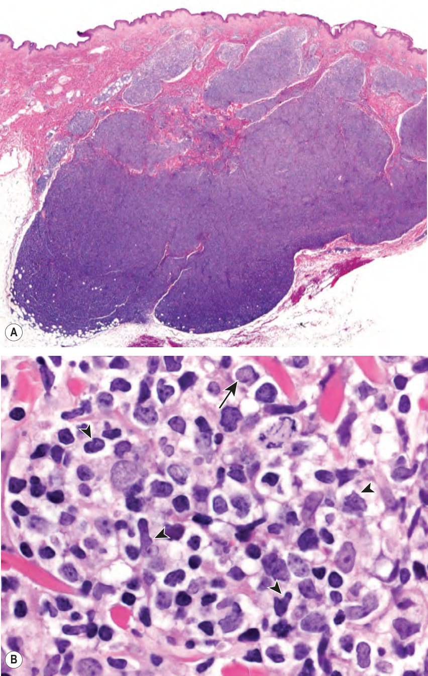
Fig. 119.6 Cutaneous follicle center lymphoma, diffuse type.A Diffuse infiltrate without a follicular pattern. B Centroblasts (arrow) and medium- and largesized centrocytes (cleaved cells; arrowheads) predominate.
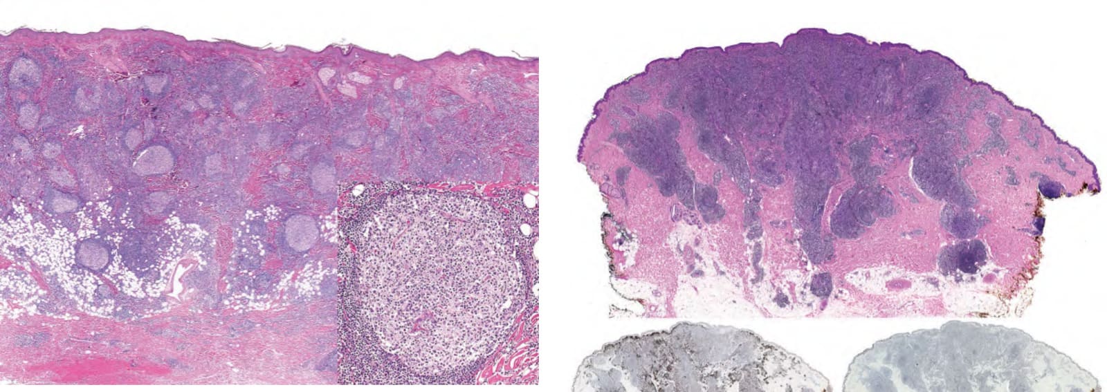
Fig. 119.7 Cutaneous follicle center lymphoma, follicular type. Neoplastic follicles with a monomorphous morphology (lack of polarization) within the entire dermis and superficial subcutis. Note the absence of dark and clear areas within the central portion of the follicle (inset).
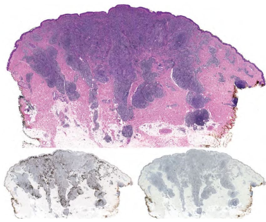
Fig. 119.8 Cutaneous marginal zone B cell lymphoproliferative disorder.
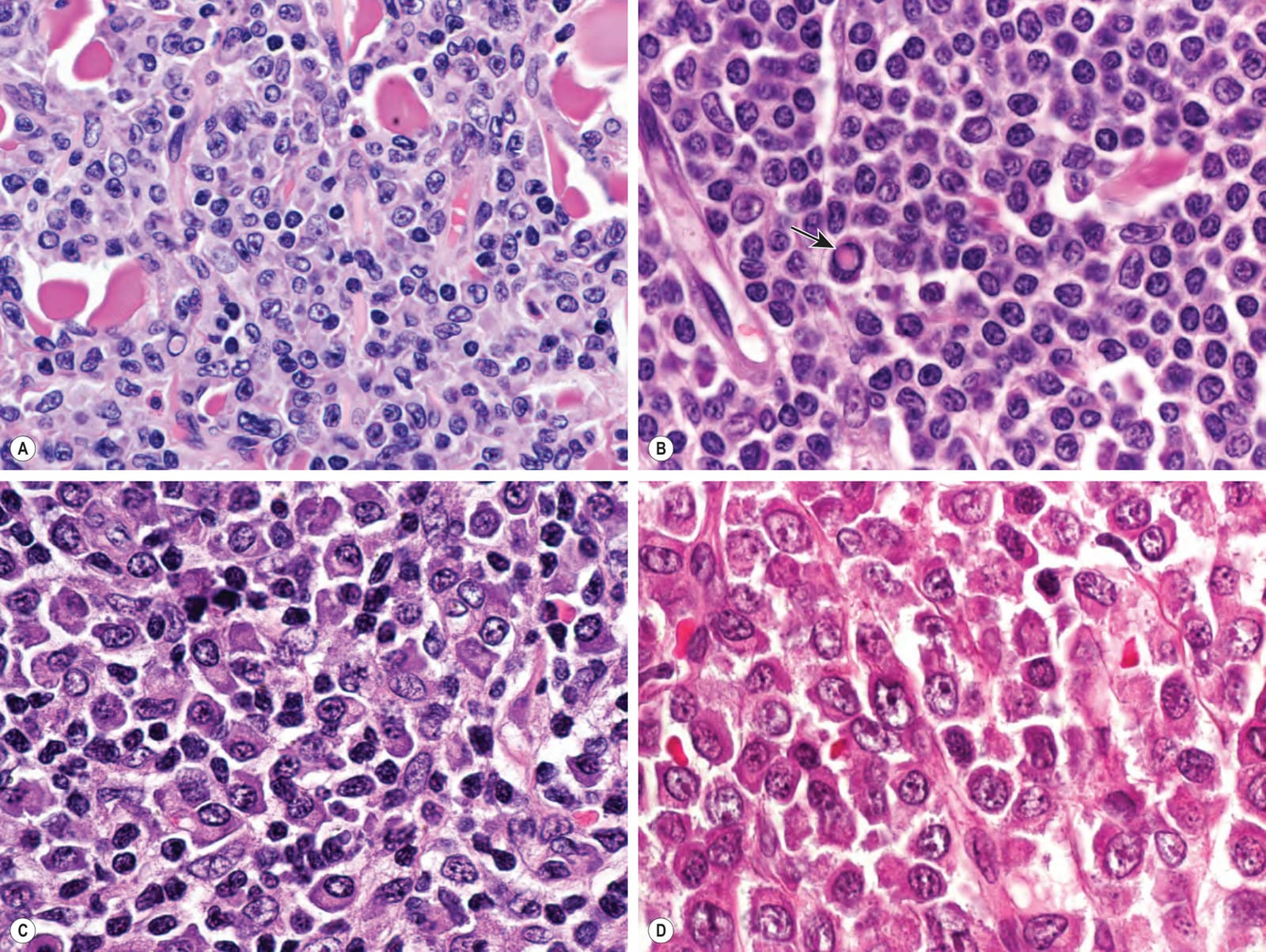
Fig. 119.9 Cutaneous marginal zone B cell lymphoproliferative disorder – spectrum of cell types.A Admixture of marginal zone cells, plasmacytoid cells, and plasma cells. B Predominance of lymphoplasmacytoid cells (formerly classified as cutaneous immunocytoma; note an intranuclear eosinophilic inclusion [Dutcher body, arrow]). C Predominance of plasma cells (formerly classified as cutaneous plasmacytoma). D Predominance of large blastoid cells similar to plasmablasts.
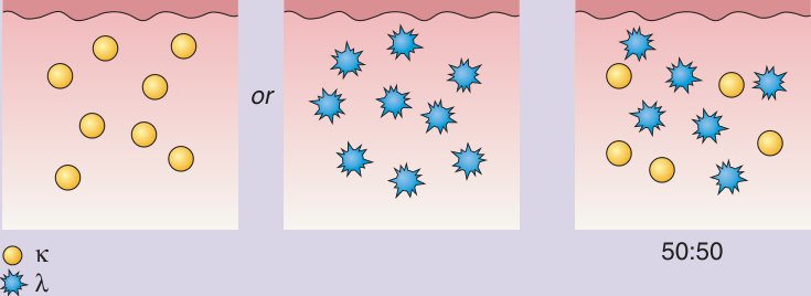
Fig. 119.10 Pattern of immunoglobulin light chain expression. Detection of monoclonal expression of immunoglobulin light chains (κ or λ) is a key diagnostic feature for primary cutaneous marginal zone B cell lymphoproliferative disorder. Monoclonality is defined as a >10/1 ratio.
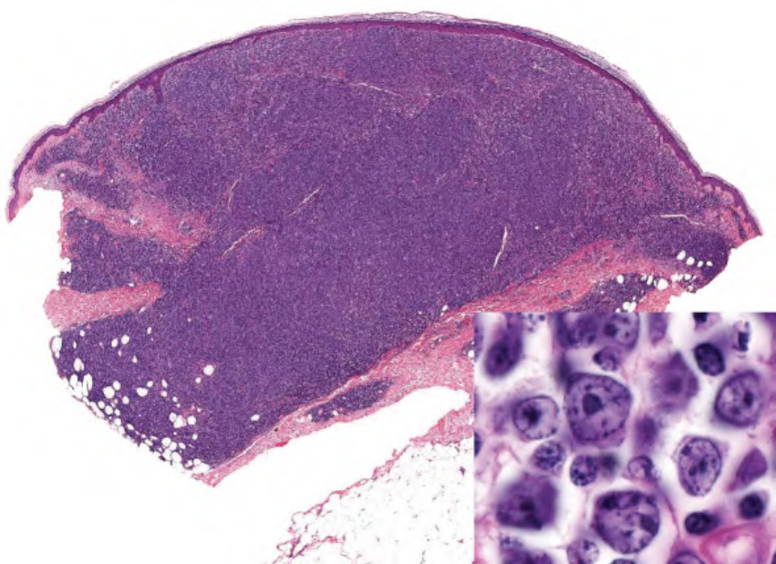
Fig. 119.11 Cutaneous diffuse large B cell lymphoma, leg type. Within the entire dermis, there is a dense, diffuse lymphoid infiltrate. Large cells with a round morphology (mainly immunoblasts) predominate (inset).
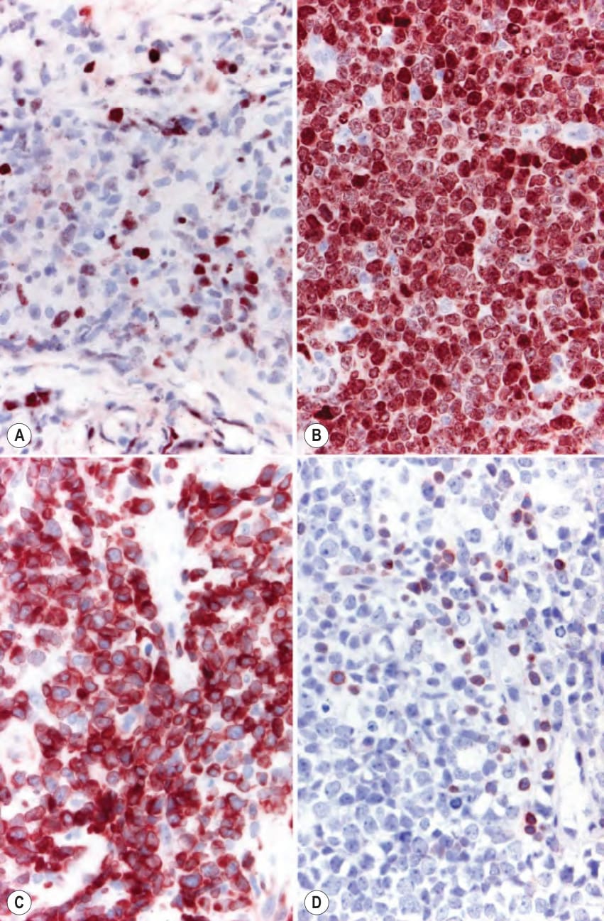
Fig. 119.12 Immunohistochemical staining with anti-Bcl-2 and anti-MUM-1 antibodies. Staining with anti-Bcl-2 antibody shows negativity (except for reactive small lymphocytes) in follicle center lymphoma, diffuse type (A) and positivity of most tumor cells in diffuse large B cell lymphoma, leg type (B). Anti-MUM-1 antibody stains virtually all the tumor cells in primary cutaneous diffuse large B cell lymphoma, leg type (C) while only a minority of cells are positive in primary cutaneous follicle center lymphoma, diffuse type (D).
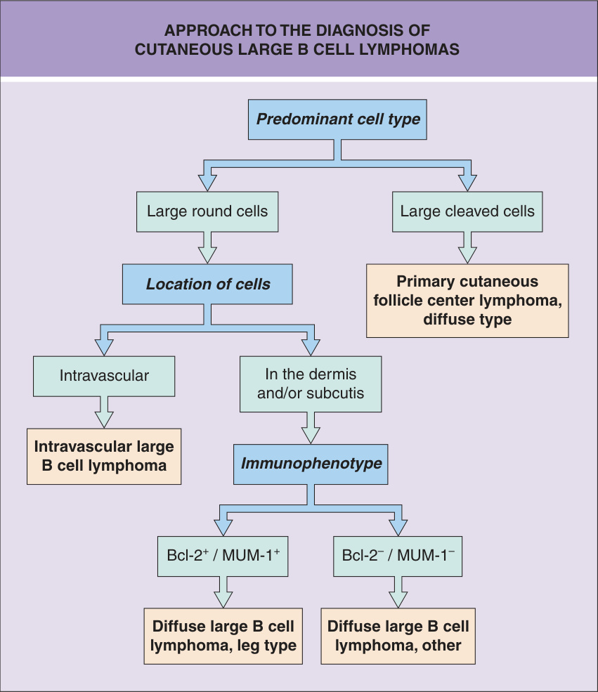
Fig. 119.13 Approach to the diagnosis of cutaneous large B cell lymphomas.
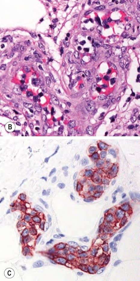
Fig. 119.14 Cutaneous diffuse intravascular large B cell lymphoma – histopathologic features.A Dermal cluster of several small blood vessels filled with large, atypical lymphocytes (B) which are positive for CD20 (C).
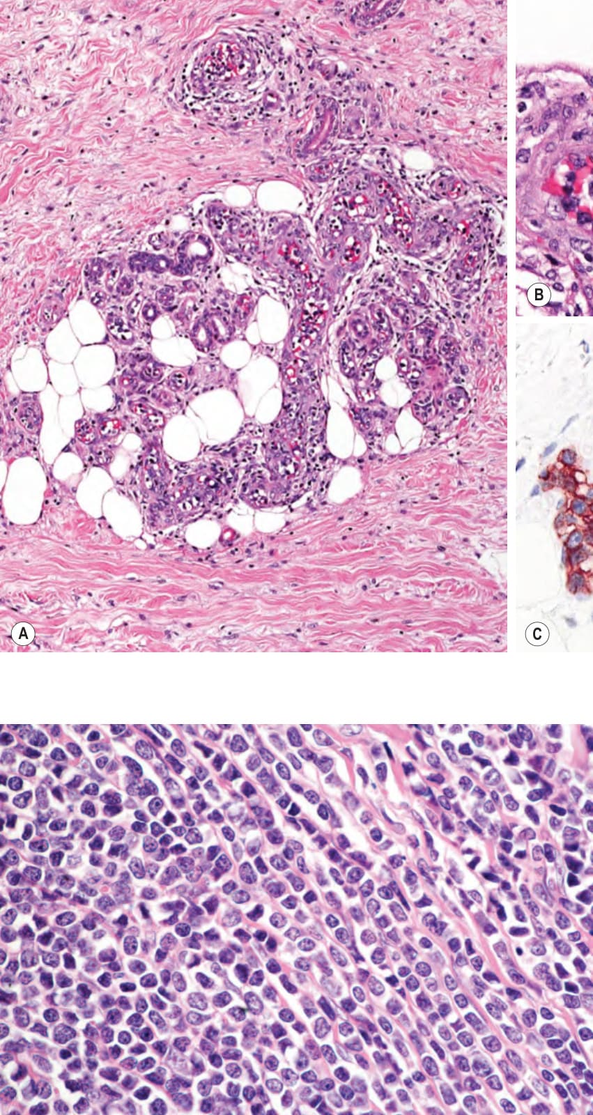
Fig. 119.15 Precursor B lymphoblastic lymphoma/leukemia. Medium-sized blasts with the characteristic “mosaic-stone” linear arrangement.
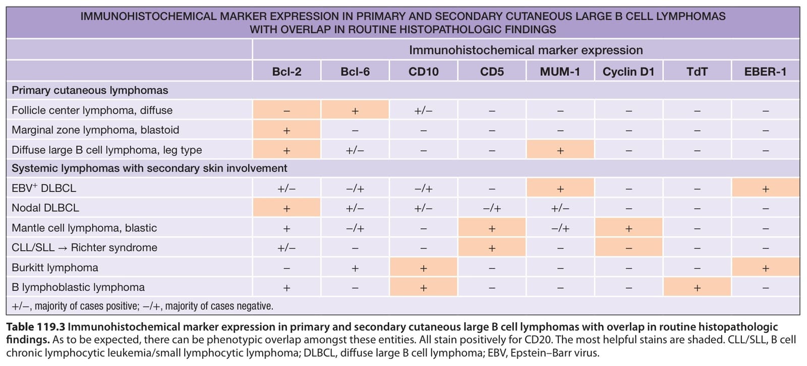
Table 119.3 Immunohistochemical marker expression in primary and secondary cutaneous large B cell lymphomas with overlap in routine histopathologic findings. As to be expected, there can be phenotypic overlap amongst these entities. All stain positively for CD20. The most helpful stains are shaded. CLL/SLL, B cell chronic lymphocytic leukemia/small lymphocytic lymphoma; DLBCL, diffuse large B cell lymphoma; EBV, Epstein–Barr virus.