TREATMENT
The most appropriate treatment modality for patients with a pCBCL is selected after exact classification of the lymphoma, analysis of results of staging investigations, and consideration of the overall condition of the patient. Those patients with secondary cutaneous lesions of extracutaneous B cell lymphoma should be treated in a hemato-oncologic, not dermatologic, setting.
Before reviewing the major therapeutic strategies, it must be emphasized that many patients with low-grade pCBCL can be managed
conservatively with a so-called watchful-waiting strategy, similar to what is often adopted for indolent B cell lymphomas and leukemias at extracutaneous sites. Follow-up examinations in these patients should be performed at least every 6 months or at the onset of new lesions and/or new symptoms, in order to treat the patient as soon as it is necessary. Many patients managed conservatively with a watchfulwaiting strategy experience a prolonged course and long survival, and they do not need aggressive treatment. For cosmesis and sometimes psychological benefit, intralesional triamcinolone can be injected into the papulonodules.
Most patients with pCBCLs that are low-grade (PCFCL, PCMZLD) and who have a solitary or few lesions can be treated by local radiotherapy, simple surgical excision, or surgical excision followed by radiotherapy of the surgical field. It has been reported that recurrences are less frequently seen in patients treated by radiotherapy with wide margins (about 10–20 cm beyond clinically apparent lesions); this approach seems to be justified for so-called Crosti’s lymphoma, a type of PCFCL arising on the back, in which erythematous nodules, papules, and patches surrounding the tumor represent specific infiltrates that can extend far beyond the main bulk of the lesion. Radiotherapy with wide margins, however, does not seem to be justified for lesions other than Crosti’s lymphoma, as margins of about 3–5 cm usually suffice (depending on the size of the lesion). Surgical excision with narrow margins is a valuable therapeutic modality for patients with solitary well-circumscribed lesions and in the authors’ opinion represents the treatment of choice for PCMZLD. In these patients, relapse rates do not seem to be higher than in patients treated with more time-intensive modalities such as local radiotherapy.
A few reports have appeared in which low-grade pCBCLs were treated with systemic antibiotics, achieving a complete resolution in at least a percentage of the patients. This type of treatment is conceptually analogous to that adopted for Helicobacter pylori-associated MALT lymphomas of the stomach, which in their early stages can be cured by eradication of H. pylori infection. Complete response to antibiotic therapy has been observed in some patients with Borrelia-associated pCBCL. Although not yet corroborated by adequate data, antibiotic treatment of patients with pCBCLs should be considered before more aggressive therapeutic options are employed, particularly in European countries endemic for Borrelia infections. It is important to treat patients at the onset of the disease, because, in later stages, lesions may no longer be sensitive to systemic antibiotics. PCR analysis of Borrelia DNA is a rapid test that should be performed in all patients with pCBCL from European endemic areas, in order to identify those who would more likely benefit from early antibiotic treatment.
Another treatment modality for low-grade pCBCL is subcutaneous or intralesional interferon, particularly interferon-α-2a. Although some favorable results have been reported, it appears that treatment with inter-feron is associated with a complete response in about 50% of patients. Therapy with interferon (or intralesional triamcinolone) should be considered for patients presenting with multiple lesions at different body sites, such that local radiotherapy becomes difficult to administer.
Intralesional or systemic injection of anti-CD20 monoclonal antibody (rituximab) has been used to induce tumor reduction in patients with indolent pCBCLs. Intralesional administration should be considered in patients presenting with a few, localized lesions. Therapy with intravenous rituximab represents a valid alternative to established treatments, especially in patients who present with disseminated skin lesions or relapse after radiotherapy. Rituximab can also be administered in combination with other treatment modalities for pCBCLs with more aggressive behavior (e.g. DLBCLLT). Additional monoclonal antibodies, bispecific T‑cell engaging antibodies (e.g. mosunetuzumab), and smallmolecule inhibitors (e.g. ibrutinib, lenalidomide) are also employed for aggressive B-cell lymphomas.
Patients with DLBCLLT and a limited number of patients with disseminated lesions of PCFCL, diffuse type, need more aggressive treatment modalities (e.g. systemic chemotherapy plus rituximab). The regimen used most frequently is cyclophosphamide, doxorubicin, vincristine and prednisone (CHOP) plus rituximab, and it represents the standard of care for DLBCLLT. In the absence of signs of transformation into high-grade lymphoma, aggressive treatment is not indicated in patients with PCMZLD. In the setting of significant comorbidities, rituximab alone or in combination with local radiotherapy may be administered. For relapsed DLBCLLT, CAR-T cell therapy is increasingly being used, especially when recurrence occurs within one year of primary therapy. In the setting of longer remissions, salvage chemotherapy followed by autologous HSCT is an option.
Systemic chemotherapy plus rituximab is the treatment of choice for patients with IVDLBCL, regardless of results of staging investigations. As with DLBCLLT, the addition of rituximab to anthracycline-based chemotherapy significantly improves outcome. Finally, patients with precursor B lymphoblastic lymphoma/leukemia should be treated as B cell acute lymphoblastic leukemia by a hematologist.
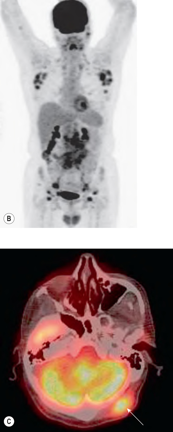
Fig. 119.17 Evaluation of the patient with a suspected diagnosis of cutaneous B cell lymphoma.A Algorithm outlining approach to the patient. B, C Positron emission tomography (PET) scans in a 66-year-old patient with stage IV B cell non-Hodgkin lymphoma. He had a 1-year history of a cutaneous nodule of the scalp that was originally diagnosed as an epidermoid inclusion cyst. Biopsy of the nodule showed a diffuse large B cell lymphoma (CD10+, CD20+, variably Bcl-2+, MUM-1−) and a PET-CT scan was performed for staging. There is evidence of widespread disease involving multiple lymph node basins in the dedicated (MIP) image (B) as well as a scalp lesion in a PET-CT fusion image (C; arrow). B,C, Courtesy Dennis Cooper, MD.
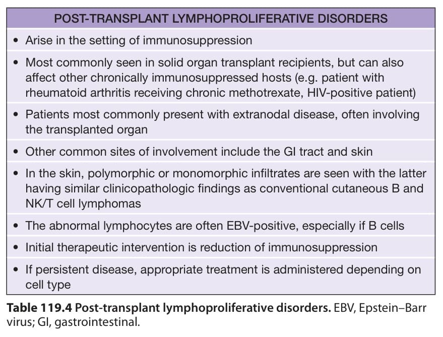
Table 119.4 Post-transplant lymphoproliferative disorders. EBV, Epstein–Barr virus; GI, gastrointestinal.