LICHEN PLANUS
Synonym: Lichen ruber planus
Key features
Idiopathic inflammatory disease of the skin, hair, nails and mucous membranes, seen most commonly in middle-aged adults
Flat-topped violaceous papules and plaques favoring the wrists, forearms, genitalia, distal lower extremities, and presacral area
Clinical variants include actinic, annular, atrophic, bullous, hypertrophic, inverse, linear, ulcerative, vulvovaginal–gingival, lichen planopilaris, lichen planus pigmentosus, and drug-induced
Some lichenoid drug eruptions have a photodistribution, while others are clinically and histologically indistinguishable from idiopathic lichen planus
The most commonly incriminated drugs include angiotensin- converting enzyme (ACE) inhibitors, thiazide diuretics, antimalarials, quinidine, and, especially in the past, gold
In most patients, the therapeutic approach to immune checkpoint inhibitor (ICI)-related lichenoid eruptions consists of topical corticosteroids and continuation of the ICI; systemic corticosteroids and a brief drug holiday may be required when the eruption is severe or recalcitrant
Histologically, there is a dense, band-like lymphocytic infiltrate and keratinocyte apoptosis with destruction of the epidermal basal cell layer
In this T cell-mediated autoimmune disorder, basal keratinocytes express altered self-antigens on their surface
Introduction
Lichen planus (LP), the prototype of lichenoid dermatoses, is an idiopathic inflammatory disease of the skin and mucous membranes. Classic LP is characterized by pruritic, violaceous papules that favor the extremities. Histologically, a dense, band-like lymphocytic infiltrate is seen underlying an acanthotic epidermis with hypergranulosis, apoptosis, and destruction of the basal cell layer. The etiology and pathogenesis of LP are not fully understood, but the disorder has been associated with multiple environmental exposures, including viral infections, medications, vaccinations, and dental restorative materials.
LP-like lesions that resemble idiopathic LP may also develop in chronic GVHD, where alloreactive T cells that recognize foreign major histocompatibility complex (MHC) molecules are central effectors (see Ch. 52). This lends support to the hypothesis that an autoimmune reaction against epitopes on lesional keratinocytes that have been modified by viral or drug antigens may be responsible for LP.
A number of clinically distinct inflammatory dermatoses have in common varying elements of a lichenoid tissue reaction, and they are referred to as lichenoid dermatoses (Table 11.1).
History
The term lichen planus was initially introduced by Erasmus Wilson in 1869 to describe the condition that had been previously named leichen ruber by Hebra.
Epidemiology
Although its incidence varies depending upon geographic locale, cutaneous LP has been reported to affect from 0.2% to 1% of the adult population, whereas oral lesions have been observed in up to 1%–4% of the population. There is no overt racial predisposition. LP most commonly has its onset during the fifth or sixth decade, with two-thirds of patients developing the disease between the ages of 30 and 60 years. It is rare in both infants and the elderly and, typically, only 1%–4% of patients are children. However, more recent studies have suggested that LP may actually be more common in children, especially in Arab populations. Oral LP is also quite uncommon in young people and it usually affects middle-aged to elderly individuals (mean age at diagnosis being 52 years). Although LP is frequently thought to have no gender predilection, some studies have found that women were affected nearly twice as often as men.
Mucosal involvement, particularly oral lesions, is observed in up to 75% of patients with cutaneous LP, but the former can be the only manifestation of the disease. Only 10%–20% of patients whose initial presentation is oral LP will eventually develop cutaneous LP. Although reports of familial LP are rare, it may occur more frequently than previously thought; for example, LP occurs in up to 10% of first-degree relatives of affected patients. Cases of familial LP tend to have an earlier age of onset, a higher relapse rate, and more frequent oral mucosal involvement. Of note, reports of concurrent LP in monozygotic twins who were living together suggest an environmental trigger. Lastly, individuals with Good syndrome, a primary immunodeficiency that is associated with thymomas, there is an increased risk of developing lichen planus.
Pathogenesis
There is a growing body of evidence that LP represents T cell-mediated autoimmune damage to basal keratinocytes that express altered self-antigens on their surface.
Target antigens
Clinical observations and anecdotal evidence have long suggested a relationship between exposure to a number of exogenous agents (e.g. viruses, medications, contact allergens) and the development of LP (see Table 11.1). In theory, T cells that normally do not respond to skin (epidermis)-restricted antigens are primed by exogenous agents when the latter possess cross-reactive antigens that can activate autoreactive T cells via molecular mimicry. In other words, the T cell receptors on the T cells that induce mucocutaneous LP could cross-react with exogenous antigens.
Of the many potential exogenous antigens, significant attention has been focused on the possible role of viruses, particularly hepatitis C virus (HCV). In several case–control studies, the prevalence of HCV
(3.5%–38%) was 2- to 13.5-fold higher in patients with LP than in controls. This association seems to be strongest in Japanese and Mediterranean populations, probably due to the high prevalence of HCV infection in these countries. In the US, one case–control study found that 12 (55%) of 22 patients with LP had anti-HCV antibodies, and this was significantly higher than the 25% of 40 psoriatic patients or the 0.17% of blood donors who tested positive. However, a systematic review and meta-analysis of existing epidemiologic studies demonstrated that an association between LP and HCV infection did exist in certain geographic regions (e.g. East and Southeast Asia, South America, the Middle East, Europe), but not in others (e.g. North America, South Asia, and Africa).
An increased frequency of the HLA-DR6 allele has been reported in Italian patients with HCV-associated oral LP, raising the possibility that CD4+ T cells activated upon recognition of HCV-encoded peptides bound to HLA-DR6 molecules could be directly involved in the pathogenesis of LP. In support of this possibility, HCV tetramer analysis has shown that HCV-specific CD4+ and/or CD8+ T cells are present with higher frequency in oral LP lesions compared with the circulating compartment, suggesting that they play a role in the pathogenesis of LP.
Of the various types of LP, it is the oral form that is most commonly viewed as a manifestation of HCV infection. By PCR, HCV RNA was detected in 93% of oral LP lesions, suggesting HCV replication within LP lesions. However, these PCR results were not confirmed by other studies. In patients receiving anti-HCV therapy, originally interferon and ribavirin and more recently direct-acting antiviral agents that target nonstructural proteins, the effects on mucocutaneous LP have varied. Lesions have improved in some patients while others experienced a flare of disease activity.
With regard to the role of other viruses in LP, human herpesvirus (HHV)- 6 and HHV-7 DNA has been detected in some LP lesions. Remission of LP was associated with decreased HHV-7 protein expression, in particular within infiltrating plasmacytoid dendritic cells. There have been multiple reports that either SARS-CoV-2 (COVID-19) infection or COVID-19 vaccines can trigger LP. Of note, in two recent systematic reviews, a stronger association was observed between LP and COVID- 19 vaccination as compared to COVID-19 infection10a,10b. There are also sporadic case reports of LP lesions developing in areas recently affected by herpes simplex virus (HSV) or varicella–zoster virus (VZV) infections (see Table 80.7). Possible explanations include a nonspecific Koebner phenomenon or isotopic response or an immune reaction to the virus. In an immunohistochemical study involving eight patients, VZV-gE antigen was detected within the eccrine epithelia of LP lesions if they were in a dermatomal (zosteriform) pattern but not if they were in a linear array along the lines of Blaschko. Two of the patients with the dermatomal pattern reported no preceding episode of herpes zoster, leading the authors to speculate if LP could be triggered by sub-clinical VZV reactivation. A link between human papillomavirus (HPV) and oral LP has also been suggested based upon the identification of a clonal expansion of HPV-16-specific CD8+ T cells within the lesions.
While measurements (via PCR) of viral genomes within lesional skin or blood have been inconclusive, as noted above, virus-specific T cells have been detected within lesions. This suggests that the pathology is a direct consequence of immune responses, perhaps to virally induced alterations in the antigenicity of epidermal cells rather than the viruses themselves. However, it is also possible that virus-specific T cells are nonspecifically trapped within sites of inflammation, having been activated systemically or locally at distant sites, and then they expand and mediate tissue damage “accidently” due to cross-reactivity with other antigens (e.g. drugs).
In addition to COVID-19 vaccines (see above), a number of reports have described the appearance of LP after administration of influenza vaccines, rabies vaccines, and different types of HBV vaccines. The time interval between the initial dose and the development of cutaneous or mucosal lesions has varied from a few days to 5 months. One recommendation is that patients who develop LP before completing their vaccination series should avoid further injections because of an increased risk of developing severe LP lesions, including the bullous variant.
Investigations regarding a relationship between bacteria and cutaneous LP have been limited. In tissue and saliva samples, the diversity and composition of microbial communities differed when patients with oral LP were compared to healthy controls, suggesting a possible role for the local microbiome.
The role of contact allergy to a variety of metals in the exacerbation or induction of oral LP has been well described, based on exposure to metallic dental restorations or constructions, positive patch test results, and then regression or complete clearing after removal of the sensitizing metal and replacement with other materials. However, improvement has also been observed after removal of metals in patch test-negative patients. The metals reported to aggravate oral LP include amalgam (mercury), copper, and gold.
Of note, the development of contact allergy to metals within dental restorations in patients with LP could be explained by easy penetration of the metal via damaged mucosa.
Cutaneous eruptions similar or even identical to LP, both clinically and histologically, have been linked to a variety of drugs. The terms “lichen planus-like” and “lichenoid” are often used to describe this phenomenon. A wide variety of drugs have been associated with lichenoid drug eruptions and the list of such drugs steadily increases (Table 11.2). However, recurrence of the lesions subsequent to drug rechallenge has not been documented for the majority of these drugs.
Autoantigens, including tumor antigens
In occasional patients, LP has been reported as a possible autoimmune reaction triggered by an underlying neoplasm. Also, a lichenoid tissue reaction is seen in patients with paraneoplastic pemphigus. The temporal relationship between LP and the underlying neoplasm in the
reported patients suggests that the neoplasms may have stimulated a cell-mediated immune response against tumor antigens that led to the generation of autoreactive T cells that cross-reacted against antigens expressed on epidermal cells.
Although numerous case reports describe patients with both LP and autoimmune diseases, studies with larger numbers of patients with LP have shown no increased incidence of autoimmune diseases. Multiple investigators have described a significant association between specific HLA antigens (e.g. HLA-DR1, -DR6, -DR9) and LP. However, a true association with a particular HLA allele has been difficult to establish because of significant geographic heterogeneity and clinical patient selection.
A murine model of LP has been established by employing autoreactive T cells capable of producing interferon-γ (IFN-γ) and tumor necrosis factor (TNF-α) (Fig. 11.1). Intradermal inoculation of CD4+ autoreactive T cell clones into the footpads of syngeneic mice can induce local histologic changes similar to LP or lichenoid skin diseases. In this model, the autoreactive T cells can respond to self MHC class II antigens constitutively expressed on macrophages and Langerhans cells, and they migrate into the epidermis, resulting in epidermal injury. These T cells, therefore, can induce LP-like lesions without any alteration in the antigenicity of the epidermis. In subsequent studies, desmoglein-specific T cells were also capable of inducing LP-like changes histologically. Thus, LP could be induced by different types of T cells – those that specifically target antigens constitutively expressed in the epidermis but also T cells that target antigens not expressed within the epidermis.
Additionally, in the natural disease process, exogenous agents (e.g. viral infections, drugs) could induce alterations in the antigenicity of epidermal cells and trigger the activation of T cells. Of note, such autoaggressive reactions could function to eliminate abnormal keratinocytes altered by these exogenous agents. However, in the situation where T cells initially responding to self-antigens modified by exogenous agents subsequently become cross-reactive with some self-epitopes, these T cells would then chronically respond to the previously ignored self-epitopes, leading to perpetuation of an autoimmune attack by such T cells rather than elimination of the abnormal keratinocytes.
Effector cells
There are conflicting data regarding the phenotype of the inflammatory infiltrate in LP lesions. Although initial immunohistochemical studies showed that the cellular infiltrate contained an increased ratio of CD4+ : CD8+ T cells, other investigators found a predominance of CD8+ T cells, particularly in older lesions. Evidence to support the crucial role of CD8+ T cells in autoimmune damage to basal keratinocytes has been provided by CD8+ T cells isolated from lesional skin; these T cells exhibited specific cytotoxic activity against autologous lesional and normal keratinocytes.
Cellular damage, as evidenced by apoptotic DNA fragments, is greatest within the basal layer of the epidermis. A possible mechanism is as follows: IFN-γ produced by CD8+ T cells upregulates Fas expression by keratinocytes, rendering them susceptible to T cell-mediated, Fas ligand-driven apoptosis. This interaction triggers a cascade of intracellular enzymatic reactions resulting in DNA fragmentation (see Ch. 107). In addition to Fas, death receptor-induced apoptosis involves signaling processes via TNF-R1, TRAIL-R1 and 2, and DR3 or DR6. Because type 1 helper T cells (Th1), such as the autoreactive CD4+ T cells in the murine model (see Fig. 11.1), can produce large amounts of IFN-γ and lymphotoxin-α (TNF-β) upon activation and thereby induce or enhance the expression of apoptosis-associated proteins such as Fas and TRAIL, they may also play a role in extensive epidermal damage by promoting apoptotic death of keratinocytes. More recent studies have shown that granule exocytosis with release of perforin and granzyme B, rather than the Fas/Fas-ligand system, is the main pathway of cytotoxicity mediated by CD4+ and CD8+ T cells in humans. However, a combination of the two mechanisms is most likely, with predominance depending upon the particular stage in the disease process.
Recent studies have provided evidence for the involvement of Toll-like receptor (TLR) signaling in the induction of autoimmunity. As depicted in Figure 4.1, certain TLRs recognize viral RNA as well as imidazoquinolones (e.g. imiquimod). Interestingly, topical application of imiquimod, which can lead to enhanced migration and maturation of dermal dendritic cells, followed by increased production of proinflammatory cytokines and activation of antigen-specific CD8+ T cells, may exacerbate lesions of LP.
In some autoimmune diseases, T cell differentiation may shift from Treg cells (immune suppression) to Th17 cells (see Ch. 4), but an increase in both populations has been found in LP lesions. However, the functionality of the Treg cells within these lesions has been questioned.
A critical event in the initiation of immune responses in LP lesions is for memory T cells to migrate from the circulation into a particular skin
site. Release of type 1 IFNs, such as IFN-α, from activated plasmacytoid dendritic cells (pDCs) and keratinocytes may be critical in inducing skin-directed migration of effector memory T cells (Fig. 11.2A). Stimulation of TLRs expressed on pDCs and keratinocytes by pathogens or endogenous ligands (released via skin damage) represents one of the earliest events and it is sufficient to induce type 1 IFN production. Type 1 IFN signaling and type 1 IFN-inducible chemokines (e.g. IP-10/ CXCL10) then serve to recruit chemokine receptor CXCR3-expressing effector memory T cells (Th1 cells) into the skin via CXCR3/IP-10 interactions (Table 11.3). A number of other molecular interactions, such as CCR4/TARC, CCR10/CTACK and LFA-1/ICAM-1 expressed on T cells and keratinocytes, respectively, have also been implicated in
the recruitment of memory T cells and pDCs to the dermal–epidermal junction. Because IFN-α from pDCs induces IFN-γ production by T cells and IFN-γ also sustains IFN-α production, a positive feedback loop may be operational within LP lesions. This ordered sequence of events provides a possible explanation for why LP lesions develop within traumatized sites and virally induced lesions.
Although chemokines produced at inflammatory skin sites are thought to regulate the composition of the Th1- or Th2-driven cellular infiltrates, memory T cells with a “surveillance” function can also migrate to skin sites under non-inflamed conditions (Fig. 11.2B). “Homeostatic” chemokines constitutively produced under non-inflamed conditions can mediate the skin-directed migration of these “immune surveillance” T cells (see Table 11.3), leading to clearance of invading pathogens such as viruses. In addition, Treg cells that have the capacity to suppress activated T cells are also able to enter inflamed skin sites. However, in LP lesions, there is no definitive means of distinguishing “immune surveillance” T cells or protective Treg cells from “pathogenic” T cells. Lastly, identification of the cells responsible for LP is further complicated by the presence of both skin tissue-resident memory T cells (Trm cells) and effector memory T cells (Tem cells) within skin lesions, with the former, whose physiologic role includes local protection against pathogens, becoming a mediator of tissue damage.
Of note, while the epidermotropic migration of effector T cells leading to epidermal damage in lichenoid tissue reactions is clearly a complicated, multistep process, the latter can be bypassed (at least in part) in the case of fixed drug eruptions (FDE). This is because effector CD8+ T cells responsible for the epidermal damage persist as a stable population within previously affected sites, even when the skin becomes normal-appearing. In particular, CD8+ Trm cells (see above) populate the epidermis of “resting” FDE lesions and although these cells are the
Skin tissue-resident memory T cells (Trm cells) and effector memory T cells (Tem cells) both play a role in producing tissue damage. In the resting stage, a stable population of CD8+ Trm cells resides within the epidermis without exerting their cytotoxic potential (the physiologic role of these cells includes local protection against pathogens). When stimulated by cross-reactive antigens such as drugs, Trm cells then display cytolytic activity towards surrounding keratinocytes. The Trm cells also release IFN-γ and TNF-α into the local environment, leading to the recruitment of CD4+ and CD8+ Tem cells from the circulation into the skin. This leads to additional tissue damage. During the resolution stage, Treg cells are recruited into the site of inflammation, in part due to the release of preformed cytokines, including IL-16, by mast cells. Although the majority of activated cell populations are removed by apoptosis (see Fig. 11.2), a proportion of Trm cells are prevented from undergoing apoptosis by IL-15 produced by surrounding keratinocytes.
principal mediators of localized tissue damage, CD4+ and CD8+ Tem cells, recruited from the circulation, also contribute to the epidermal damage (Fig. 11.3).
Sweating disturbance
Sweat contains interleukin (IL)-1, IL-6, IL-8, and TNF-α, proinflammatory cytokines that have been implicated in the induction of T cell recruitment into the skin. In theory, extravasated sweat could explain the syringotropic T cell migration occasionally seen in the early lesions of LP. Recent studies have shown that leakage of sweat into the dermal–epidermal junction initiates a complex sequence of events which leads to syringotropic T cell migration.
Clinical Features
The characteristic primary lesion of LP is a small, polygonal-shaped, violaceous, flat-topped papule (Fig. 11.4); occasionally papules are umbilicated. The surface is slightly shiny or transparent, and a network of fine white lines called “Wickham striae” or small gray–white puncta are also seen. The latter correspond histologically to focal thickening of the granular layer. Wickham striae are readily apparent by dermoscopy (see Ch. 0). Postinflammatory hyperpigmentation is also a common finding (Fig. 11.5).
The papules of LP may be widely dispersed or they may cluster or coalesce into larger plaques. LP is usually pruritic. Although the Koebner phenomenon (i.e. isomorphic response) is commonly seen in LP (Fig. 11.6), excoriations and impetiginization are unusual. A linear array of lesions can be seen as a consequence of the Koebner phenomenon or an isotopic response at the site of healed herpes zoster, as well as in the linear variant of LP which follows the lines of Blaschko.
The most common sites of involvement are the flexor wrists and forearms, the dorsal hands, the shins, and the presacral area. Mucous membranes, especially the oral mucosa (see below), are affected in more than half of patients, and this is often the only site of disease. Lesions are also commonly seen on the glans penis (Fig. 11.7A), where they can have an annular or figurate configuration and may become erosive.
The duration of the disease can vary depending upon the LP variant. While the lesions of exanthematous LP typically resolve within a year, hypertrophic, oral, and nail LP tend to be more persistent. In particular, ulcerative oral LP may be a lifelong affliction.
Actinic LP
This variant is reported under a variety of names, including LP actinicus, LP subtropicus, LP tropicus and lichenoid melanodermatitis.
Although the majority of reported patients have been from Middle Eastern countries, this variant has been observed worldwide. Most patients are young adults or children, but there is no sex predilection. Lesions primarily involve sun-exposed skin of the face, followed by the neck and dorsal surfaces of the hands and arms. They usually consist of red–brown plaques with an annular configuration, but melasma-like hyperpigmented patches have been observed. In temperate climates, spontaneous improvement may occur during the winter months.
Acute (exanthematous) LP
Because lesions are usually widely distributed and disseminate rapidly, this form is also known as exanthematous or eruptive LP. The commonly affected areas include the trunk (Fig. 11.8), the inner aspects of the wrists and the dorsal feet. Reports in the literature of this variant probably include lichenoid drug eruptions. The clinical course is usually self-limited, and in general lesions resolve with hyperpigmentation within 3 to 9 months.
Annular LP
This form is thought to occur when papules spread peripherally and the central area resolves. The annular edge is slightly raised and typically
Violaceous papules and plaques with white scale and Wickham striae on the dorsal foot. B Note the flat-topped (lichenoid) nature of the violaceous papules on the penis. C–E While violaceous is the most common color, lesions can vary in color from pink to dark purple– black. B, Courtesy Louis A. Fragola, Jr, MD; C, Courtesy Julie V. Schaffer, MD; D, Courtesy Jeffrey M. Cohen, MD; E, Courtesy Kalman Watsky, MD
Lesions also appeared where Steri-Strips™ had been applied.
Atrophic LP
Atrophic LP may represent a resolving phase of LP, given the history of the lesions: papules coalesce to form larger plaques that over time become atrophic centrally, with residual hyperpigmentation. The clinical appearance of atrophic LP is likely a result of thinning of the epidermis rather than degeneration of elastic fibers, and the epidermal atrophy may be accentuated by the use of potent topical corticosteroids. Common sites of involvement include the intertriginous zones and the lower extremities (Fig. 11.9A).
Bullous LP and LP pemphigoides
In bullous LP, vesicles and bullae develop within pre-existing lesions of LP as a result of intense lichenoid inflammation and significant epidermal damage (Fig. 11.9B). This leads to epidermal–dermal separation, i.e. exaggerated Max-Joseph spaces. In contrast, patients with LP pemphigoides have circulating IgG autoantibodies directed against the 180 kDa BP antigen (BP180; BPAG2), as in idiopathic BP. However, compared to BP, LP pemphigoides has a younger age of onset. In this variant, bullae can arise either within LP lesions or previously uninvolved skin (Fig. 11.9C), but the diagnosis of LP usually precedes the LP pemphigoides. These findings suggest that damage to the basal layer by a lichenoid infiltrate may expose hidden antigens to the autoreactive T cells, leading to the formation of autoantibodies and subepidermal bullae. Indeed, LP pemphigoides evolving into pemphigoid nodularis has been described. There are conflicting results regarding the autoantigen in LP pemphigoides, but one study suggested a novel epitope within the C-terminal NC16A domain of BP180 (see Fig. 31.9).To date, no reactivity against the 230 kDa BP antigen, type VII collagen, or the laminin-5 subunits has been detected. An additional difference between these two disorders is the finding of abundant VZV antigen-bearing monocytes and mast cells in the upper dermis of bullous LP lesions as opposed to such cells being rare in LP pemphigoides.
Hypertrophic LP
This variant is also referred to as LP verrucosus (Fig. 11.10). Extremely pruritic, thick, hyperkeratotic plaques are seen primarily on the shins or dorsal aspect of the feet and may be covered by a fine adherent scale. The lesions are usually symmetric and tend to be chronic because of repetitive scratching. The average duration of hypertrophic LP in patients whose lesions had cleared was reported to be 6 years. Chronic venous stasis frequently contributes to the development of this condition. As in hypertrophic LE, squamous cell carcinoma (SCC) may arise within longstanding hypertrophic LP, and distinguishing pseudoepitheliomatous hyperplasia from SCC can be difficult. In one retrospective review, the presence of an increased number of eosinophils pointed to the diagnosis of hypertrophic LP.
Inverse LP
In this unusual variant, an inverse distribution pattern is observed. Pink to violaceous papules and plaques appear in intertriginous zones (axillae > inguinal and inframammary folds) and less often in the popliteal
LP pigmentosus
LP pigmentosus typically presents as brown to gray–brown macules in sun-exposed areas of the face and neck, usually with no preceding erythema and often evolving into diffuse or reticulated pigmentation (see Ch. 67). This variant favors individuals with skin types III and IV, in particular those from South Asia, Latin America, and the Middle East. Involvement of intertriginous sites is occasionally observed (Fig. 11.12), and a linear distribution following the lines of Blaschko has also been described. In the photodistributed form, topical application of mustard oil, which contains a potential photosensitizer, has been implicated as a possible trigger.
Given the similarity of histologic findings, distinction from erythema dyschromicum perstans (EDP) is based primarily upon clinical features.
In EDP, there is often truncal involvement, a younger mean age of onset, and lack of either diffuse pigmentation or the occasional coexisting LP lesions. Of note, small nests of keratinocytes can be seen within the basal layer and these may be confused with nests of melanocytes.
Lichen planopilaris
In lichen planopilaris, involvement of the hair follicle is observed, both clinically and histologically. This variant is also called follicular LP and LP acuminatus. Multiple, keratotic plugs surrounded by a narrow violaceous rim are observed primarily on the scalp, although other hairbearing areas can also be affected (Fig. 11.13). The inflammatory process may result in scarring and loss of follicular structure, i.e. a permanent alopecia. Over time, the areas of involvement often “burn out” centrally and are indistinguishable from other causes of “end-stage” cicatricial alopecia (see Ch. 69). However, examination of the periphery may reveal the primary lesions. Women are more frequently affected than men, and this form may occur alone or with typical LP lesions elsewhere.
A variant of lichen planopilaris known as Graham-Little–Piccardi– Lassueur syndrome is characterized by the triad of: (1) non-cicatricial loss of pubic and axillary hairs and disseminated spinous or acuminated follicular papules (see Fig. 11.13B); (2) typical cutaneous or mucosal LP; and (3) scarring alopecia of the scalp with or without atrophy (see Fig. 11.13C). These features need not be present simultaneously. Another more recently recognized variant of LP of the scalp is frontal fibrosing alopecia which occurs more commonly in older women and can affect the eyebrows as well (see Ch. 69); patients may also have LP pigmentosus and papules on the face.
Linear LP
Although linear lesions frequently occur in sites of scratching or trauma in patients with LP as a result of the Koebner phenomenon, the term linear LP (Fig. 11.14) is usually reserved for lesions that appear spontaneously within the lines of Blaschko (see Fig. 11.13A). This form has also been referred to as zosteriform, but with the exception of LP developing within the site of previous herpes zoster, the distribution pattern of linear LP is generally not dermatomal. The possibility exists that when LP has a strictly dermatomal pattern, it may have been preceded by “zoster sine herpete”. As noted previously (see Pathogenesis), in a case series, VZV antigen was detected in the zosteriform variant of LP but not in the linear variant. Attempts to determine genetic differences between involved and uninvolved skin are ongoing.
Discoid lupus erythematosus/lichen planus
Patients whose lesions have overlapping features of both LP and lupus erythematosus (LE) have been reported. These lesions are preferentially located in acral sites, particularly the palms and soles. Histologic and
direct immunofluorescence (DIF) microscopic findings show features of both LP and LE. Whether systemic immunologic abnormalities such as high titers of ANA are present in these patients is controversial, but case reports suggest that some patients have disease at the chronic cutaneous LE end of the clinical spectrum while others meet the criteria for systemic LE (see Ch. 41).
Nail LP
The nails are affected in ~10% of patients with LP and usually several nails are affected (see Ch. 71). The characteristic nail abnormalities include lateral thinning (Fig. 11.15A), longitudinal ridging, and fissuring (Fig. 11.15B). These changes are manifestations of matrix damage, which can lead to scarring and dorsal pterygium formation if left untreated (Fig. 11.15C). Nonspecific changes in the nail bed include yellow discoloration, onycholysis, and subungual hyperkeratosis. In some patients, twenty-nail dystrophy may represent a variant of LP. Nail LP presenting as twenty-nail dystrophy is much more common in
children than in adults, although other forms of nail involvement are extremely rare in children.
Oral LP
Oral LP can appear in at least seven forms, which occur separately or simultaneously: atrophic, bullous, erosive, papular, pigmented, plaquelike, and reticular. The most common and characteristic form of oral LP is the reticular pattern (Fig. 11.16A). It is characterized by slightly raised, whitish linear lines in a lace-like pattern or in rings with short radiating spines. This form is usually asymptomatic and the most common site of involvement is the buccal mucosa; lesions are often bilateral and symmetric. Gingival involvement is common, and oral LP affecting the gingivae exclusively is seen in ~10% of cases. It typically presents as chronic desquamative gingivitis (see Fig. 72.7). Gingivitis also occurs as a component of the “vulvovaginal–gingival syndrome” (see below).
Atrophic, erosive, and bullous lesions are associated with symptoms ranging from mild discomfort to severe pain (Fig. 11.16B). There is a higher incidence of plaque-like lesions among tobacco smokers. For unknown reasons, oral LP is very uncommon in young patients, and in some studies, women have been affected about twice as often as men.
Patients with oral LP should be questioned about symptoms related to esophageal involvement and examined for other mucosal lesions, particularly genital lesions, and vice versa, because ~70% of patients with mucosal vulvovaginal LP have clinical signs of oral LP. Esophageal LP has been increasingly recognized. It tends to be chronic and is associated with the development of dysphagia, strictures, stenosis, and even SCC.
Reportedly, the erosive or ulcerative type of oral LP is less frequently associated with cutaneous LP than are all other types of oral LP. Such mucous membrane lesions are more therapy-resistant and less likely to
spontaneously remit than cutaneous lesions. Malignant transformation of longstanding, non-healing oral LP has been reported, but it must be distinguished from pseudoepitheliomatous hyperplasia. The WHO has defined oral LP as a “premalignant condition” because of this risk.
Several studies have reported a relationship between oral LP and chronic liver disease, particularly that due to HCV infection (see Pathogenesis). In the HCV-positive oral LP group, oral lesions were more frequently located on the tongue, labial mucosa, and gingivae.
Ulcerative LP
Ulcerations can occur within palmoplantar lesions of LP, particularly those on the soles. Palmoplantar LP is not as rare as was once thought and usually appears between the third and fifth decade of life. Although palmoplantar LP is more common in men than in women, ulcerative LP prevails in female patients. Typical LP lesions may be present in additional sites of the body. The ulcers are intensely painful and often recalcitrant to conventional therapy. Chronic ulcerative lesions are at risk of developing SCC.
Vulvovaginal LP
LP of the vulva can present with several clinical variants, but the most common appears to be erosive disease. Vaginal involvement occurs in up to 70% of women with erosive vulvar LP, and because there is often oral mucosal involvement as well, the term “vulvovaginal–gingival syndrome” has been introduced. The differential diagnosis of vulvar and vulvovaginal LP includes lichen sclerosus and blistering diseases, respectively (see Ch. 73). Since scarring may be a sequela of vulvovaginal LP, patients should be carefully monitored for the development of SCC, including after the resolution of active disease. Less often an ano-gingival variant is observed.
Lichenoid drug eruption (drug-induced LP)
Despite the significant overlap between LP and lichenoid drug eruption (Fig. 11.17), there are both clinical and histologic clues that favor one diagnosis over the other (Table 11.4).
There is usually a latent period of several months from drug introduction to the appearance of the cutaneous eruption. In a series of 17 patients with lichenoid eruptions induced by a variety of drugs, the mean latent period was 12 months. The latent period varies depending not only on the offending drug, but also on other factors such as the frequency of drug administration, the dosage, and the intensity of the patient’s individual reaction to the offending drug. For example, the latent period has been reported to be between 2 months and 3 years for penicillamine, ~1 year for β-blockers, and 4–6 weeks for quinacrine. In contrast to morbilliform eruptions, the time course between drug cessation and lesional resolution can be several weeks to months.
A wide range of cutaneous eruptions have been observed in patients receiving TNF inhibitors; they include new onset or paradoxical worsening of psoriasis (including palmoplantar pustulosis), eczematous eruptions, interstitial granulomatous dermatitis, cutaneous small vessel vasculitis, cutaneous LE, and lichenoid eruptions. The latter are not limited to any particular TNF inhibitor, and while some patients have clinical features similar to idiopathic LP, others have less specific skin lesions clinically, but on histologic examination lichenoid interface changes are seen. The time interval between initiation of TNF blockade and appearance of the lichenoid eruption varies from 3 weeks to 16 months. In some patients, complete resolution or significant improvement followed discontinuation of the TNF inhibitor, whereas in others, clearance occurred despite continued administration of the drug. In that TNF inhibitors are used to treat severe LP, a possible explanation for the paradoxical induction of lichenoid eruptions is a tipping of the balance between Th1 vs Th2 immune responses, Treg vs Th17 cellular responses, or IFN-α vs IFN-γ production. Perhaps inhibition of TNF allows for upregulation of other cytokines such as IFN-α.
“Lichenoid granulomatous dermatitis,” also referred to as “giant cell lichenoid dermatitis,” is usually drug-related and is characterized by a lichenoid dermatitis with a granulomatous infiltrate composed of histiocytes and multinucleated giant cells. In addition to being
Photodistributed lichenoid eruption due to hydrochlorothiazide (note sparing under watchband). B Lichenoid dermatitis secondary to anti-PD-1 antibody therapy for non-small cell lung cancer that is characterized by pink–violet papules and plaques with white scale and crusts on the back. B, Courtesy Jonathan S. Leventhal, MD.
drug-induced, it can be associated with cutaneous T cell lymphoma, rheumatoid arthritis, sarcoidosis, and viral infections. Patients present with red–purple papules and plaques, and the diagnosis is based upon histologic features. The implicated drugs include antibiotics, ACE inhibitors, β-blockers, lipid-lowering agents, phenolphthalein, and non-steroidal anti-inflammatory drugs (NSAIDs).
Immune checkpoint inhibitors (ICIs) include monoclonal antibodies that bind cytotoxic T lymphocyte-associated antigen 4 (CTLA-4), programmed cell death protein 1 (PD-1), or programmed cell death ligand 1 (PD-L1). They are increasingly being employed for a wide range of malignancies, with the development of immune-related adverse events (irAEs) correlating with therapeutic response. Cutaneous irAEs occur in at least 50% of patients and they can appear early on, i.e. within 1 to 2 months. In addition to pruritus, maculopapular, psoriasiform, eczematous, and lichenoid eruptions are the most frequently observed cutaneous irAEs. In some patients, lichenoid eruptions arise de novo while in others they evolve from a maculopapular rash, typically after several months of ICI therapy. Of note, eruptions can have a delayed onset and may even appear after discontinuation of the ICI.
ICI-induced lichenoid eruptions range from classic LP to exanthematous. Histologically, compared to classic LP, there are eosinophils and a variable number of histiocytes. Given the clinical scenario, the goal is to use topical and systemic corticosteroids, phototherapy, or other medications (e.g. acitretin, methotrexate, apremilast) to allow either continuation of the ICI or a brief drug holiday. If a severe cutaneous adverse reaction develops, e.g. Stevens–Johnson syndrome, the ICI is discontinued.
Pathology
Despite its different clinical manifestations, the histopathology of LP is relatively uniform. The primary features are hyperkeratosis without parakeratosis, focal increases in the granular cell layer, irregular acanthosis with a “sawtooth” appearance, vacuolar degeneration of the basal cell layer, and a band-like lymphocytic infiltrate at the dermal–epidermal junction (Fig. 11.18). Colloid bodies representing apoptotic or dyskeratotic keratinocytes (also referred to as Civatte, hyaline, or cytoid bodies) are usually present in the lower levels of the epidermis and the superficial dermis. Vacuolar changes within the basal cell layer may become confluent and result in small separations between the epidermis and the dermis, termed “Max-Joseph spaces”. There is often incontinence of pigment with multiple dermal melanophages.
The number of epidermal Langerhans cells is usually increased in active lesions. The majority of cells in the inflammatory infiltrate are lymphocytes, mainly CD3+ T cells (see Pathogenesis). However,
exocytosis of lymphocytes into the epidermis is usually not abundant in LP. Plasma cells, macrophages, eosinophils, and mast cells may be seen within the dermis, but not frequently. Occasionally, inflammation and destruction of the distal and proximal ends of the sweat ducts are also seen.
Oral LP lesions often show parakeratosis rather than hyperkeratosis, and the epithelium may become atrophic in late stages. Lesions of lichen planopilaris are characterized by an inflammatory cell infiltrate around the hair follicles even at an early stage (see Ch. 69). Most of the inflammation involves the upper half of the follicle, with the isthmus affected in one-third of patients. Destruction of the follicles with fibrosis represents a later stage. In LP pemphigoides, a primary histologic feature of the bullous lesions is a subepidermal separation with an abundance of eosinophils (occasionally neutrophil-rich infiltrates), whereas the papular lesions have the usual features of LP.
In a nonspecific “sponge-like” manner, colloid bodies often stain for IgM, IgA, IgG or C3, and this is the characteristic finding by DIF microscopy. In lichen planopilaris lesions, IgM, IgG and IgA are found in varying combinations along the follicle–dermal interface.
The histologic changes in lichenoid drug eruption are very similar to those in idiopathic LP (see Table 11.4). In a retrospective study of 15 patients with idiopathic LP and 15 with a lichenoid drug eruption, no single histologic feature was significantly more likely to occur in the latter than in the former. Although eosinophils were observed exclusively in the lichenoid drug eruptions, they were found in only 2 of the 15 cases. These results indicate that the absence of eosinophils cannot be interpreted as a sign of idiopathic LP. In addition, a few eosinophils may occasionally be seen in conventional LP. Therefore, it would be illadvised to make the diagnosis of lichenoid drug eruption solely on the basis of histologic criteria.
Differential Diagnosis
LP can be viewed as one of the major reaction patterns in response to various exogenous agents such as drugs, viruses, and contact allergens. Before labeling patients as having “idiopathic LP”, an in-depth search should be undertaken to identify any such inducing factors, because there are no definite criteria (either clinical or histologic) for differentiating drug-induced LP from idiopathic LP. Patients with oral LP lesions in apposition to metallic dental restorations should undergo patch testing with relevant metals (see Ch. 14). These metals include amalgam (mercury), copper, and gold. A lichenoid “id” reaction can be seen in children with contact dermatitis to nickel, e.g. in metal snaps in pants.
Other inflammatory conditions that are in the clinical differential diagnosis include LE and erythema dyschromicum perstans, as well as other papulosquamous disorders including pityriasis rosea, psoriasis and secondary syphilis and other lichenoid dermatoses including lichen nitidus, lichen striatus, keratosis lichenoides chronica, lichenoid GVHD, and in the anogenital region, lichen sclerosus. Hypertrophic LP needs be distinguished from lichen simplex chronicus, but in some patients the latter may be superimposed on the former. Patients with paraneoplastic pemphigus can have clinical features of a mucocutaneous lichenoid eruption (see Ch. 29). Multiple lichenoid keratoses arising in sun-damaged skin may be misdiagnosed as a lichenoid drug eruption.
Distinguishing LP from LE can be difficult, especially when there are only oral or scalp lesions, and it may require longitudinal observation, additional biopsies, and DIF studies. Granular or homogeneous bands of immunoglobulin in the basement membrane zone have been demonstrated in lesional mucosa in those with systemic LE (100%) or discoid LE (73%), but only rarely in LP (4%). The situation becomes even more complicated when there is a flare of disease in a patient with cutaneous LE who is receiving an antimalarial drug, one of the more common causes of a lichenoid drug eruption (see Table 11.2).
When LP affects the mucosal surface of the vulva, the lesions may be difficult to differentiate clinically or histologically from other inflammatory diseases, especially lichen sclerosus. While LP typically involves the inner aspects of the labia minora and consists of a glazed erythema that may easily bleed on touching, lichen sclerosus is typically observed on the outer aspects of the labia minora, and the vagina and oral mucosa are usually not involved (see Ch. 73). This mucosal vulvar variant of LP affects adult women and has not been reported before puberty, while lichen sclerosus is frequently seen in children. Both lichen sclerosus and LP can involve perianal skin.
Treatment
It is difficult to evaluate the efficacy of different forms of therapy in LP because the majority of therapeutic recommendations are based upon small case series or anecdotes (Table 11.5). In addition, spontaneous remission of cutaneous and oral LP can occur after varying amounts of time. For example, spontaneous remission of cutaneous LP has been observed in up to two-thirds of patients after 1 year, whereas the reported mean duration of oral LP is about 5 years and the erosive form rarely spontaneously resolves.
In all instances, the possibility that LP is drug-induced should be considered before embarking on therapy. The diagnosis of lichenoid drug eruption is confirmed by withdrawal of the suspected drug followed by gradual disappearance of the lesions and their recurrence upon re-exposure to the drug. However, rechallenge is often not performed. There are reports of patch testing and in vitro tests that involve incubation with the offending drug (e.g. lymphocyte stimulation and macrophage migration inhibition tests) being used to identify the causative drugs, but they remain of limited value in confirming the diagnosis.
The next step is to determine which drug – or drugs, if multiple drugs are prescribed – should be withdrawn. The degree of difficulty depends upon the ability to substitute an unrelated but equally effective drug. Lastly, both the patient and the physician who prescribed the medication must be advised that withdrawal of the causative drug may not lead to quick resolution of the skin eruption and that therapies utilized for idiopathic LP may be required.
Well-described therapies for LP include topical, intralesional and systemic corticosteroids, systemic retinoids, narrowband UVB, PUVA, topical calcineurin inhibitors, antimalarials and, for severe or treatment-resistant cases, oral immunosuppressive agents (see Table 11.5). For mild disease, symptomatic treatment includes topical corticosteroids and oral antihistamines for reducing pruritus. Topical corticosteroids are particularly popular for children. Hypertrophic LP lesions may benefit from intralesional corticosteroids or topical corticosteroids under occlusion. Vulvovaginal LP is best treated with topical super potent (class I) corticosteroids and calcineurin inhibitors plus intra-vaginal corticosteroids, but if they fail, oral immunosuppressive drugs (e.g. methotrexate, azathioprine, mycophenolate mofetil) and/or low-dose oral prednisone may be required. Inhaled forms of corticosteroids are sometimes used for oral LP. Of note, despite their worldwide use as a mainstay of therapy, the efficacy of topical corticosteroids in cutaneous LP has not been systematically evaluated.
In several studies, tacrolimus 0.1% ointment was found to be effective in reducing the symptoms of oral LP unresponsive to more conventional therapies, including erosive disease. Most patients experienced symptomatic improvement in less than 1 month following twice-daily application. However, in one study, the median relapse time was 5 weeks (range, 2 to 20 weeks), and patients often require intermittent therapy as maintenance to prevent subsequent flares. Symptoms of burning did resolve with continued use of the ointment. To date, serious adverse effects from long-term use (>1 year) have not been reported. However, given the FDA warning regarding topical calcineurin inhibitors and a possible increased risk of cutaneous carcinomas plus the increased risk of developing SCC in the setting of erosive oral and vulvar LP, continued safety monitoring is recommended.
In a more recent small case series, topical rapamycin (sirolimus) resulted in a complete (4/6 patients) or partial (2/6 patients) remission by 3 months in individuals with refractory erosive oral LP. In an openlabel, randomized, controlled trial, topical calcipotriene (calcipotriol) and betamethasone valerate had equal efficacy. There are also reports of improvement with an oral suspension of apremilast (two 30 mg tablets crushed; swish and swallow). Maintaining oral hygiene is essential for patients with oral LP (see Ch. 72).
Systemic therapies
In severe, acute LP, systemic corticosteroids remain a commonly employed intervention. Although different dosage regimens have been proposed, the minimal effective daily dose of prednisone is usually 15 to 20 mg; treatment is continued for 2–6 weeks and then gradually tapered
over several weeks. Rebound and relapses may occur, but long-term maintenance therapy with systemic corticosteroids should be avoided. One study demonstrated that the median time to clearing was 18 weeks in the corticosteroid-treated group and 29 weeks in the placebo group.
Acitretin is the only systemic retinoid that has a relatively good level of evidence regarding its efficacy in the treatment of cutaneous LP.
A therapeutic regimen consisting of acitretin 30 mg/day for 8 weeks resulted in significant improvement or remission in 64% of those in the treatment group, compared with 13% in the placebo group. In case reports, alitretinoin, up to 25 mg/day for 4 weeks, was reported to clear oral, esophageal, and cutaneous LP.
Long-term (3 to 6 months) administration of griseofulvin was reported to result in a complete response in 86% of patients with LP. In particular, oral erosive lesions have been reported to respond favorably to this drug. As a result, griseofulvin is often tried in such patients. A complete response or significant improvement (79%) of generalized LP (mean duration of disease, 3.5 months) was observed with metronidazole, 500 mg twice daily for 20 to 60 days. More recently, in a retrospective study, hydroxychloroquine (200 mg twice daily) was shown to improve both lichen planopilaris and frontal fibrosing alopecia; the latter is also treated with oral 5-α-reductase inhibitors, tetracyclines, and mycophenolate mofetil. Oral sulfasalazine administered in increasing doses from 1.5 to 3 g/day for at least 4 weeks was reported to be effective for cutaneous LP but not for mucosal LP. A randomized, double-blind, placebo-controlled trial utilizing a maximum of 2.5 g/day of sulfasalazine observed cutaneous improvement (>50%) in 82.6% vs 9.6% of patients at six weeks.
Based upon a case series, weekly low-dose methotrexate was noted to improve oral LP. Efficacy of methotrexate for generalized LP has also been described.
Based on a small case series, oral cyclosporine was reported as useful in inducing a remission in severe cases of LP resistant to systemic retinoids and corticosteroids. Complete responses were observed with doses of cyclosporine ranging from 1 to 6 mg/kg/day. The majority of patients experienced a relapse during follow-up periods of several months, and a rebound in disease activity can occur upon discontinuation of cyclosporine. Similar results were described in lichen planopilaris; alleviation of symptoms, resolution of clinical activity, and halting the progression of hair loss was achieved within 3 to 5 months. However, long-term use of cyclosporine can be associated with renal toxicity, hypertension, and an increased risk of developing cutaneous SCCs (see Ch. 130).
Mycophenolate mofetil, an immunosuppressive agent which specifically and reversibly inhibits the proliferation of activated T cells, was reported to be effective in the management of disseminated, erosive, hypertrophic and bullous variants of LP as well as lichen planopilaris. It may be preferable to other immunosuppressive drugs such as cyclosporine because of its safer side-effect profile. Thalidomide has also been reported to be an effective therapy.
TNF inhibitors have been used in scattered patients with severe recalcitrant LP. Paradoxically, TNF inhibitors can induce LP-like eruptions (see above), usually within two months of initial administration. However, resolution was observed in a few patients despite continued use of the drug.
An open-label pilot study of apremilast, an oral phosphodiesterase IV inhibitor, in 10 patients with moderate to severe LP demonstrated statistically significant clinical improvement in all of the patients after 12 weeks. In a patient with steroid-refractory oral LP, improvement was observed after 2 cycles of IVIg (0.4 g/kg/day for 5 days), in combination with oral prednisolone (20 mg/day). Janus kinase (JAK) inhibitors, both topical and systemic, are emerging as a new therapeutic tool for cutaneous diseases53a. Oral JAK inhibitors, including tofacitinib, upadacitinib and baricitinib, have resulted in marked improvement in erosive LP, as well as nail LP, lichen planopilaris, and hypertrophic LP54–57,57a.
Phototherapy
In patients with resistant longstanding LP, significant improvement has been observed after bath or systemic PUVA. However, the risk of promoting carcinogenesis, especially in patients with skin types I and II, has to be balanced against the benefits. The usefulness of PUVA prompted evaluation of extracorporeal photopheresis (ECP) for recalcitrant LP. One case series demonstrated that erosive oral LP cleared in all seven patients after an average of 24 sessions of ECP (two consecutive days per month) and follow-up at 24 months revealed no recurrences. The use of narrowband UVB for recalcitrant LP has also been reported. In an open prospective trial in 10 patients with recalcitrant LP, complete clearance occurred after 30 exposures (mean cumulative dose, 17.7 J/cm) in five patients, while partial responses were observed in the remainder. The excimer laser (308 nm) was used to treat oral LP unresponsive to conventional therapies and was reported to produce excellent results.
Remission times ranged from 2 to 17 months. Of note, the only poor responder in the study had chronic active HCV infection.

Fig. 11.1 Mouse model for the lichenoid tissue reaction. Because in humans all the analyses are performed on existing skin lesions (after the inflammatory response is underway), it is difficult to provide insight into initiating events and to establish whether the T cells present are indeed relevant to the pathogenesis. This experimentally induced animal model has the advantage that the orchestrated series of events resulting in epidermal injury can be examined from the onset and over time. Of note, injection of suboptimal or supraoptimal doses of T cells can lead to the histologic features of fixed drug eruption or toxic epidermal necrolysis, respectively.
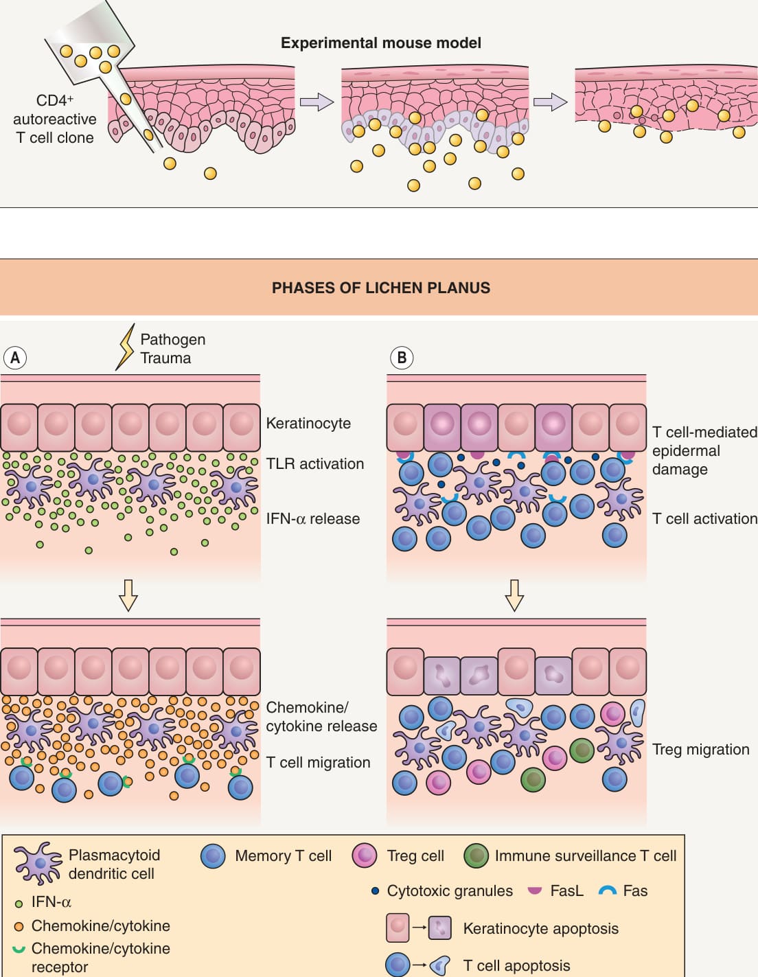
Fig. 11.2 Phases of lichen planus.A In the induction phase, keratinocytes and plasmacytoid dendritic cells (pDCs), upon stimulation of their Toll-like receptors (TLRs) by pathogens or endogenous ligands, can release type 1 IFNs (e. g. IFN-α); this represents an early event in the cascade leading to T cell-mediated epidermal damage. Activated keratinocytes, via production of IL-1β and TNF-α, can induce activation and migration of DCs. Chemokines, such as IP-10/ CXCL10, released locally by pDCs, serve to attract CXCR3-expressing CD8+ or CD4+ effector memory T cells (which have differentiated from naive T cells within lymph nodes following presentation by DCs of self-peptides modified by exogenous antigens [viruses, medications and contact allergens]). Additional chemokine and chemokine receptor pairs have also been implicated in this process (see Table 11.3), and the precise mix of chemokines and cytokines released into the tissue plays an important role in determining the composition of the inflammatory infiltrates. B In the evolution phase, effector T cells (Te) that come to express skin-homing receptors (E-selectin ligands) migrate into the inflammatory site and upon recognition of antigens, are activated and release proinflammatory cytokines and cytotoxic granules, which in turn cause epidermal injury. In addition, Fas/FasL interactions can trigger cell death of both lymphocytes and keratinocytes with possible elimination of potentially harmful autoaggressive T cells. “Inflammatory” and “homeostatic” chemokines produced by keratinocytes direct the traffic of not only “pathogenic” T cells (Te) but also “immune surveillance” T cells (Ts) or regulatory T cells (Treg) into the sites; the relative balance of chemokines produced may determine the outcome of the T cell-mediated immune responses. FasL, Fas ligand.
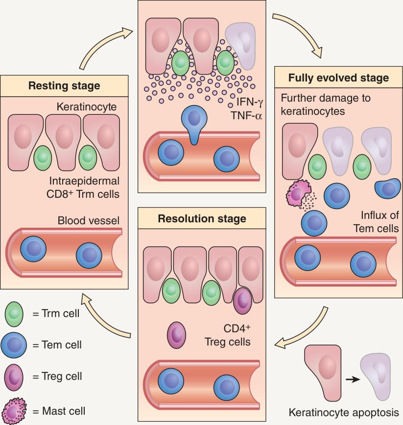
Fig. 11.3 Proposed cascade of events within lesions of fixed drug eruption.
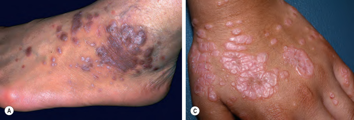
Fig. 11.4 Lichen planus.A
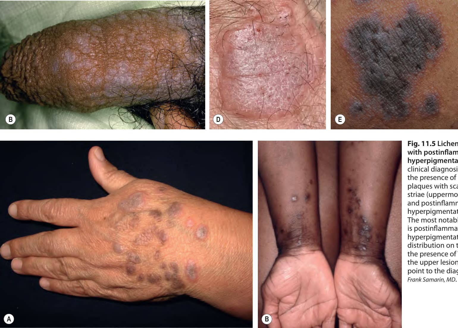
Fig. 11.5 Lichen planus with postinflammatory hyperpigmentation.A The clinical diagnosis is based upon the presence of violaceous plaques with scale, Wickham striae (uppermost lesion), and postinflammatory hyperpigmentation. B The most notable finding is postinflammatory hyperpigmentation, but the distribution on the flexor wrists and the presence of Wickham striae in the upper lesion on the right arm point to the diagnosis. A, Courtesy Frank Samarin, MD
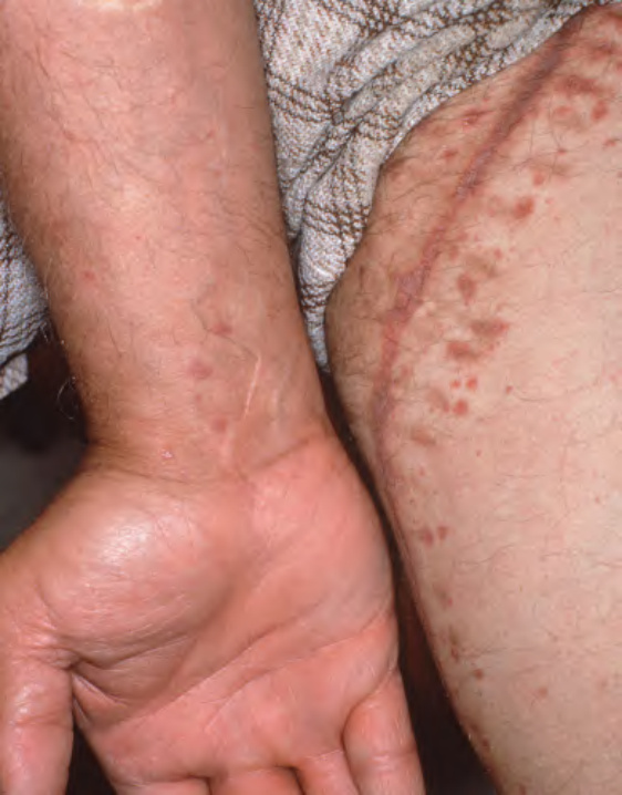
Fig. 11.6 Koebnerization of lichen planus into the site of the excision of the saphenous vein.
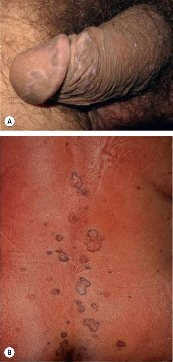
Fig. 11.7 Annular lichen planus.A On the penis, the lesions have a figurate outline with a thin pale violet border and central hyperpigmentation. B On the trunk, the lesions have a thin hyperpigmented rim. A, Courtesy Frank Samarin, MD purple to white in color, while the central portion is hyperpigmented or skin-colored (see Fig. 11.7). Lesions can resemble porokeratosis, tinea or if there is minimal scale, granuloma annulare. Annular lesions occur in ~10% of patients with LP and are usually scattered among more typical lesions (see Fig. 11.4C), but the former may represent the pre-dominant finding. The most common site of involvement is the axilla, followed by the penis, extremities, and groin. While some have pruritus, most patients are asymptomatic.
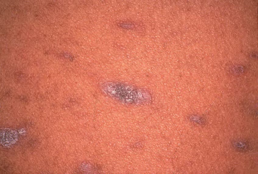
Fig. 11.8 Exanthematous lichen planus. Papulosquamous lesions on the back.
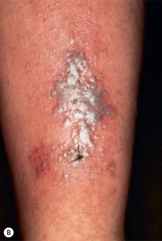
Fig. 11.9 Unusual variants of lichen planus.A Atrophic lichen planus of the lower extremities. B Bullous lichen planus on the shin. C Lichen planus pemphigoides in a patient with anti-BP180 autoantibodies.
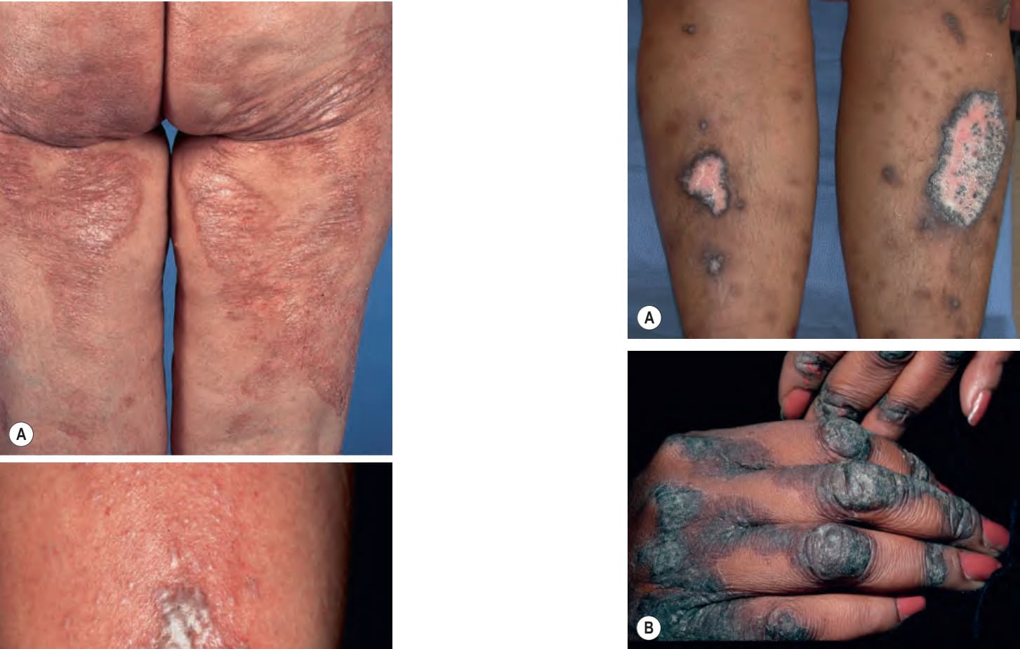
Fig. 11.10 Hypertrophic lichen planus.A On the shins, very thick discrete plaques with dyspigmentation are admixed with smaller linear plaques and areas of postinflammatory hyperpigmentation. B On the dorsal digits, thin violaceous plaques in addition to thick keratotic plaques that favor the knuckles. Courtesy Joyce Rico, MD
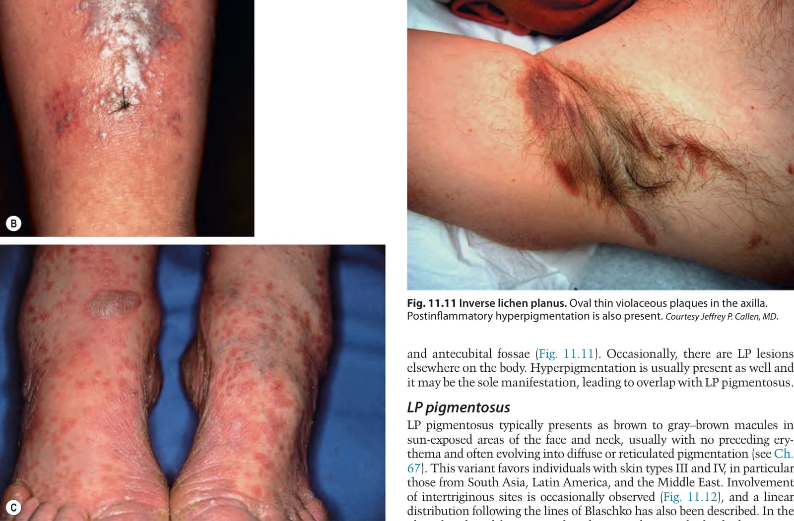
Fig. 11.11 Inverse lichen planus. Oval thin violaceous plaques in the axilla. Postinflammatory hyperpigmentation is also present. Courtesy Jeffrey P. Callen, MD and antecubital fossae (Fig. 11.11). Occasionally, there are LP lesions elsewhere on the body. Hyperpigmentation is usually present as well and it may be the sole manifestation, leading to overlap with LP pigmentosus.
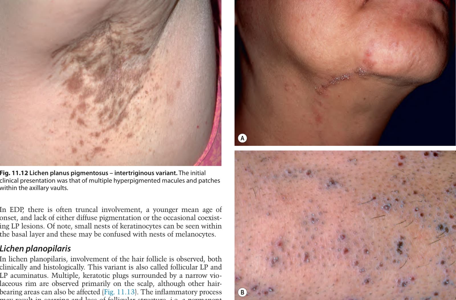
Fig. 11.12 Lichen planus pigmentosus – intertriginous variant. The initial clinical presentation was that of multiple hyperpigmented macules and patches within the axillary vaults.
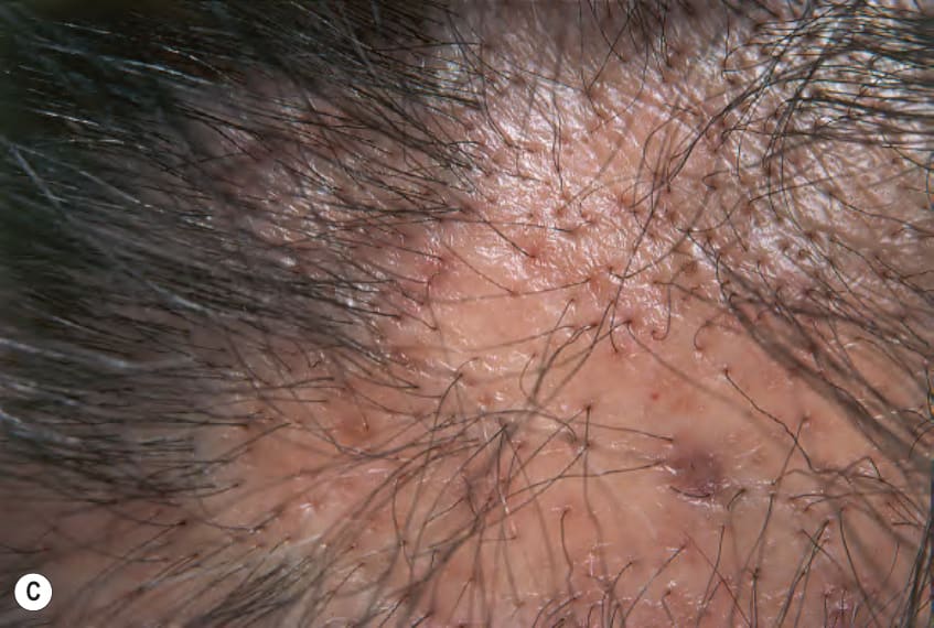
Fig. 11.13 Lichen planopilaris.A Keratotic spines surrounded by a violaceous rim in the linear variant. B Multiple clusters of follicular keratotic plugs on the leg admixed with small violaceous papules, some of which are folliculocentric. C Cicatricial alopecia with “end-stage” changes centrally, but perifollicular inflammation at the margins.
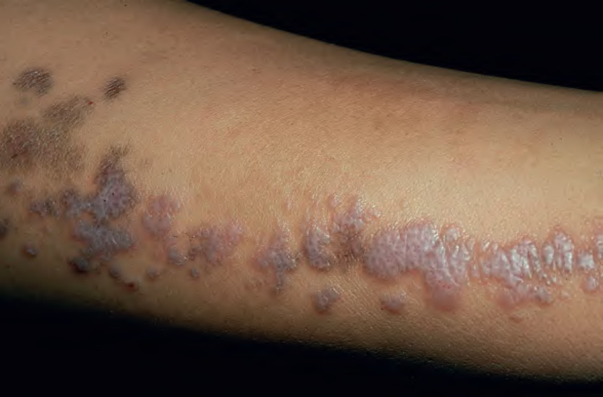
Fig. 11.14 Linear lichen planus. Coalescence of violaceous lesions with Wickham striae along the lines of Blaschko on an extremity. Note the postinflammatory hyperpigmentation proximally. Courtesy Joyce Rico, MD.
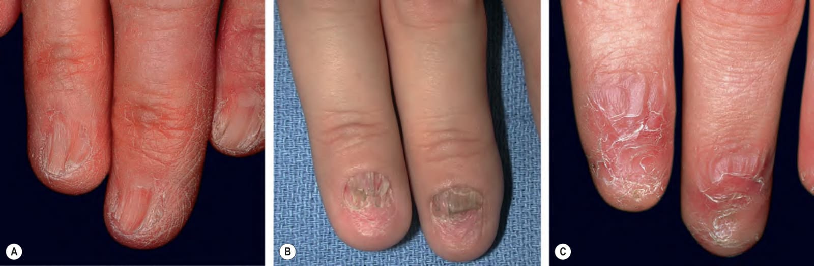
Fig. 11.15 Nail lichen planus.A Thinning of the nail plate with lateral loss. B Longitudinal fissuring of shortened nail plates. C Violaceous discoloration of the periungual area with pterygium formation.
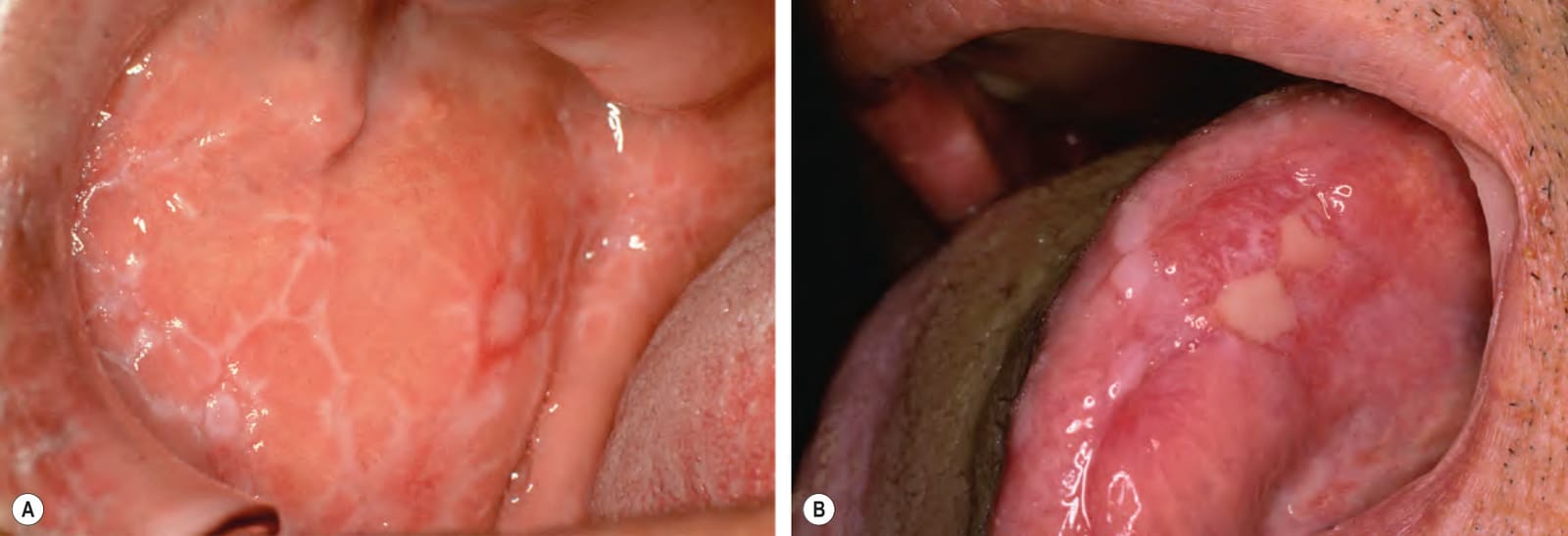
Fig. 11.16 Oral lichen planus.A White lacy pattern and an erosion on the buccal mucosa, the most common location for the reticular form. Note the ring configuration with short radiating spines. B Erosions on the lateral aspect of the tongue in addition to lacy white plaques and scarring. B, Courtesy Louis A. Fragola, Jr, MD.
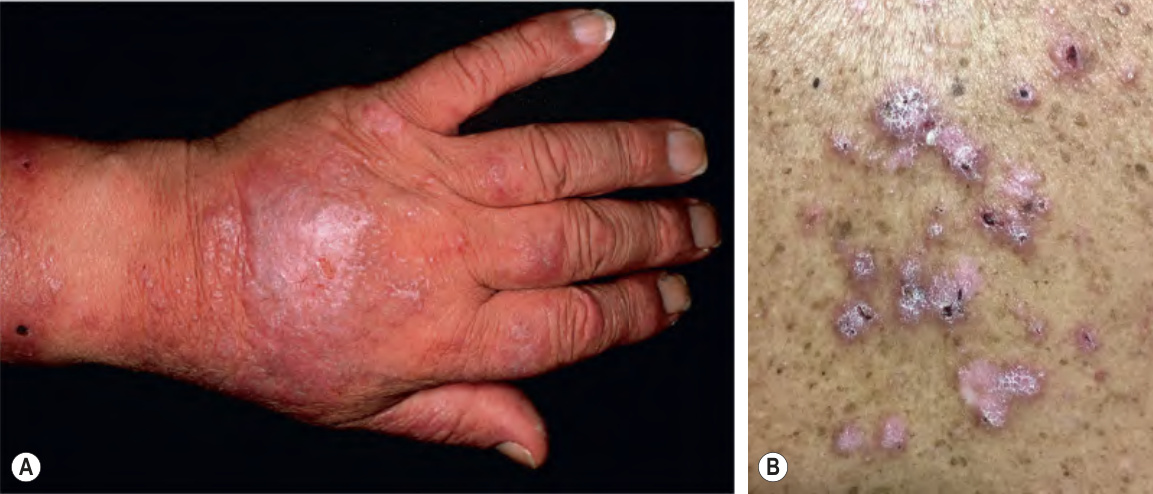
Fig. 11.17 Lichenoid drug eruption.A
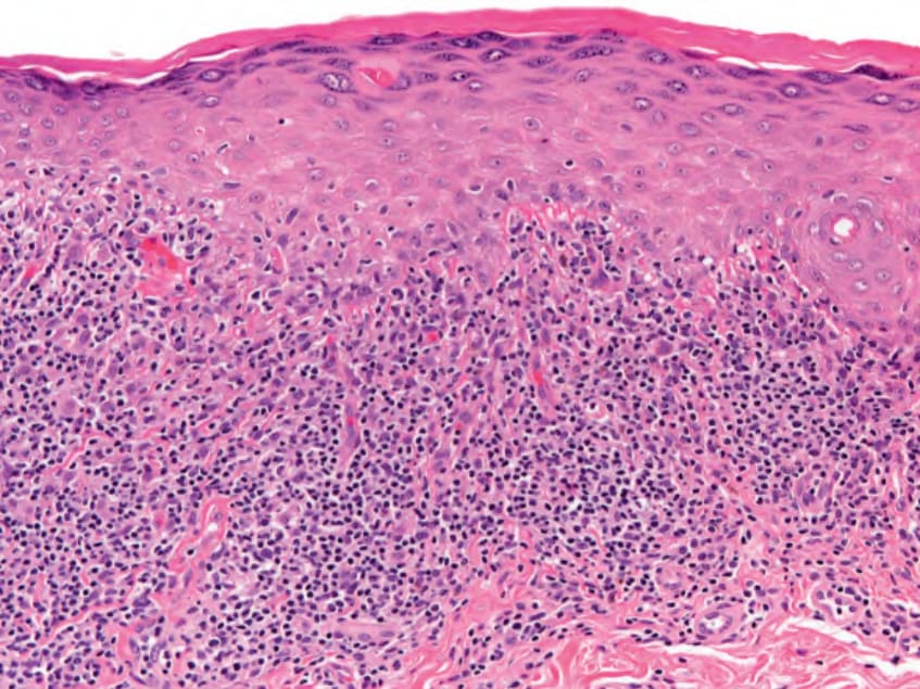
Fig. 11.18 Lichen planus – histopathologic features. Hyperkeratosis, focal increase in the granular layer, sawtoothing of the epidermis with keratinization of the basal layer, and a lichenoid infiltrate. Apoptosis of keratinocytes is also present. Courtesy Lorenzo Cerroni, MD.
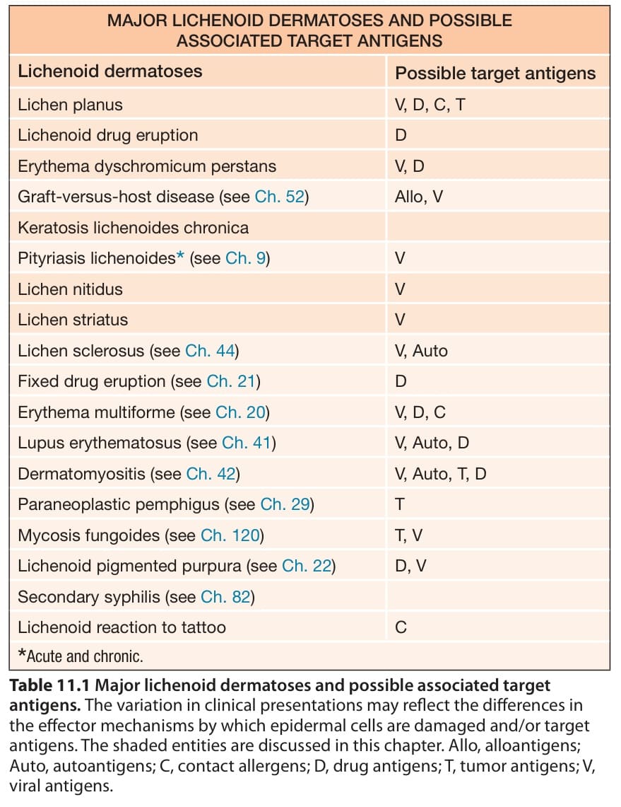
Table 11.1 Major lichenoid dermatoses and possible associated target antigens. The variation in clinical presentations may reflect the differences in the effector mechanisms by which epidermal cells are damaged and/or target antigens. The shaded entities are discussed in this chapter. Allo, alloantigens; Auto, autoantigens; C, contact allergens; D, drug antigens; T, tumor antigens; V, viral antigens.
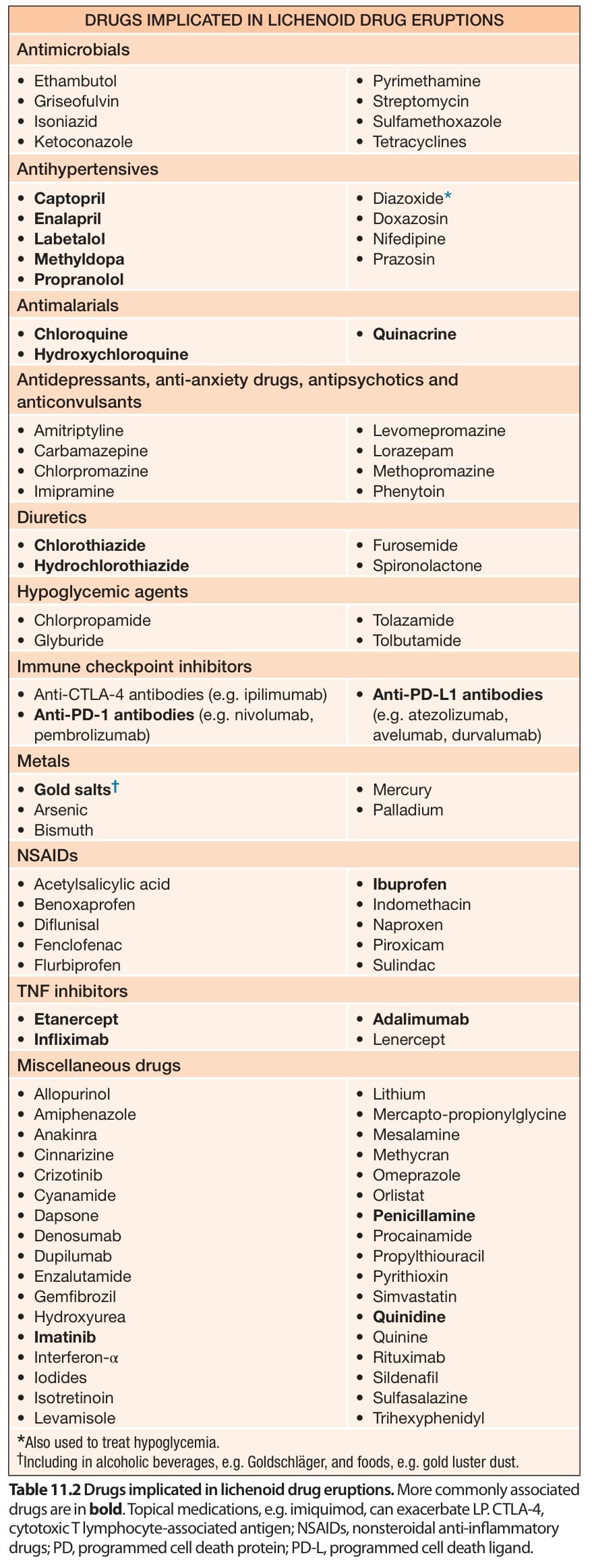
Table 11.2 Drugs implicated in lichenoid drug eruptions. More commonly associated drugs are in bold. Topical medications, e.g. imiquimod, can exacerbate LP. CTLA-4, cytotoxic T lymphocyte-associated antigen; NSAIDs, nonsteroidal anti-inflammatory drugs; PD, programmed cell death protein; PD-L, programmed cell death ligand.
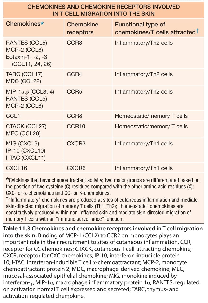
Table 11.3 Chemokines and chemokine receptors involved in T cell migration into the skin. Binding of MCP-1 (CCL2) to CCR2 on monocytes plays an important role in their recruitment to sites of cutaneous inflammation. CCR, receptor for CC chemokines; CTACK, cutaneous T cell-attracting chemokine; CXCR, receptor for CXC chemokines; IP-10, interferon-inducible protein 10; I-TAC, interferon-inducible T cell α-chemoattractant; MCP-2, monocyte chemoattractant protein 2; MDC, macrophage-derived chemokine; MEC, mucosal-associated epithelial chemokine; MIG, monokine induced by interferon-γ; MIP-1α, macrophage inflammatory protein 1α; RANTES, regulated on activation normal T cell expressed and secreted; TARC, thymus- and activation-regulated chemokine.
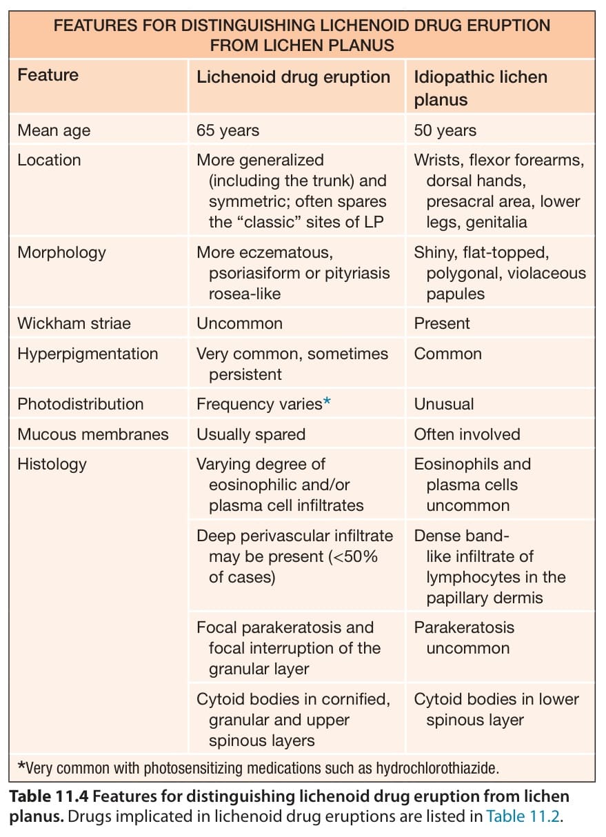
Table 11.4 Features for distinguishing lichenoid drug eruption from lichen planus. Drugs implicated in lichenoid drug eruptions are listed in Table 11.2
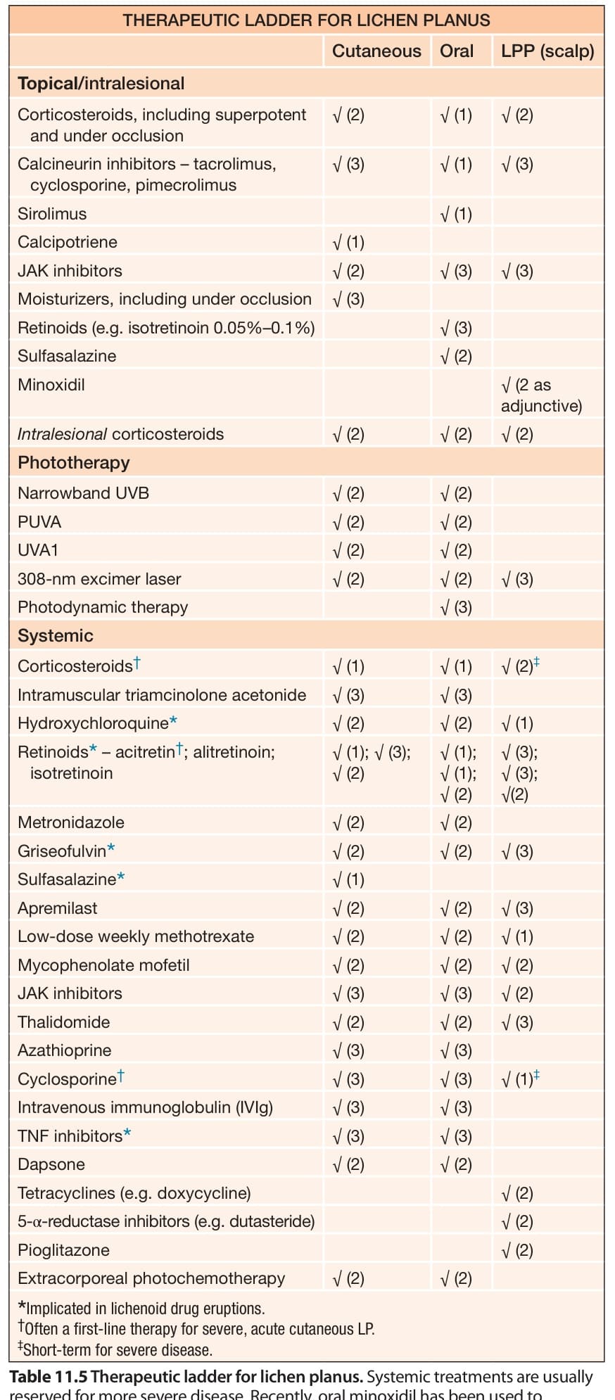
Table 11.5 Therapeutic ladder for lichen planus. Systemic treatments are usually reserved for more severe disease. Recently, oral minoxidil has been used to increase the thickness of remaining scalp hairs in lichen planopilaris (LPP). Key to evidence-based support: (1) prospective controlled trial; (2) retrospective study or large case series; (3) small case series or individual case reports.