LICHEN STRIATUS
Synonyms: Linear lichenoid dermatosis Blaschko linear acquired inflammatory skin eruption (BLAISE)
Key features
An asymptomatic, linear dermatosis that primarily affects children
The primary lesion is a small, flat-topped papule that ranges in color from pink to skin-colored to tan (hypopigmented)
Multiple lesions appear over the course of days to weeks along the lines of Blaschko and usually on an extremity
Spontaneously resolves over months to a few years
Digital involvement may result in nail dystrophy
Introduction
Lichen striatus is an asymptomatic, uncommon, self-limited, linear dermatosis of unknown etiology that generally affects children. The diagnosis is usually made clinically based upon the appearance of the primary lesions and the distinctive developmental pattern. Its distribution along the lines of Blaschko plus the age of the patient usually narrows the differential diagnosis rather quickly. Occasionally, there is overlap with linear LP and “blaschkitis” (see Ch. 62).
History
In 1898, Balzer and Mercier first described a peculiar linear papular eruption that they termed “lichenoid trophoneurosis”. Forty years later, Senear and Caro proposed the name “lichen striatus”. Ever since the condition was first described, the pathogenesis of its linearity has been the subject of debate.
Epidemiology
Lichen striatus is seen primarily in children between the ages of 4 months and 15 years, although the disorder occasionally occurs in adults. The median age of onset is 2 to 3 years and the vast majority of cases occur in preschool-age children. The reported female : male ratio has varied from 1.6 : 1 to 2 : 1.
Pathogenesis
Although the distribution of lichen striatus along the lines of Blaschko points to somatic mosaicism (see Ch. 62), neither the gene(s) involved nor the triggering factors are known. Environmental agents, particularly viruses, have been implicated, given the predominance of the disorder in young children and its seasonal variation (it appears more commonly during spring and summer). However, to date, a viral association has not been proven via serologic testing or cultures. Concurrent familial occurrence of lichen striatus in a mother and child has been touted as evidence for a viral trigger.
In theory, during early fetal development, aberrant clone(s) of epidermal cells produced by somatic mutation migrate out along the lines of Blaschko. Exposure to an infectious agent (e.g. virus, BCG vaccine) or other precipitant could then break previous tolerance to the aberrant clone by inducing a novel membrane antigen. The presence of CD8+ T cells scattered or in clusters around necrotic keratinocytes supports a cell-mediated immunologic reaction by which cytotoxic T cells would attack and eliminate the mutated or virally modified keratinocyte clones. A similar mechanism has been proposed for linear LP, and a post-transplant loss of tolerance could explain linear GVHD.
Lichen striatus may represent a manifestation of an atopic diathesis with the abnormal immune responses usually associated with atopy being a predisposing factor. The timing and relative infrequency of lichen striatus suggests that an infectious agent acts as a trigger in genetically predisposed individuals. Lastly, there are scattered reports of lichen striatus occurring at sites of injury (e.g. the periphery of a burn scar) rather than along the lines of Blaschko, but this could also be explained by a break in tolerance.
Clinical Features
Lichen striatus is typically asymptomatic, but intense pruritus can occasionally occur. The eruption consists of a continuous or inter-rupted band composed of discrete or clustered pink, skin-colored or tan (hypopigmented) papules that are flat-topped, smooth or scaly, and range in size from 2 to 4 mm. Infrequently, vesicles may be present. Often, there is a single, unilateral streak on an extremity along the lines of Blaschko (Fig. 11.19); occasionally, there is a bilateral distribution pattern and/or multiple parallel bands. It is uncommon for lichen striatus to involve the trunk or head and neck region. There are reports of the eruption spreading distally from the trunk down an extremity, as well as observations of proximal extension along an
extremity. Although lichen striatus does not usually recur, relapses may occasionally occur, either in the same site or on the same side of the body.
The eruption usually appears suddenly, develops fully over days to weeks, and after several months to a year or more, undergoes spontaneous resolution. Postinflammatory hypopigmentation may be seen, particularly in those with more darkly pigmented skin. In the latter individuals, the eruption is often first noticed as linear hypopigmentation. When lesions involve the nail apparatus, onycholysis, splitting, fraying and total nail loss may result.
Pathology
The histologic features of lichen striatus are variable and depend upon the age of the lesion at the time the biopsy is performed. In addition, different sites within the same streak can have different findings. In general, there is a lichenoid tissue reaction in addition to varying degrees of involvement of the hair follicles and sweat glands and ducts (Fig. 11.20). Even though the lichenoid inflammation that may be present around hair follicles is indistinguishable from that seen in lichen planopilaris, sweat gland and hair follicle involvement can still be a helpful diagnostic feature of lichen striatus. Exocytosis, parakeratosis, dyskeratosis, and focal or diffuse vacuolar degeneration can be seen in the epidermis overlying the lichenoid infiltrate. Occasionally, older lesions may have features similar to LP.
By immunohistochemistry, CD3+ T cell infiltrates were noted in which CD8+ T cells surrounded necrotic keratinocytes and infiltrated vesicles filled with Langerhans cells. Depending upon the age of the lesions, Langerhans cells were either decreased (earlier lesions) or increased (later phase due to an influx of precursor cells).
Differential Diagnosis
Although the differential diagnosis includes other inflammatory diseases that can assume a linear pattern, such as linear porokeratosis, linear psoriasis, inflammatory linear verrucous epidermal nevus, linear lichen sclerosus and linear Darier disease, the primary differential
diagnosis is linear LP, blaschkitis, and linear GVHD. The latter occurs in a specific clinical setting, while blaschkitis favors the trunk, is usually seen in adults, often consists of multiple streaks, recurs, and can have features of dermatitis. Although linear LP and lichen striatus can occasionally be indistinguishable histologically, the primary lesions usually differ in size and color, and hypopigmentation is a frequent sequela of lichen striatus, while, in general, hyperpigmentation appears in the wake of LP. In lichen nitidus, linear lesions reflect previous traumatic injury to the skin.
Treatment
Treatment of lichen striatus is usually not needed because it is self-limited, usually resolving within 1 to 2 years. Topical corticosteroids under occlusion can be used to hasten spontaneous resolution. In scattered case reports, topical calcineurin inhibitors have also been reported to be effective, including for the nail dystrophy. Obviously, with any purported therapy, the natural history of lichen striatus must be kept in mind.
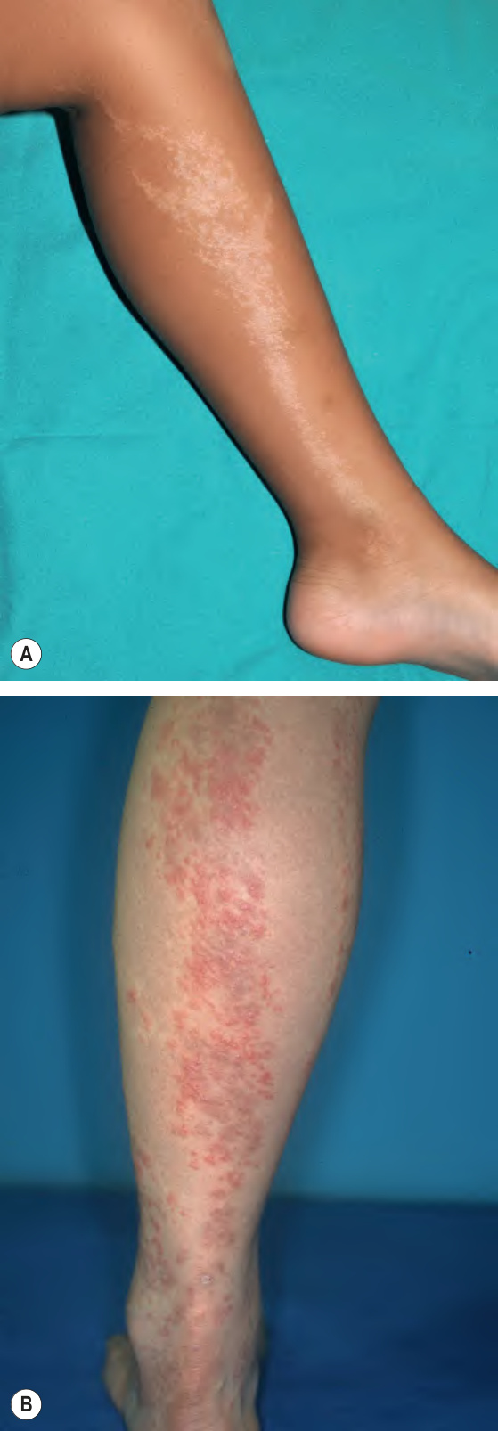
Fig. 11.19 Lichen striatus.A Linear streak on the leg that follows the lines of Blaschko. It is composed of numerous small, tan (hypopigmented), flat-topped papules. B Three linear streaks on the lower extremity composed of multiple pink papules, some of which are flat-topped with scale. The primary differential diagnosis would be blaschkitis. A, Courtesy Antonio Torrello, MD.
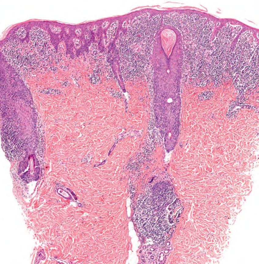
Fig. 11.20 Lichen striatus – histopathologic features. In addition to hyperkeratosis with focal parakeratosis, both a lichenoid and a perivascular and periadnexal lymphocytic infiltrate extending into the deeper dermis is seen. Courtesy Lorenzo Cerroni, MD.