LICHEN NITIDUS
Key features
The eruption consists of multiple, tiny, discrete, shiny papules, often in clusters
Favored sites of involvement include the flexor aspects of the upper extremities, the genitalia, and the anterior trunk
Linear arrays of papules occur secondary to the Koebner phenomenon
The histologic correlate of the papule is a well-circumscribed infiltrate composed of lymphocytes and epithelioid cells confined to the width of two to three dermal papillae
Introduction
Lichen nitidus is an uncommon chronic eruption consisting of multiple, tiny, discrete, skin-colored papules that are often arranged in larger clusters. The papules have a distinctive histology with a dense, well-circumscribed, lymphohistiocytic infiltrate closely apposed to the epidermis. There has been considerable debate, however, as to whether lichen nitidus represents a distinct and separate entity or should be regarded as an unusual variant of LP; their coexistence has certainly been reported and progression from lichen nitidus to LP has also been observed. It is generally accepted that lichen nitidus has no relationship to any systemic illness. Only a few authors believe that it may be a cutaneous manifestation of Crohn disease.
History
In 1901, Pinkus first described a peculiar papular eruption he termed “lichen nitidus” and suggested that it was a distinct entity histologically.
Epidemiology
Reliable epidemiologic data are difficult to accumulate because of the relative rarity of lichen nitidus. In a US study of 43 patients, primarily Caucasians and African-Americans, the disorder was not found to be restricted to any specific population group based on race, sex, or age. Other authors have observed that lichen nitidus is more prevalent among children or young adults, and a female predominance has been described in the generalized variant. It has also been reported in patients with Down syndrome. Familial lichen nitidus, including in identical twins, is very rare.
Pathogenesis
Although much attention has been focused on the relationship of lichen nitidus to LP, there has been limited recent research regarding its pathogenesis. Lichen nitidus was initially thought to represent a tuberculous lesion or tuberculid because of its histologic features. However, no causative infectious agents have ever been demonstrated, even by repeated inoculations of tissue into animals.
Even though LP and lichen nitidus can coexist in the same patient and they share some similar clinical features (Table 11.6), most authors believe that lichen nitidus is a separate entity because of its distinctive clinical and histologic features. The possibility exists that the two conditions represent different responses to a similar triggering factor, e.g. hepatitis B vaccine. An additional example would be a patient who had Crohn disease in whom LP and lichen nitidus developed over different time frames.
Clinical Features
Lichen nitidus is characterized by numerous tiny, discrete, skin-colored, uniform, pinhead-sized papules (Fig. 11.21A). Individual papules are usually flat with a shiny surface (Fig. 11.21B); occasionally they have a central depression. There may be pruritus, but it is an uncommon feature. Although most often skin-colored, the papules can exhibit a variety of hues, from pink or yellow to red–blue or brown. In darkly pigmented individuals, the papules tend to be hypopigmented, but sometimes they are hyperpigmented; marked hyperpigmentation has rarely been reported.
The lesions are usually distributed on the flexor aspects of the upper extremities as well as the chest, abdomen, dorsal aspects of the hands, and genitalia (Fig. 11.21A,C). Less commonly, the face, neck, lower extremities, palms, soles, and mucous membranes are involved. Oral lesions are thought to be rare but are probably underestimated; they appear as minute, flat, gray–white papules on the soft mucosa or white plaques on the tongue and hard palate. Nail involvement is observed in up to 10% of patients (primarily adults) and the changes include pitting, rippling, longitudinal ridging, terminal splitting, and increased longitudinal linear striations.
Lichen nitidus is one of several diseases that exhibit the Koebner phenomenon. Vesicular and hemorrhagic lesions are found in some patients, and they are often admixed with the more typical papules. Several other clinical variants have been described, including palmoplantar, linear, and follicular spines. A perforating variant has also been reported, usually in association with LP. In the generalized form, lesions occasionally coalesce. According to one study, the duration in two-thirds of the patients was 1 year or less and the longest duration was 8 years.
Pathology
The constellation of histologic features in lichen nitidus is very distinctive. As a result, the clinical diagnosis can be confirmed histologically.
A well-circumscribed infiltrate composed of lymphocytes, epithelioid cells, and occasional Langhans giant cells is typically “clutched” by the surrounding hyperplastic rete ridges in a “ball and claw” configuration (Fig. 11.22). In most lesions, the infiltrate is confined to the width of one to three dermal papillae.
The overlying epidermis is usually atrophic and frequently exhibits a parakeratotic “cap” centrally. Absence or thinning of the overlying granular layer is seen and vacuolar degeneration of the basal layer is consistently observed, often accompanied by incontinence of melanin pigment. Civatte bodies are occasionally seen. A Max-Joseph-like space, i.e. focal separation of the epidermis and dermis secondary to vacuolar degeneration, is sometimes observed. The lichenoid infiltrate closely apposes the epidermis and is thought to play a role in inducing reactive epidermal hyperplasia of the surrounding rete ridges. In perforating lichen nitidus, transepidermal elimination is observed (see Ch. 96).
The predominant cell types in the dermal infiltrate are lymphocytes and epithelioid histiocytic cells, and in some cases, epithelioid cells pre-dominate. Immunohistochemical studies have shown a marked pre-dominance of CD4+ T cells over CD8+ T cells, as well as the presence of large numbers of CD1+ Langerhans cells in the dermal infiltrates. Of note, the pattern in lichen nitidus lesions of a mixed cellular infiltrate characterized by macrophages and a helper T cell response with few cutaneous lymphocyte antigen-positive (CLA+) cells is different from that seen in LP.
Differential Diagnosis
The differential diagnosis includes LP, guttate lichen sclerosus, lichen spinulosus, frictional lichenoid dermatitis (elbows and knees), and papular eczema (especially in skin phototypes V and VI), as well as verruca plana, lichen scrofulosorum, lichen striatus, papular sarcoidosis, and the lichenoid variant of secondary syphilis. All of these diagnoses, except LP and perhaps lichen scrofulosorum, can usually be excluded relatively easily on the basis of clinical plus histologic findings. It is not always possible to differentiate early tiny papules of LP because they may be clinically and histologically indistinguishable from lichen nitidus (see Table 11.6). Furthermore, lesions identical to lichen nitidus can be found in 25% to 30% of patients with LP. In such cases, the lack of variation in size, the absence of a violaceous color or Wickham striae, and no deposits of immunoglobulin in dermal papillae would suggest the diagnosis of lichen nitidus.
Periadnexal inflammation is more typical of lichen striatus, but patients with the latter have been described where both periadnexal inflammation and superficial features of lichen nitidus were present, suggesting a morphologic spectrum. Lichen spinulosus is characterized by follicular keratotic papules, 1–3 mm in size with a central horny spine, that are grouped into patches on the neck, arms, abdomen and buttocks. The possibility of the spinous follicular variant of lichen
nitidus should be kept in mind when a patient presents with spiny keratotic papules plus typical histologic features of lichen nitidus. Lastly, lichen nitidus-like lesions can be seen in a photodistribution in summertime actinic lichenoid eruption, also referred to as actinic lichen nitidus (see below).
Treatment
Since the majority of patients experience spontaneous clearing within one or several years, treatment is primarily symptomatic. When significant pruritus is present, topical corticosteroids and oral antihistamines may be helpful. Anecdotally, topical calcineurin inhibitors have been reported as an effective therapy in children with lichen nitidus. Generalized lichen nitidus that had failed to respond to topical corticosteroids has been successfully treated with either narrowband UVB or PUVA. One patient with peripheral CD4+ T cell lymphocytopenia and lichen nitidus who was treated with topical dinitrochlorobenzene (DNCB) developed a pruritic erythematous eruption followed by resolution of the lesions of lichen nitidus.
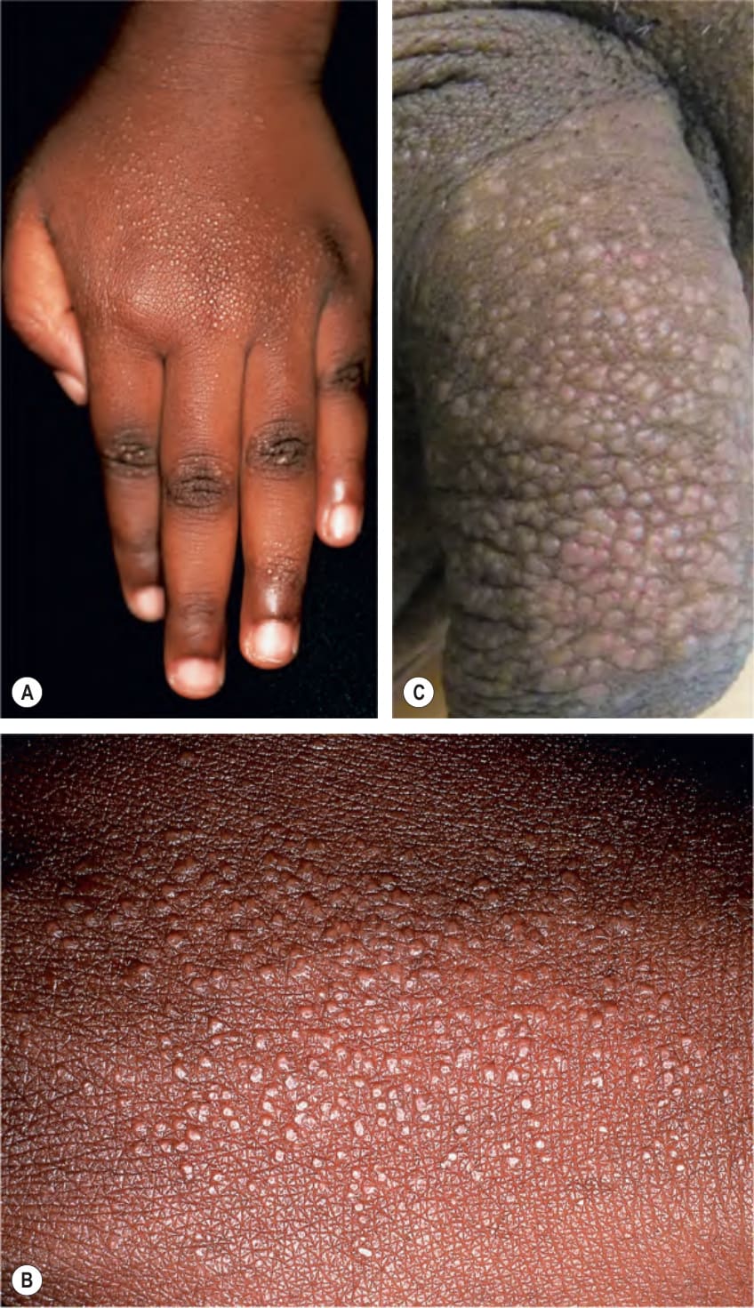
Fig. 11.21 Lichen nitidus. A Numerous tiny flat-topped papules on the hand. B A close-up view shows the shiny surface. C Numerous pink lesions on penis. C, Courtesy Kiran Motaparthi, MD.
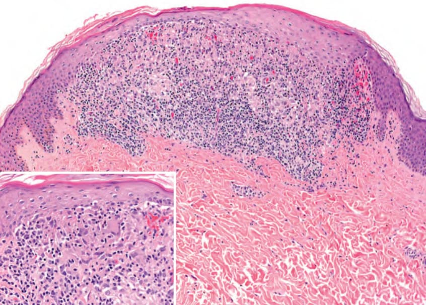
Fig. 11.22 Lichen nitidus – histopathologic features. Note the typical “ball and claw” configuration for the infiltrate and epidermis, respectively. There is thinning of the epidermis that overlies the infiltrate. Several Langhans giant cells are within the infiltrate (inset). Courtesy Lorenzo Cerroni, MD.
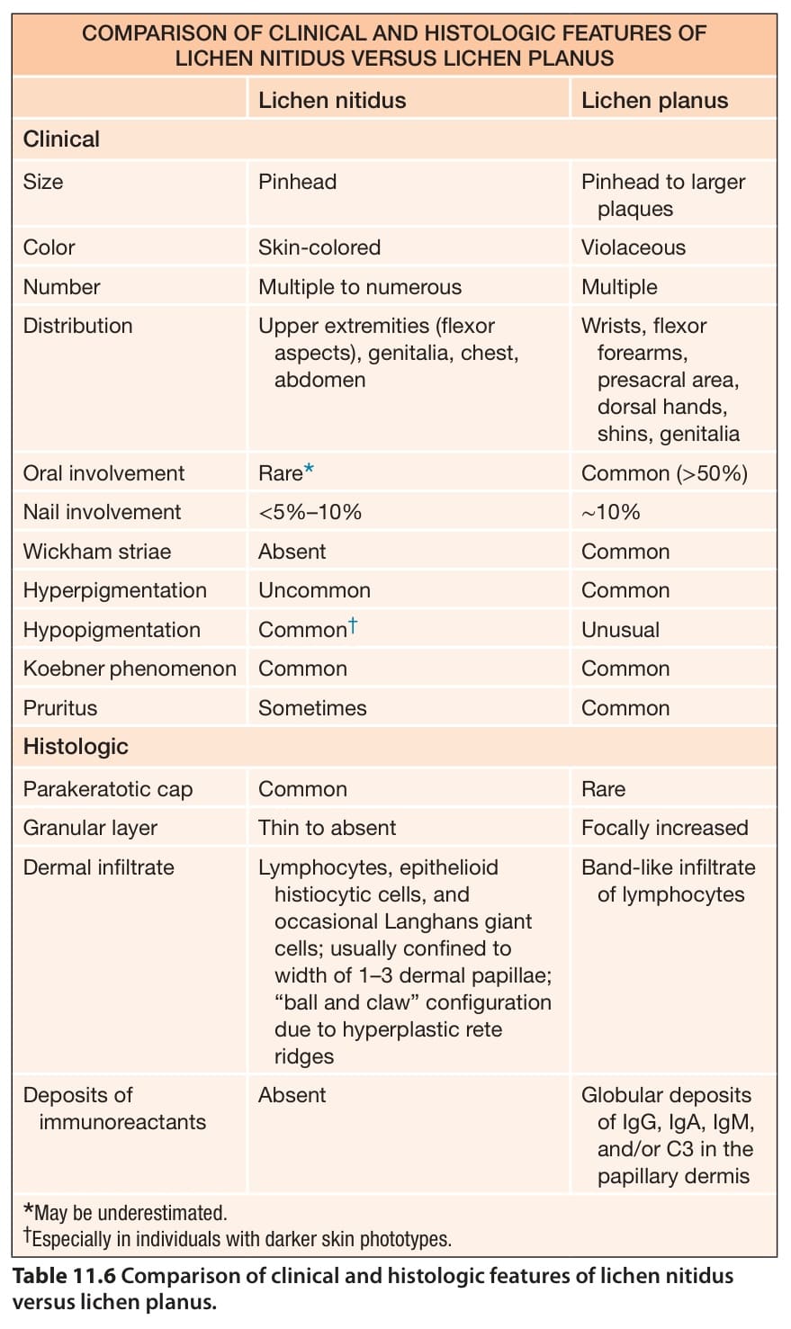
Table 11.6 Comparison of clinical and histologic features of lichen nitidus versus lichen planus.