MYCOSIS FUNGOIDES
Definition
Mycosis fungoides (MF) represents the most common type of CTCL and accounts for ~50% of all primary cutaneous lymphomas (see Table 120.1). The term MF should be restricted to the classic “Alibert– Bazin” type characterized by the typical evolution of patches, plaques, and tumors, or for clinicopathologic variants showing a similar clinical course.
Epidemiology
MF has an incidence of about 0.4 per 100 000 inhabitants per year in the US. MF typically affects older adults (median age at diagnosis: 55–60 years), but it may occur in children and adolescents as well. Men are affected more often than women, with a male-to-female ratio of 1.6–2.0 : 1.
Pathogenesis
The etiology and the pathogenetic mechanisms involved in the development and stepwise progression of MF are largely unknown. Genetic, environmental, and immunologic factors have all been considered.
Genetic factors
Lymphomagenesis is considered to be a multifactorial process, in which a stepwise accumulation of genetic abnormalities may result in clonal proliferation, malignant transformation, and ultimately, progressive and widely disseminated disease. Although the successive clinical steps of tumor progression were described more than a century ago, the molecular events underlying the different steps of tumor progression have not been fully identified. In gene expression studies of early stage MF, an overexpression of the transcription factor TOX and PDCD1 (PD-1) was observed and noted to be a useful diagnostic marker. Advanced stages of MF usually have complex karyotypes with somatic copy-number variants constituting the vast majority of all driver mutations.
In tumor stage MF, integrated whole-genome sequencing and RNA sequencing have shown rearrangements in multiple genes involved in transcriptional regulation and signal transduction. Deletions of HNRNPK and SOCS1, which encode signaling inhibitors of the JAK-STAT pathway, were the most frequent genetic alterations in MF after deletion of CDKN2A. Deletion or inactivation of CDKN2A has been associated with a shorter survival in patients with tumor stage MF while deletion of HNRNPK and SOCS1 may contribute to constitutive activation of STAT3.
Staphylococcus aureus and its toxins can also activate STAT3 signaling and increase expression of the interleukin-2 receptor (IL-2R) in tumor cells and nonmalignant T cells, thereby stimulating proliferation of tumor cells in CTCL. Aggressive antibiotic treatment can effectively eradicate this stimulus, normalize the tumor microenvironment, inhibit STAT3 signaling and cellular proliferation within lesional skin, and provide significant clinical improvement in MF and Sézary syndrome. Staphylococcus aureusα-toxin also blocks CD8+ T cell-mediated killing of malignant T cells, allowing immune evasion and continued proliferation.
Environmental factors
Persistent antigenic stimulation has been demonstrated to play a crucial role in the development of various malignant lymphomas, including mucosa-associated lymphoid tissue (MALT) lymphomas (Helicobacter pylori infection), CBCL (Borrelia burgdorferi infection), and enteropathy-type T cell lymphoma (celiac disease). In MF, persistent antigenic stimulation has also been proposed as an initial event, but the nature of the antigen(s) involved is unknown. Large case–control studies have suggested a relationship with industrial or environmental exposures, but their role in the development of MF remains controversial. Whereas the etiologic roles of human T cell leukemia virus 1 (human T cell lymphotropic virus 1; HTLV-1) in adult T cell leukemia/ lymphoma and EBV in nasal NK/T cell lymphoma have been firmly established, conclusive evidence for a primary etiologic role of these and other viruses in MF is lacking.
Immunologic factors
CD8+ cytotoxic T cells (CTL) are thought to play a crucial role in the antitumor response in MF. A relationship between high percentages of CD8+ CTL in the dermal infiltrates and improved survival has been described. These CD8+ T cells exert their antitumor effect both by a direct cytotoxic effect and by the production of cytokines, particularly interferon (IFN)-γ. They can mediate tumor cell lysis via exocytosis of cytotoxic granules containing perforin, granzymes, and T cell-restricted intracellular antigen (TIA-1), and by expression of Fas ligand (FasL), which interacts with Fas (CD95; APO-1) on the neoplastic T cells. Both pathways ultimately lead to activation of caspase 3 and tumor cell death. Loss of Fas expression or function by the neoplastic T cells is one of the many mechanisms by which tumor cells can escape from an effective antitumor response.
In most patients, the neoplastic T cells in SS and tumor stage MF are derived from CD4+ T cells with a Th2 cytokine profile (production of IL-4, IL-5, and IL-10), whereas the cytotoxic T cells are the main producers of IFN-γ, which plays an important role in augmenting T celland NK cell-mediated killing. In accordance with this concept, a gradual shift from a predominantly type 1 cytokine profile in MF patches and plaques to a predominantly type 2 cytokine profile in MF tumors has been suggested. Increased levels of Th2 cytokines may impair the Th1 cell-mediated antitumor response and contribute to the immunosuppression seen in patients with advanced MF.
Clinical Features
Characteristically, patients with classic MF progress from patch stage to plaque stage and finally to tumor stage disease, and they have a protracted clinical course over years or even decades. Before a definite diagnosis is made, patients generally have many years of nonspecific eczematous or psoriasiform skin lesions and non-diagnostic biopsies. The median duration from onset of skin lesions to the diagnosis of MF is 4–6 years, but it may vary from several months to more than five decades.
Early patch stage MF is characterized by the presence of variably sized erythematous, finely scaling lesions, which may be mildly pruritic (Fig. 120.2A). These early lesions may show variable degrees of atrophy, and a poikilodermatous variant consisting of patches with mottled hyper- and hypopigmentation, atrophy, and telangiectasia has been described (formerly called poikiloderma vasculare atrophicans). Generalized hypopigmented lesions may be observed in patients with
1A).A Patches on the buttocks involving less than 10% of the skin surface. B Few atypical T cells in the basal layer of the epidermis. Immunohistochemical staining for CD3 more clearly demonstrates many atypical lymphocytes in a characteristic linear configuration along the epidermal basal layer (inset).
darkly pigmented skin (Fig. 120.3), and this is also a common presentation of juvenile-onset MF.
The initial skin lesions have a predilection for the buttocks and other covered sites of the trunk and limbs. With progression, more infiltrated reddish-brown, scaling plaques develop, which gradually enlarge and may have an annular, polycyclic, or typical horseshoe-shaped configuration (Fig. 120.4A). It should be stressed that many patients never progress beyond the plaque stage of the disease. However, a minority of patients may develop nodules or tumors. These patients with tumor stage MF characteristically show a combination of patches, plaques, and tumors (Fig. 120.5A); the latter often show ulceration.
If only skin tumors are present without preceding or concurrent patches or plaques, a diagnosis of MF is highly unlikely and another type of CTCL should be considered. The risk of developing extracutaneous disease correlates with the extent and type of skin lesions. It is exceedingly rare in patients with limited patch/plaque stage disease, relatively uncommon in patients with generalized plaques, and most likely in patients with skin tumors or erythroderma. Initially extracutaneous dissemination most often involves the regional lymph nodes draining areas of extensive skin involvement. Visceral involvement may develop subsequently and can involve any organ. The bone marrow is rarely involved.
(stage 1B). Generalized hypopigmented lesions as well as pink to pink– brown plaques.
Pathology
Early patch lesions in MF show superficial band-like or lichenoid infiltrates, consisting primarily of lymphocytes. Atypical cells, with small to medium-sized, highly convoluted (cerebriform) and sometimes hyperchromatic nuclei, are few in number and are mostly confined to the epidermis (epidermotropism). These lymphocytes characteristically colonize the basal layer of the epidermis as single cells surrounded by vacuolated halos, often in a linear configuration (Fig. 120.2B).
In plaques, epidermotropism is generally more pronounced (Fig. 120.4B). The presence of intraepidermal nests of atypical cells (referred to as Pautrier microabscesses, although the initial description was by Darier) is a highly characteristic feature, but is observed in only a minority of cases. The epidermis may show acanthosis and elongated rete ridges, but spongiosis is generally mild or absent. The dermal infiltrates are also more pronounced, and they may contain a higher number of atypical cells with cerebriform nuclei and occasional blast cells, as well as admixed eosinophils and plasma cells. Rarely, a predominantly inter-stitial infiltrate that may resemble morphea is observed (interstitial MF).
With progression to tumor stage MF, the dermal infiltrates can involve the entire dermis and extend into the subcutaneous tissue. Epidermotropism may no longer be present. The tumor cells increase in number and size, showing variable proportions of small, mediumsized, or large cells with cerebriform nuclei, blast cells with prominent nuclei, and intermediate forms (Fig. 120.5B). Large cell transformation, defined by the presence of CD30-negative or CD30-positive large cells exceeding 25% of the infiltrate or forming microscopic nodules, may occur and is generally associated with a poor prognosis. However, within this group, CD30-positive patients have a much better prognosis than CD30-negative patients.
Immunophenotype
The neoplastic cells in MF have a mature CD3+, CD4+, CD45RO+, CD8− memory T cell phenotype. In a minority of patients with otherwise classic MF, a CD3+, CD4−, CD8+ mature T cell phenotype or more rarely a γ/δ T cell phenotype (βF1−, TCRγ/δ+, CD3+, CD4−, CD8+) may be seen. These patients have the same clinical behavior and prognosis as CD4+ cases and should not be considered separately. Demonstration of an aberrant phenotype (e.g. loss of pan-T cell antigens such as CD2, CD3, and CD5) is an important adjunct in the diagnosis of MF, but is uncommon in the early stages of MF. A CD8+ T cell phenotype is more common in pediatric MF and in some variants of MF including hypopigmented, hyperpigmented, or poikilodermatous MF.
Differential Diagnosis
Regarding the differential diagnosis of MF, three categories should be considered. The first category contains a diverse group of benign
(stage 1B).A Extensive patches and plaques with scale involving more than 10% of the skin surface. B Epidermotropism with the formation of small nests of atypical cells (Pautrier microabscesses). The majority of the dermal infiltrate is composed of small reactive lymphocytes. A, Courtesy Lorenzo Cerroni, MD.
dermatoses, which early MF may resemble clinically, and it includes several types of eczema, psoriasis, superficial fungal infections, and drug reactions. These specific diagnoses can generally be excluded by histologic and other standard dermatologic examinations. This category may also include patients with large plaque parapsoriasis (parapsoriasis en plaque) who show slightly scaly, sometimes atrophic, erythematous patches or plaques, which are commonly located on the trunk and buttocks (see Ch. 9). Whereas large plaque parapsoriasis cannot be distinguished clinically from early patch or plaque stage MF, the histologic features are often not consistent with MF. Long-term follow-up studies have documented progression of large plaque parapsoriasis to overt MF in ~10% of cases. However, one prevailing opinion is that large plaque parapsoriasis should be considered a form of MF, rather than a potential precursor of MF, but the authors feel there is no consensus. There is more consensus that small plaque parapsoriasis does not represent MF.
A second category includes several benign conditions with histologic features highly suggestive of MF. Examples of these are lymphomatoid contact dermatitis, lymphomatoid drug reactions, and actinic reticuloid. Apart from subtle histologic differences (e.g. the predominance of atypical T cells in the dermal infiltrates rather than in the epidermis), careful evaluation of the clinical features, which are generally not consistent with MF, often results in a correct diagnosis.
The third category includes other types of (epidermotropic) CTCL, which may resemble MF histologically. Diagnostic features of these entities are presented in Table 120.2.
Staging Systems and Staging Procedures
Staging patients with MF and SS is important, since it determines management and treatment and has prognostic significance. In 2007, a revised clinical staging system for MF and SS was proposed, which is based on the TNM (tumor–node–metastasis) classification system and takes into account the type and extent of skin lesions (T1–4) as well as the presence or absence of nodal (N0–3), visceral (M0–1), and peripheral blood involvement (B0–2) (Table 120.3 & 120.4).
Evaluation of patients suspected of having MF should include a thorough physical examination with special attention to the type and extent of skin lesions and the presence of palpable lymph nodes, as well as skin biopsies, complete blood counts, and serum chemistries. Enlarged lymph nodes should be biopsied. Histologically, distinction can be made between lymph nodes showing dermatopathic lymphadenopathy without involvement by MF (N1), dermatopathic lymphadenopathy with early MF involvement (N2), and lymph nodes showing effacement of the normal lymph node architecture by neoplastic T cells (N3). The prognostic significance of such a subdivision has been well established.
No further examinations are recommended for patients with stage IA–B disease. CT or PET-CT scans of the chest and abdomen are recommended in patients in whom extracutaneous disease is suspected, but they are less useful in patients with limited patches
and/or plaques without lymphadenopathy. Examination of other organs, including the bone marrow, should only be performed if clinically indicated.
Treatment
The choice of an initial treatment in MF depends on the stage of the disease and the general condition and age of the patient. Given the chronic and recurrent nature of MF, treatment should be aimed at improving symptoms while limiting toxicity. Following the traditional “stage-based” approach, skin-directed therapies are preferred in the
early stages of MF (stages IA–IIA) and even in patients with limited tumor stage MF (IIB) (Table 120.5). These skin-directed therapies include topical or intralesional corticosteroids, topical cytotoxic agents (e.g. mechlorethamine [nitrogen mustard]), phototherapy, and radiotherapy. In patients with stage IA disease even expectant management with active surveillance can be considered. The efficacy of skin-directed therapies in MF is explained by the preferential localization of the neoplastic skin-homing T cells to the epidermis and superficial dermis. Systemic multi-agent chemotherapy is not useful in these early stages, since it does not improve survival and is associated with considerable morbidity.
In patients with refractory or progressive skin disease, skin-directed therapies can be combined with IFN-α or systemic retinoids. Alternatively, novel agents such as denileukin diftitox, mogamulizumab, or histone deacetylase inhibitors (HDACi) such as vorinostat and romidepsin can be used, before systemic chemotherapy is considered. HDACi and denileukin diftitox have been approved by the FDA for patients with relapsed and refractory CTCL, but they have not been registered for CTCL in Europe. While denileukin diftitox was withdrawn from the market in 2014, a revised version is currently under investigation. In general, systemic chemotherapy is only indicated in advanced stages when there is nodal or visceral involvement or in patients with rapidly progressive tumors unresponsive to less aggressive therapies.
Skin-directed therapies
In many MF patients with only patches and thin plaques, application of topical corticosteroids is effective in controlling disease activity. In patients with limited patch/plaque stage disease (mainly patches), complete remissions in up to 60% of patients have been reported. In more advanced stages, they continue to be an important adjuvant therapy.
Topical application of mechlorethamine has proven to be an effective treatment for early stage MF. Mechlorethamine, either dissolved in water or compounded in an ointment- or gel-based preparation (see Ch. 129), results in complete remissions in approximately 60%–80% of patients with stage IA–B disease. Side effects include skin irritation, allergic contact dermatitis, and an increased risk for the development of skin cancer related to long-term use.
Total skin electron beam irradiation (TSEB) with an energy of 4–6 MeV is a highly effective treatment in patients with skin-limited MF (see Ch. 139). The total dose is traditionally 36 Gy administered in fractions of 1.5–2 Gy over an 8- to 10-week period. Recently, lower total doses (10–12 Gy) have been employed, with the advantages of a shorter duration of treatment, fewer side effects, and opportunity for re-treatment. TSEB is most effective in patients with stage IA–B disease, with complete response rates of >80%. However, in most centers, such patients are treated with phototherapy or topical chemotherapy. TSEB is particularly useful in patients with tumor stage MF, where complete
response rates of ~40% have been reported. Side effects are generally mild and include erythema, scaling, and temporary loss of hair, nails, and sweat gland function.
Local radiotherapy with X-ray or preferably electron beam may be considered for single tumors in patients with plaque stage disease, either in combination with other modalities (e.g. PUVA) as an alternative to TSEB, or for new tumors following TSEB. A dose of ≥8 Gy suffices. In patients with unilesional MF, local radiotherapy may be curative.
Several types of phototherapy can be used in the treatment of MF, in particular PUVA therapy and narrowband UVB therapy (Ch. 134). Extracorporeal photopheresis (ECP) may be effective in patients with erythrodermic MF (see the section on Sézary syndrome).
PUVA treatment has become a standard therapy for the early stages of MF. In patients with stages IA–IIA, complete response rates of 80%–90% have been reported. In many centers, maintenance PUVA therapy (every 2 to 4 weeks) is given to prolong remission. Although sustained complete remissions have been reported, most patients will relapse after cessation of PUVA therapy or during maintenance treatment. Recurrent or persistent lesions particularly favor UV-shielded areas, such as the inner thighs and the gluteal cleft. In tumor stage MF, PUVA therapy alone is unlikely to result in complete responses, but favorable results may be achieved when combined with systemic retinoids, IFN-α, or radiotherapy.
In patients with only patches, narrowband UVB therapy (311 nm) represents first-line therapy.
Systemic therapies (other than chemotherapy)
The most commonly prescribed biological response modifier has been interferon-alpha (IFN-α). In most centers, IFN-α is administered subcutaneously in doses of 3 to 9 million units three times a week. Side effects include flu-like symptoms, hair loss, nausea, depression, and bone marrow suppression and although they are generally mild and reversible, dose reductions are commonplace. The overall response rate of IFN-α, when used as a single agent, is ~50%, with 17% representing complete remissions. The combination of PUVA and IFN-α appears to produce higher response rates than PUVA alone, and this combination may also be considered in patients with early tumor stage disease, when PUVA therapy alone is insufficient.
When used as a single agent, the overall and complete response rates of several first- and second-generation oral retinoids (isotretinoin, acitretin) and a novel RXR-selective retinoid (bexarotene) are roughly similar to those of IFN-α. A combination of retinoids (including bexarotene) plus PUVA (RePUVA) produces response rates similar to those of PUVA alone, although patients treated with RePUVA require fewer treatments and a lower cumulative UVA dose. In many centers, bexarotene has replaced the earlier-generation retinoids (see Table 126.4), but comparative studies have never been performed. In patients with patches or thin plaques, topical retinoids (bexarotene 1% gel [FDA approved for CTCL], tazarotene 0.1% gel, alitretinoin 0.1% gel) may be considered, but skin irritation is a limiting factor.
Denileukin diftitox is a fusion protein, in which diphtheria toxin is linked to IL-2. It binds to the high-affinity IL-2 receptor expressed by the neoplastic T cells in MF, and internalization of the toxin results in inhibition of protein synthesis and cell death. Overall and complete clinical response rates are ~30% and 10%, respectively. Denileukin diftitox can have substantial side effects, including capillary leak syndrome, fever, and fluid retention. A revised version is currently under investigation (see above).
HDACi, such as vorinostat and romidepsin, represent a novel class of drugs used in cancer therapy. Inhibition of the enzyme HDAC affects the expression of many genes (and their protein products) that are involved in cellular proliferation, differentiation, migration, and apoptosis. Studies of both vorinostat and romidepsin report overall response rates of ~35% in patients with MF and SS, but complete responses are rare. The most common side effects are fatigue, gastrointestinal symptoms, and reversible thrombocytopenia. It remains to be determined which patients are most likely to benefit from this therapy.
Discussed in SS treatment section.
Systemic chemotherapy
In patients with advanced and refractory disease, gemcitabine or liposomal doxorubicin may be considered, but responses are generally short-lived. More recently, high response rates have been reported with brentuximab vedotin (an anti-CD30 monoclonal antibody coupled to the anti-tubulin agent monomethyl auristatin E) in patients with advanced, CD30-expressing MF/SS. Systemic multi-agent chemotherapy should only be used in patients with unequivocal lymph node or visceral involvement, or in patients with progressive skin tumors that have failed to respond to other therapies. In many centers, the standard treatment in such cases was the administration of six cycles of CHOP (cyclophosphamide, hydroxydaunomycin [doxorubicin], Oncovin® [vincristine] and prednisone). However, with this and other combination regimens, high response rates can be achieved for extracutaneous involvement, but as with single-agent chemotherapy, the responses are generally short-lived. Moreover, concurrent patches and plaques are often less responsive, and may require additional treatment with PUVA or mechlorethamine.
In young patients with refractory, progressive MF and SS, an allogeneic hematopoietic stem cell transplantation (HSCT) may be considered. Using reduced-intensity conditioning regimens, durable responses have been reported, but the optimal conditioning regimen and optimal timing for the transplant are still a matter of debate. Prior to transplantation, patients may benefit from tumor debulking via TSEB or brentuximab vedotin. Results with autologous HSCT in MF and SS have been disappointing, suggesting the need for a graft-versustumor response.
Prognosis
The prognosis of patients with MF is dependent on the stage, and in particular the type and extent of skin lesions and the presence of extracutaneous disease. Patients with limited patch/plaque stage MF have a similar long-term life expectancy as an age-, sex-, and race-matched control population. The disease-related 10-year survival is 96% for stage IA, 77%–83% for stage IB, 42% for stage IIB, but only 20% for stage IV. Patients usually die of systemic involvement or infections.
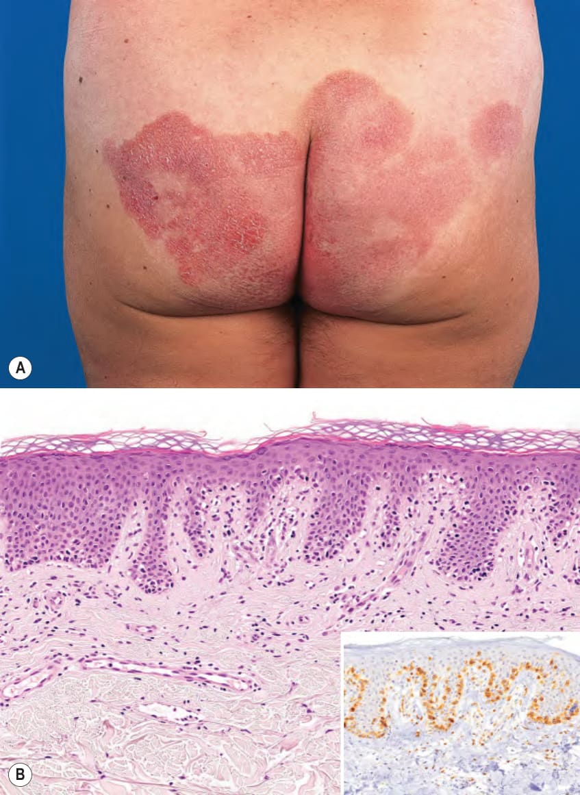
Fig. 120.2 Mycosis fungoides, limited patch/plaque stage disease (stage
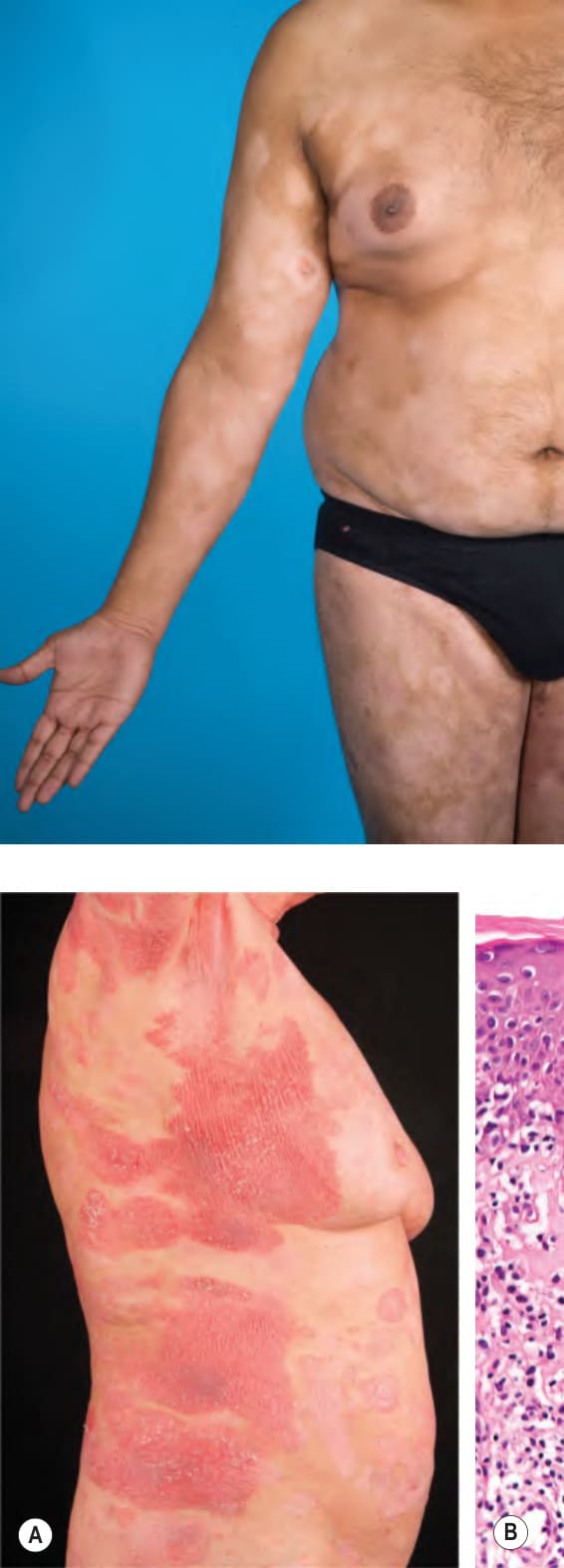
Fig. 120.3 Hypopigmented mycosis fungoides
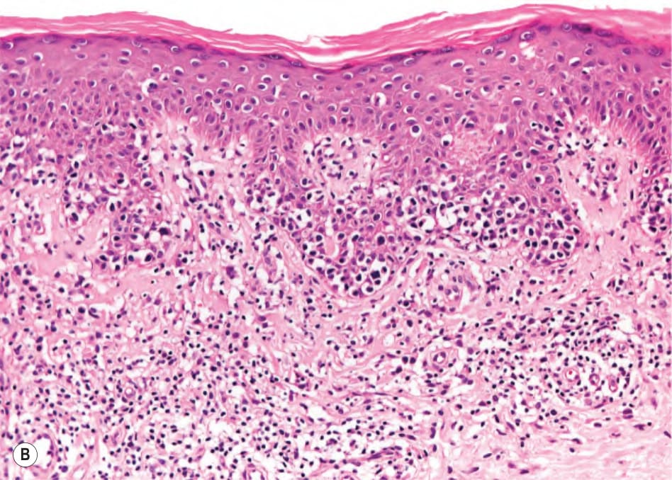
Fig. 120.4 Mycosis fungoides, generalized patch/plaque stage disease
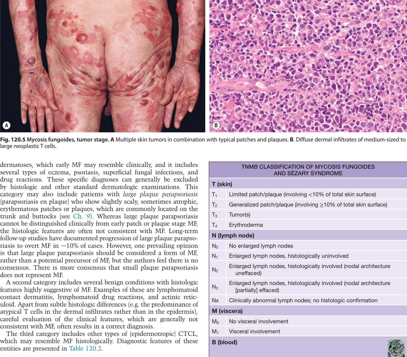
Fig. 120.5 Mycosis fungoides, tumor stage.A Multiple skin tumors in combination with typical patches and plaques. B. Diffuse dermal infiltrates of medium-sized to large neoplastic T cells.
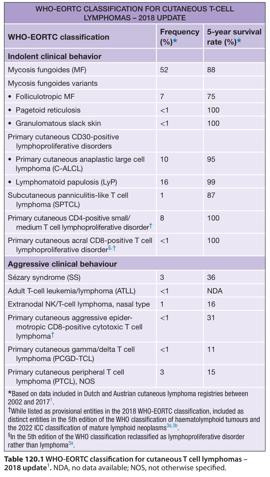
Table 120.1 WHO-EORTC classification for cutaneous T cell lymphomas –
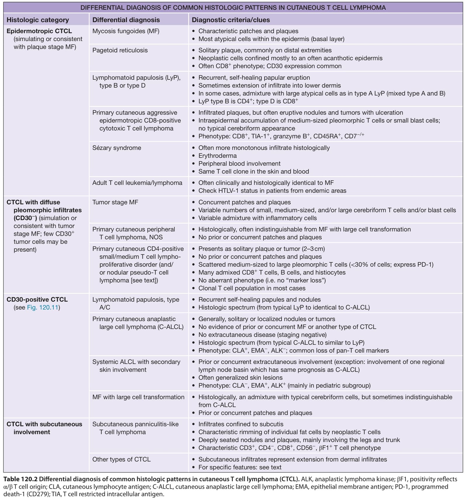
Table 120.2 Differential diagnosis of common histologic patterns in cutaneous T cell lymphoma (CTCL). ALK, anaplastic lymphoma kinase; βF1, positivity reflects α/β T cell origin; CLA, cutaneous lymphocyte antigen; C-ALCL, cutaneous anaplastic large cell lymphoma; EMA, epithelial membrane antigen; PD-1, programmed death-1 (CD279); TIA, T cell restricted intracellular antigen.
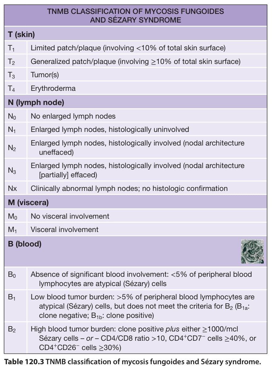
Table 120.3 TNMB classification of mycosis fungoides and Sézary syndrome.
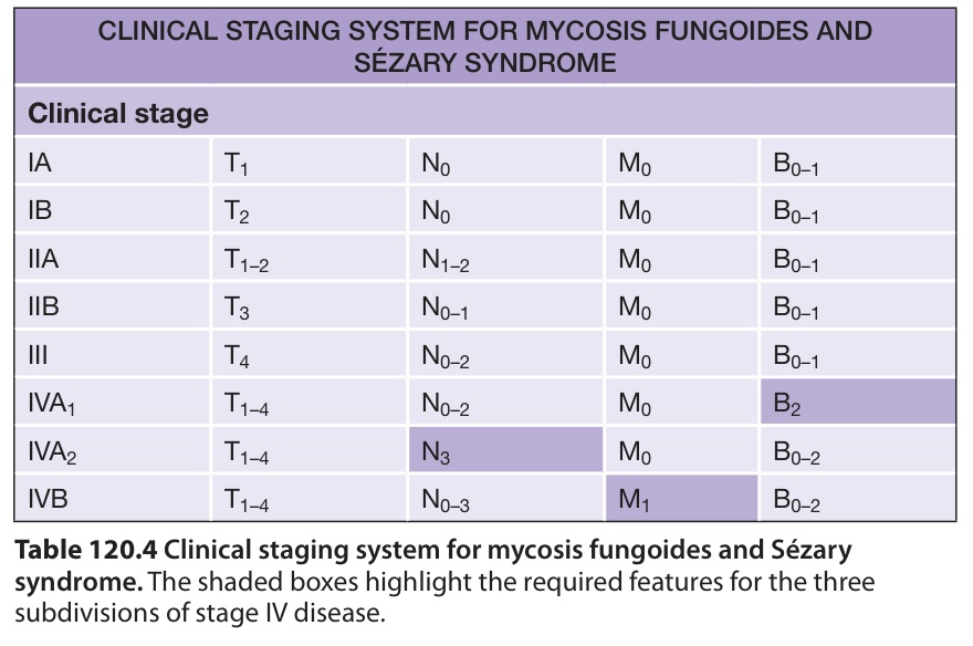
Table 120.4 Clinical staging system for mycosis fungoides and Sézary syndrome. The shaded boxes highlight the required features for the three subdivisions of stage IV disease.
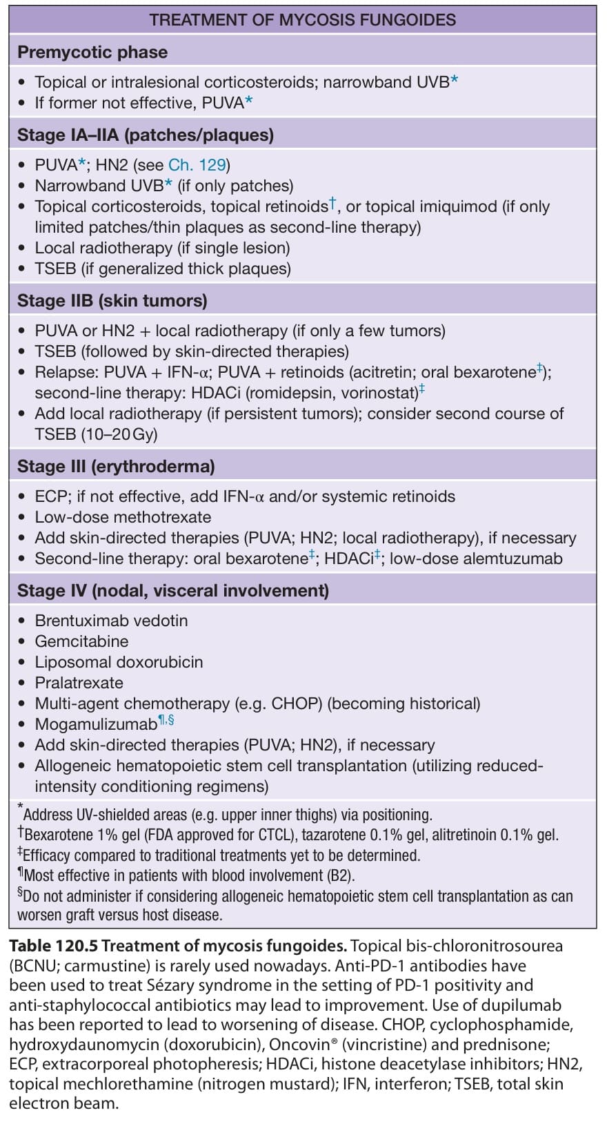
Table 120.5 Treatment of mycosis fungoides. Topical bis-chloronitrosourea (BCNU; carmustine) is rarely used nowadays. Anti-PD-1 antibodies have been used to treat Sézary syndrome in the setting of PD-1 positivity and anti-staphylococcal antibiotics may lead to improvement. Use of dupilumab has been reported to lead to worsening of disease. CHOP, cyclophosphamide, hydroxydaunomycin (doxorubicin), Oncovin® (vincristine) and prednisone; ECP, extracorporeal photopheresis; HDACi, histone deacetylase inhibitors; HN2, topical mechlorethamine (nitrogen mustard); IFN, interferon; TSEB, total skin electron beam.