SÉZARY SYNDROME
Definition
Sézary syndrome (SS) is defined historically by the triad of erythroderma, generalized lymphadenopathy, and the presence of neoplastic T cells (Sézary cells) in the skin, lymph nodes, and peripheral blood (see Table 120.4). Criteria for the diagnosis of SS include demonstration of a T cell clone in the peripheral blood (preferably the same T cell clone as in the skin), in combination with either an absolute Sézary cell count ≥1000 cells/mcl or immunophenotypic abnormalities including an expanded CD4+ T cell population resulting in a CD4/ CD8 ratio >10, CD4+CD7− cells ≥40%, or CD4+CD26− cells ≥30%. While SS is often designated as a leukemic phase or variant of MF, more recent studies have revealed major genomic and phenotypic differences between these two entities, suggesting that SS and MF should
be considered as separate lymphomas arising from distinct functional T cell subsets.
Epidemiology
SS is a rare disease accounting for <5% of all CTCL (see Table 120.1). It occurs exclusively in adults.
Clinical Features
SS is characterized by erythroderma, which may be associated with marked exfoliation, edema, and lichenification; it is intensely pruritic (Fig. 120.9A). Lymphadenopathy, alopecia, onychodystrophy, and palmoplantar hyperkeratosis are common findings. The overt clinical picture may be preceded by a non-diagnostic dermatitis. The prognosis is generally poor, with an overall 5-year survival of ~35%. Most patients die of opportunistic infections due to immunosuppression.
Pathology
The histologic features in SS may be similar to those in MF. However, the cellular infiltrates in SS are more often monotonous, and epidermo tropism may be absent. In up to one-third of skin biopsies from patients with otherwise classic SS, the histologic findings may be nonspecific. Involved lymph nodes characteristically show a dense, monotonous infiltrate of Sézary cells with effacement of the normal lymph node architecture. The bone marrow may be involved, but the infiltrates are often sparse and mainly interstitial. The neoplastic T cells have a CD3+, CD4+, CD8− phenotype, characteristically lack CD7 and CD26 expression, and express programmed death-1 (PD-1; CD279) in almost all cases (Fig. 120.9B).
Pathogenesis
The pathogenesis of SS is unknown. Conclusive evidence of an etiologic role for HTLV-1 is lacking. Previous studies suggested that
Erythroderma with diffuse scaling. B Strong expression of PD-1 by neoplastic T cells within the dermal infiltrate and Pautrier microabscesses.
the neoplastic CD4+ T cells in SS patients were derived from central memory T cells and the neoplastic cells in MF from effector memory T cells. However, more recent studies found that Sézary cells may also be derived from other T cell subsets. In addition, tumor cell populations in individual SS patients had a high degree of single-cell heterogeneity, and these distinct subpopulations of malignant T cells may differ in their sensitivity to treatments.
Studies of genetic alterations in SS have shown extensive genetic instability and complex structural and numerical alterations. Chromosomal aberrations include deletions involving chromosomes 10q22–25 (harbors PTEN) and 17p12–13 (includes TP53) and gains involving chromosomes 8q22–24 (includes MYC), 10p11.2, and 17q22–25. Gene expression studies found increased expression of PLS3 (T-plastin), TWIST1, DNM3, EPH4 and CD158k/KIR3DL2, which might serve as diagnostic markers. With next generation sequencing, alterations in genes that play a role in T cell activation, NF-κB and JAK-STAT signaling, apoptosis control, chromatin remodeling, and DNA damage response were identified.
Differential Diagnosis
Differentiation between SS and non-neoplastic forms of erythroderma may be extremely difficult. The differential diagnosis includes erythroderma secondary to psoriasis, atopic dermatitis, or other forms of dermatitis, pityriasis rubra pilaris, and drug reactions as well as idiopathic erythroderma (see Ch. 10). Demonstration of an identical T cell clone in skin and peripheral blood is an important diagnostic criterion favoring SS. New biomarkers supporting a diagnosis of SS include PD-1 (CD279), TOX, and KIRDL2 (CD158k). Demonstration of a predominant CD3+, CD4−, CD8+ T cell population in the skin and peripheral blood is highly suggestive of actinic reticuloid (see Ch. 87).
Treatment
Being a systemic disease (leukemia) by definition, systemic treatment is required. Skin-directed therapies like PUVA or potent topical corticosteroids may be used as adjuvant therapy. Extracorporeal photopheresis (ECP), either alone or in combination with other treatment modalities, has been suggested as the treatment of choice in SS and erythrodermic MF, with overall response rates of 30%–80%, and complete response rates of 14%–25%. This great variation in response rates may reflect differences in patient selection and/or concurrent therapies. The suggested superiority of ECP over traditional low-dose chemotherapy regimens has not yet been substantiated by randomized controlled trials. Beneficial effects have also been reported with IFN-α (either alone or in combination with PUVA therapy) as well as prolonged treatment with methotrexate, but complete responses are uncommon.
Low-dose alemtuzumab (10 mg SC thrice weekly for 12 weeks), single-agent chemotherapy (gemcitabine, liposomal doxorubicin), or multi-agent chemotherapy (CHOP or CHOP-like regimens) have been recommended as second-line treatments for SS, but responses are generally short-lived. Allogeneic HSCT may have curative potential, even in patients with advanced disease (see MF treatment). The monoclonal antibody mogamulizumab targets the CC chemokine receptor 4 (CCR4), which is consistently expressed on malignant T cells in MF/SS as well as on Th2, Th17, and regulatory T cells, resulting in their destruction via antibody-dependent cellular cytotoxicity. While mogamulizumab has shown significant clinical efficacy in both MF and SS, it is most effective in patients with blood involvement. Patients may develop a mogamulizumab-associated rash (MAR) that often favors the head and neck and may mimic MF39a. Clinical presentation is variable and may include folliculotropic MF-like scalp lesions with alopecia, photodistributed dermatitis, a morbilliform eruption, and erythroderma. Histologically, psoriasiform, spongiotic, lichenoid, and/ or granulomatous patterns can be seen. Demonstration of an inverted or normalized CD4:CD8 ratio of intraepidermal T cells and an absence of clonal T cells may be useful criteria for differentiating MAR from recurrent MF/SS. Treatment options include high potency topical corticosteroids, systemic corticosteroids, and/or methotrexate; increasing the dosing interval or discontinuation of mogamulizumab may be considered39b.
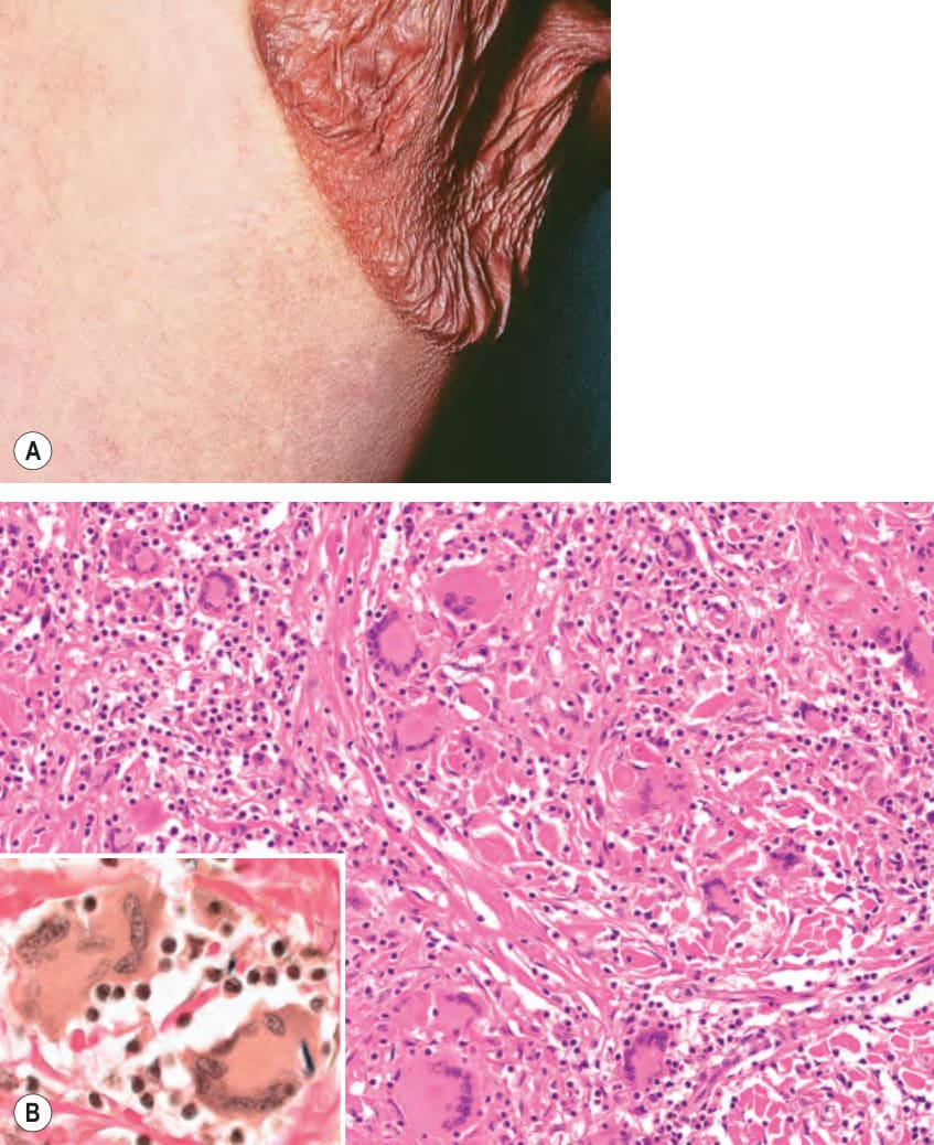
Fig. 120.8 Granulomatous slack skin.A Pendulous fold of atrophic lax skin in the right inguinal area. B Multinucleated giant cells with intracellular leukocytes, surrounded by a dense leukocytic infiltrate. Inset: Elastic tissue stain showing a multinucleated giant cell containing an elastic fiber (elastophagocytosis).
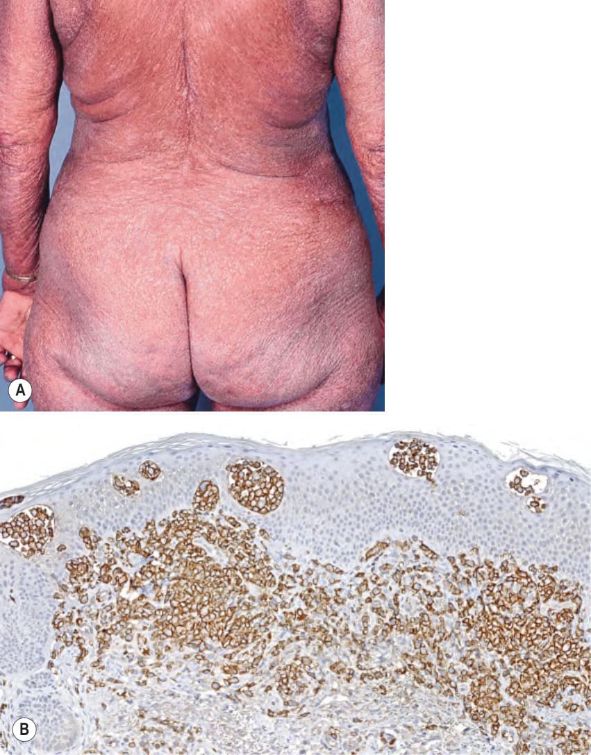
Fig. 120.9 Sézary syndrome.A
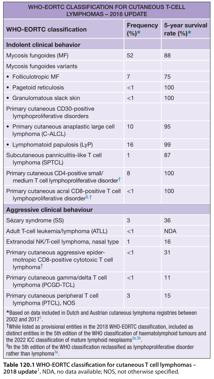
Table 120.1 WHO-EORTC classification for cutaneous T cell lymphomas –
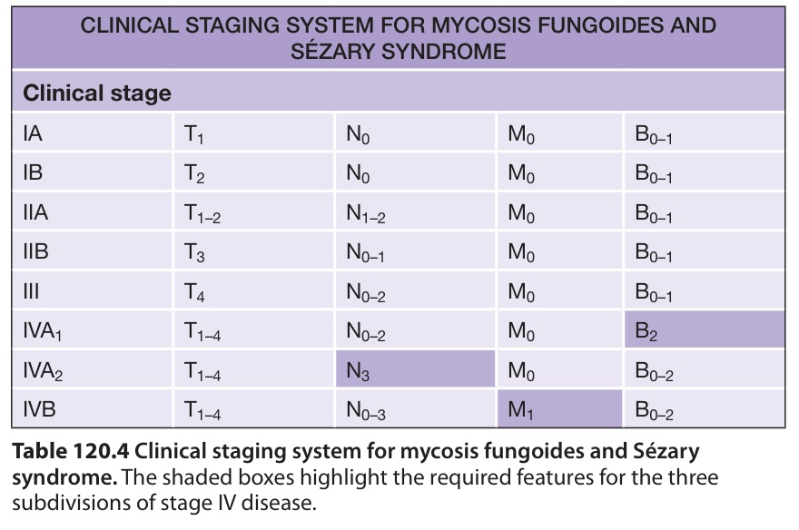
Table 120.4 Clinical staging system for mycosis fungoides and Sézary syndrome. The shaded boxes highlight the required features for the three subdivisions of stage IV disease.