Leukemia Cutis
Key features
Pink to red–brown or purple papules and nodules; hemorrhagic ulcers
Diffuse dermal infiltrate of neoplastic leukocytes
Monomorphic immature precursor cells in some types
Epidemiology
Multiple types of leukemia exist, each with its own epidemiologic characteristics. Acute lymphoblastic leukemia (ALL) tends to be a neoplasm of childhood. Acute myelogenous leukemia (AML) and chronic myelogenous leukemia (CML) occur primarily in adults, and chronic lymphocytic leukemia (CLL) and hairy cell leukemia are most common in elderly patients. Development of CML-associated leukemia cutis can be a harbinger of blast transformation, while the development of leukemia cutis in a patient with myelodysplastic syndrome (MDS) points to progression to AML. In some patients with MDS, cutaneous lesions with an accumulation of benign-appearing dendritic and myeloid cells and a chronic course have been described. Leukemia cutis is most commonly seen in patients with NPM1-mutated AML and less often in CLL9,28,29a,29b.
Pathogenesis
Specific chromosomal abnormalities are associated with particular subtypes of leukemia. The Philadelphia chromosome, the primary diagnostic feature of CML, represents a translocation between chromosomes 9 and 22 [t(9;22)]. This translocation results in fusion of the two genes BCR and ABL, with constitutive activation of the tyrosine kinase activity of the fusion oncoprotein. It is this kinase activity that is selectively inhibited by imatinib mesylate (Gleevec®). Additionally, 95% of patients with promyelocytic leukemia have a t(15;17) translocation, which leads to abnormal expression of a fusion retinoic acid receptor that is the target of all-trans retinoic acid therapy.
A variety of non-random chromosomal abnormalities exists in ALL and AML that can influence prognosis, and in AML mutations in particular genes can be favorable (e.g. CEBPA) or unfavorable (e.g. FLT3- ITD)29a,29b. NPM1 is the most frequently mutated gene in patients with AML and normal cytogenetics; as noted previously, NPM1- mutated AML is the subtype most commonly associated with leukemia cutis29a,29b. Table 121.5 outlines the relationship between specific abnormalities and prognosis in CLL.
Clinical features
Leukemia can lead to nonspecific reactive skin lesions (Table 121.6) or specific infiltrates. The latter most commonly present as multiple, generalized or localized, firm papules and nodules that vary in color from pink to red–brown to purple. The papulonodules often become hemorrhagic and thrombocytopenia usually plays a role in this associated hemorrhage. Ulcerative and rarely bullous lesions occur. Leukemia cutis can develop in any location, but the head, neck, and trunk are most commonly involved (Fig. 121.8A). Leukemic infiltrates may arise at sites of trauma or scars.
Rarely, myelogenous leukemias may present with dermal or subcutaneous nodules known as myeloid sarcomas or chloromas. These skin lesions may precede the development of systemic leukemia by months.
Gingival hyperplasia, secondary to leukemic infiltrates, favors acute monocytic or acute myelomonocytic leukemia, especially those with NPM1 mutations.
In one series, cutaneous eruptions of leukemia accounted for 30% of all skin biopsy specimens in patients with leukemia. Other diagnoses included GVHD, drug eruptions, infectious processes, purpura, and small vessel vasculitis. In most patients with acute leukemia, cutaneous lesions are present at the time of diagnosis or recurrence, but occasionally, skin involvement precedes the appearance of leukemia in the peripheral smear (“aleukemic” leukemia cutis). Rarely, leukemia cutis predates apparent bone marrow involvement by months or even years.
Of note, asymptomatic dermal leukemic infiltrates of CLL may be evident in biopsy specimens of primary cutaneous neoplasms (e.g. squamous cell carcinoma) and inflammatory or infectious disorders such as herpes simplex and herpes zoster viral infections. This phenomenon indicates that these neoplastic cells retain the capability to respond to chemotactic stimuli and migrate into tissues (inflammatory oncotaxis).
The characteristics of the different forms of leukemia cutis are summarized in Table 121.7.
Pathology
Histologic features of leukemia cutis vary with the type of leukemia. The neoplastic infiltrates may be perivascular, interstitial, nodular, and/ or diffuse; occasionally, they densely surround the eccrine glands. Diagnosis is more difficult when the perivascular and periadnexal infiltrates are sparse and mimic inflammatory disorders.
AML represents a neoplastic proliferation of immature myeloid cells and encompasses multiple subtypes29a,29b. In general, the neoplastic cells recapitulate myeloid precursor cells in their immature stages of development (Fig. 121.8B). In many cases, cytoplasmic granules can be found. The most common forms of AML that involve the skin and present de novo (without previous evidence of systemic disease) are the more differentiated subtypes – acute myelomonocytic and acute monocytic leukemia. Immunohistochemically, the infiltrates of AML are positive for myeloid markers such as myeloperoxidase, CD33, CD34, and CD68 and are usually negative for CD1239,29a,29b.
In CML, there is a range of myeloid precursors, including promyelocytes, metamyelocytes, bands, and mature neutrophils. Although cutaneous lesions of CML are rare, they are typically seen in patients with chronic myelomonocytic leukemia and are associated with blast transformation and a poor prognosis9,29a,29b.
ALL represents a clonal expansion of immature lymphocytes, most commonly of B cell lineage and less commonly T cell lineage. Rarely, cytoplasmic granularity may be present, and this corresponds with a precursor B cell immunophenotype. Cells of ALL may be difficult to identify in skin biopsy specimens and immunostaining for terminal deoxytransferase may prove helpful.
Cutaneous CLL is characterized by a dense infiltrate of uniformappearing, small, round lymphocytes of B cell lineage (Fig. 121.9). The cells appear mature, but the monomorphous nature and paucity of other cell types within the infiltrate suggest a leukemic infiltrate. The lymphocytes often surround adnexal and vascular structures. Immunohistochemically, CLL shows positivity for LEF1, CD5 (variable), CD20, and CD23.
Differential diagnosis
The clinical differential diagnosis includes cutaneous lymphoma, cutaneous lymphoid hyperplasia, infectious emboli, vasculitis, panniculitis, and drug eruptions. The histologic differential diagnosis of leukemia cutis depends upon the type of leukemia present. Distinguishing some cases of leukemia cutis from lymphomas involving the skin can be difficult. For myeloid leukemias, the differential diagnosis includes mainly extramedullary hematopoiesis (see above), diffuse large cell lymphomas, and histiocytoid Sweet syndrome. Of note, in some cases of histiocytoid Sweet syndrome neoplastic cells of AML may be present within the infiltrate.
Treatment
No specific treatment for leukemia cutis currently exists. The cutaneous eruptions typically resolve following successful treatment of the leukemia.
Numerous violet–brown papules and plaques on the back of a patient with acute myelogenous leukemia. B Histologically, an infiltrate of atypical monocytoid cells characterized by nuclear pleomorphism and relatively abundant cytoplasm is seen.
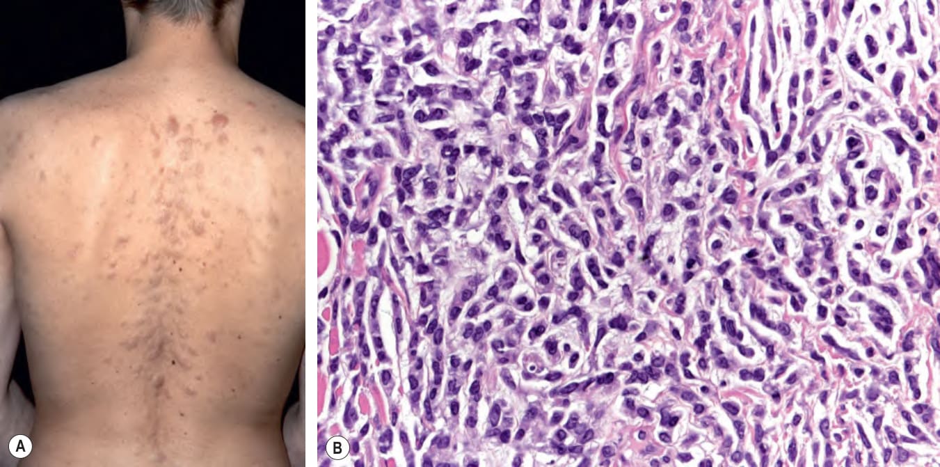
Fig. 121.8 Leukemia cutis.A
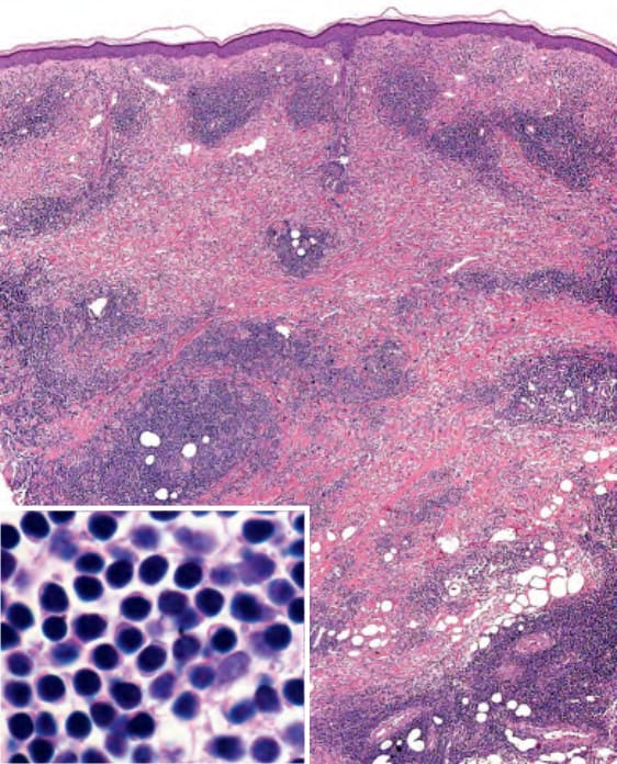
Fig. 121.9 Chronic lymphocytic leukemia
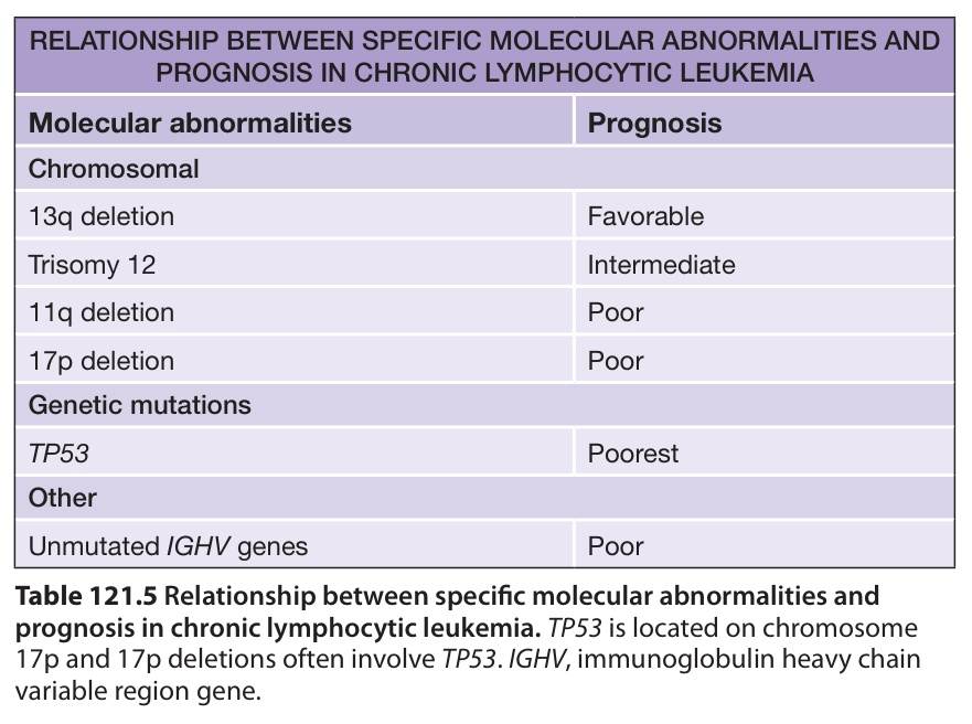
Table 121.5 Relationship between specific molecular abnormalities and prognosis in chronic lymphocytic leukemia.TP53 is located on chromosome 17p and 17p deletions often involve TP53. IGHV, immunoglobulin heavy chain variable region gene.
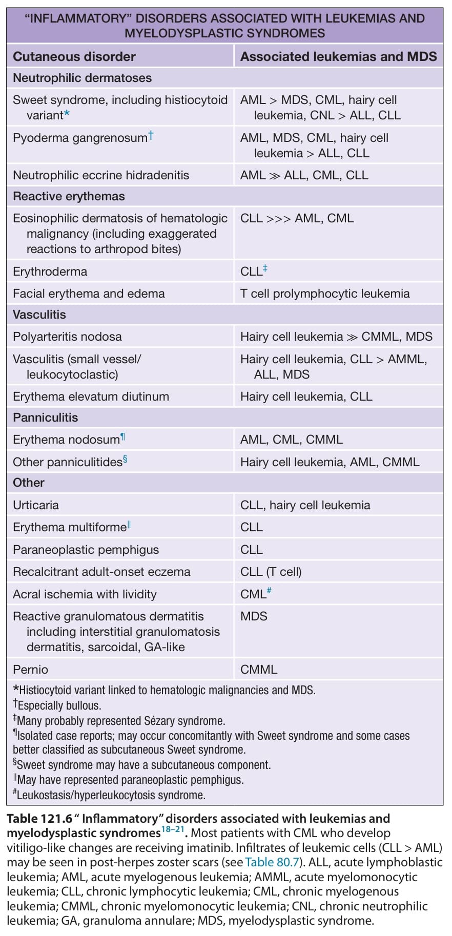
Table 121.6 “ Inflammatory” disorders associated with leukemias and myelodysplastic syndromes. Most patients with CML who develop vitiligo-like changes are receiving imatinib. Infiltrates of leukemic cells (CLL > AML) may be seen in post-herpes zoster scars (see Table 80.7). ALL, acute lymphoblastic leukemia; AML, acute myelogenous leukemia; AMML, acute myelomonocytic leukemia; CLL, chronic lymphocytic leukemia; CML, chronic myelogenous leukemia; CMML, chronic myelomonocytic leukemia; CNL, chronic neutrophilic leukemia; GA, granuloma annulare; MDS, myelodysplastic syndrome.
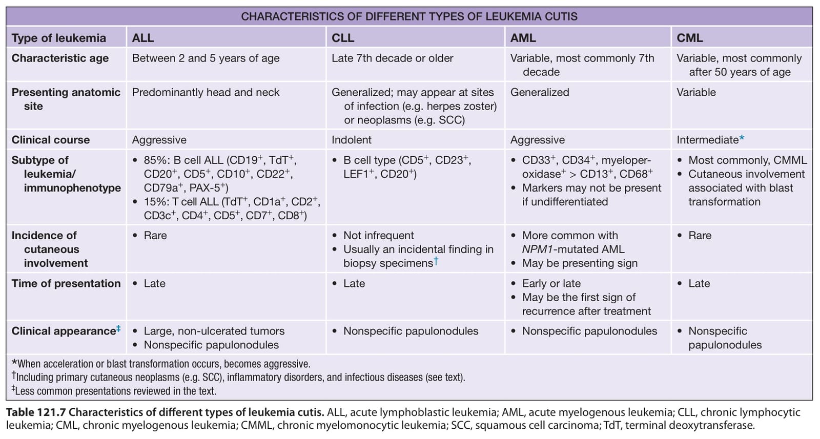
Table 121.7 Characteristics of different types of leukemia cutis. ALL, acute lymphoblastic leukemia; AML, acute myelogenous leukemia; CLL, chronic lymphocytic leukemia; CML, chronic myelogenous leukemia; CMML, chronic myelomonocytic leukemia; SCC, squamous cell carcinoma; TdT, terminal deoxytransferase.