Lymphomatoid Granulomatosis
Key features
EBV-driven angiocentric/destructive lymphoproliferative disorder
Presents with cough, dyspnea, chest pain, and constitutional symptoms
Cavitary pulmonary nodules on chest radiograph
Nodular or ulcerative cutaneous lesions in 25%–50% of patients
History
Lymphomatoid granulomatosis (LG) was first described in 1972 as a pulmonary angiitis/granulomatosis that mimicked granulomatosis with polyangiitis. Initially thought to represent a reactive process, subsequent studies demonstrated a malignant clonal B cell population admixed with an abundant, polyclonal, reactive T cell population.
Epidemiology
LG is a rare disease of adults, usually presenting in the fifth to sixth decade of life. The male-to-female ratio is approximately 2 : 1. The disease can affect children, most commonly in the setting of immunodeficiency syndromes.
Pathogenesis
The pathogenesis of LG is often related to EBV infection, occasionally in combination with immunosuppression. Factors associated with LG include use of immunosuppressive medications (e.g. azathioprine, methotrexate, imatinib), HIV infection, Wiskott–Aldrich syndrome, X-linked lymphoproliferative syndrome, and additional lymphoproliferative disorders. In the WHO classification of hematopoietic tumors, LG is categorized as an angiocentric/angiodestructive lymphoproliferative disease with variable histological grades.
Clinical features
Patients with LG present with symptoms related to pulmonary involvement, such as cough, dyspnea, and chest pain. Constitutional symptoms include fever, weight loss, malaise, arthralgias, and myalgias. Cutaneous lesions develop in 25%–50% of patients and usually present as nodules or ulcerated plaques (Fig. 121.12). Rarely, LG may present as a maculopapular exanthem. The kidney, brain, and gastrointestinal tract may be affected. In contrast to angioimmunoblastic T cell lymphoma, the lymph nodes and spleen are rarely involved.
Pathology
In LG, there are three histopathologic grades: (1) grade 1 – polymorphous lymphoid infiltrate without cytologic atypia, along with a few large cells, and few cells are positive for EBV-encoded RNA 1 (EBER-1) and latent membrane protein via in situ hybridization and immunohistochemistry, respectively; (2) grade 2 – polymorphous inflammatory background with scattered large cells and more abundant EBER-1+ cells; and (3) grade 3 – sheets of large B cells and numerous EBER-1+ cells predominate over the inflammatory background. The amount of necrosis increases with the grade and angiocentricity/angiodestruction is consistently present. Granulomatous changes are often present. The neoplastic cells stain positively for CD20 and CD79a, and in most high-grade lesions there is a monoclonal rearrangement of the JH genes.
Differential diagnosis
The clinical differential diagnosis includes various forms of cutaneous lymphoma, as well as infectious and inflammatory disorders (e.g. medium-sized vessel vasculitis, pyoderma gangrenosum). The histopathologic differential diagnosis is broad and encompasses most of the lymphoproliferative disorders mentioned above, including B cell and T cell lymphomas. Other disorders with histologic evidence of granulomatosis include granulomatosis with polyangiitis, sarcoidosis, and infectious processes. Diagnosis is based upon histopathologic findings plus immunohistochemical and molecular analyses. Because the existence
of skin-limited disease without pulmonary involvement is debatable, such a diagnosis requires compelling evidence.
Treatment
LG follows an aggressive course, with the 5-year mortality ranging from 60% to 90%. For higher-grade disease, treatment commonly involves
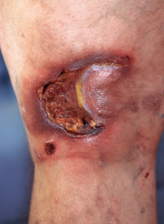
Fig. 121.12 Lymphomatoid granu- lomatosis. Ulcerated violaceous plaque in the popliteal fossa. Courtesy Jean L. Bolognia, MD.
multidrug chemotherapy and/or rituximab (anti-CD20 monoclonal antibody), and, for grade 1 disease, IFN-α. Reversal of exogenous immunosuppression may also lead to clinical improvement.