CLINICAL FEATURES
The clinical characteristics of cutaneous metastasis can provide important clues. When a patient with a known history of cancer presents with an extremely firm and rapidly growing erythematous nodule or an eruption of multiple skin nodules in close proximity to the primary tumor (Table 122.2), the diagnosis of cutaneous metastasis is relatively straightforward. However, the skin lesions may grow more slowly, and while metastases typically appear within several years of the diagnosis of the primary malignancy, they can present decades later.
The color of cutaneous metastases varies, from skin-colored to pink–red to blue–black, with the latter most commonly observed in metastatic melanoma (Fig. 122.3). In general, dermal and subcutaneous metastases are firmer on palpation than lipomas or epidermoid and tricholemmal (pilar) cysts. Cutaneous metastases can arise from hematogenous or lymphatic spread as well as direct extension. In the case of the latter two scenarios, as stated previously, proximity of the
metastasis to the primary malignancy serves as a diagnostic clue. An example of lymphatic spread is in-transit metastases of melanoma while direct extension is most commonly observed with breast carcinoma and squamous cell carcinoma of the head and neck.
There are additional examples of anatomic localization, including the predilection of gastrointestinal malignancies to metastasize to the periumbilical region, referred to as a Sister Mary Joseph nodule (Fig. 122.4). Occasionally, implantation of tumor occurs at the time of a surgical procedure, e.g. chest tube placement for a malignant pleural effusion due to lung cancer. Although the distribution of cutaneous metastases is usually more random in the case of hematogenous spread, the scalp can be a preferred site for some malignancies, e.g. renal cell carcinoma.
A Pink nodule on the scalp with associated alopecia due to a metastasis from lung carcinoma (alopecia neoplastica). B Red–purple eroded nodule due to a metastasis from renal cell carcinoma; such lesions may be confused with vascular tumors. C Multiple small dark purple to black papules on the left neck and chest due to metastatic melanoma. D Pink papules and plaques on the chest due to metastases from adenocarcinoma of the colon. The patient was referred for treatment of dermatitis. A, Courtesy Lorenzo Cerroni, MD; B, Courtesy Edward Cowen, MD; C, Courtesy Chris Bunick, MD; D, Courtesy Kalman Watsky, MD.
Of all the carcinomas that metastasize to the skin, breast cancer may be the one with the widest range of clinical presentations (Table 122.3), varying from papulonodules to patches of erythema mimicking erysipelas (inflammatory carcinoma) to woody induration with a peau d’orange appearance (Fig. 122.5). The latter is often referred to as “en cuirasse” due to its clinical resemblance to the leather armor of a soldier (cuirassier). In addition, intravascular metastases can lead to erythematous papules that may resemble vascular proliferations, including the angiosarcomas that can follow breast irradiation (see Ch. 114).
Less common presentations include the dermatitis-like appearance of Paget disease, both mammary and extramammary (see Table 122.3). Metastases of renal cell carcinoma can resemble pyogenic granulomas, including a tendency to bleed (see Fig. 122.3B). Rarely, intravascular metastases lead to a reticulated vascular pattern that is reflective of a vaso-occlusive process or the cutaneous metastases assume an unusual distribution pattern, e.g. zosteriform.
In addition to metastases from solid organs, leukemias and systemic lymphomas can also involve the skin (see Chs. 119–121). The lesions are often pink–violet to red–brown papulonodules and therefore may be clinically indistinguishable from cutaneous metastases due to solid organ malignancies. Skin involvement by leukemia or lymphoma is classically not referred to as “metastases”.
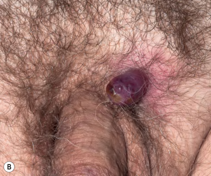
Fig. 122.3 Cutaneous metastases.
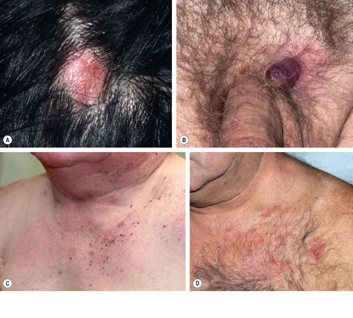
Fig. 122.4 Cutaneous metastasis of colon carcinoma. An umbilical Sister Mary Joseph nodule presenting as a pink plaque with scalecrust. Courtesy Stuart Lessin, MD.
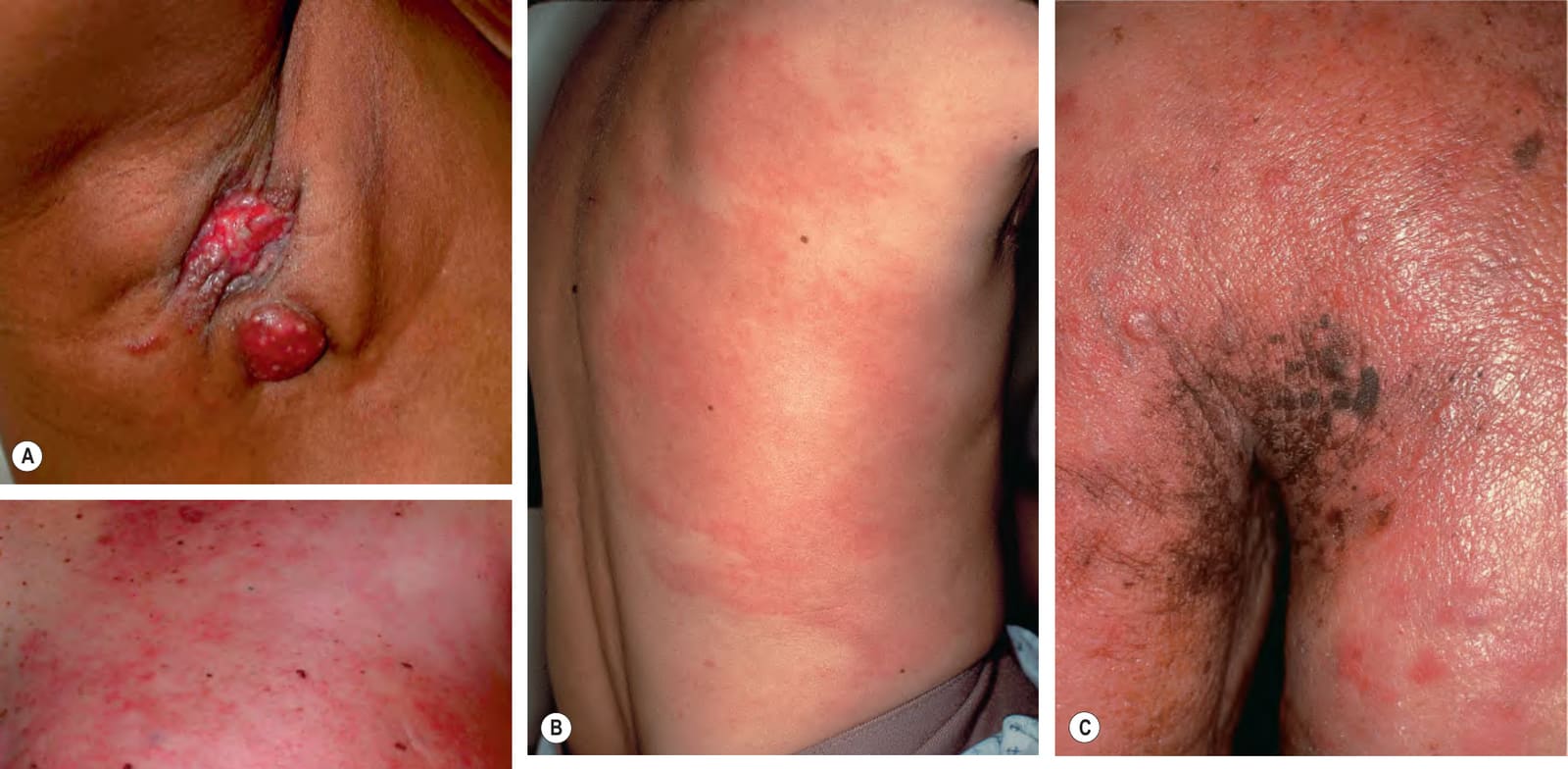
Fig. 122.5 Various presentations of cutaneous metastases of breast carcinoma.A Eroded erythematous nodules in the axilla. B Inflammatory form (carcinoma erysipeloides) with patches of erythema that may initially be misdiagnosed as infectious cellulitis. C Primarily en cuirasse form with obvious induration and peau d’orange appearance in addition to papulonodules. D Mixed pattern – reticulated erythema of carcinoma erysipeloides as well as peau d’orange appearance near the areola. A, D, Courtesy Stuart Lessin, MD.
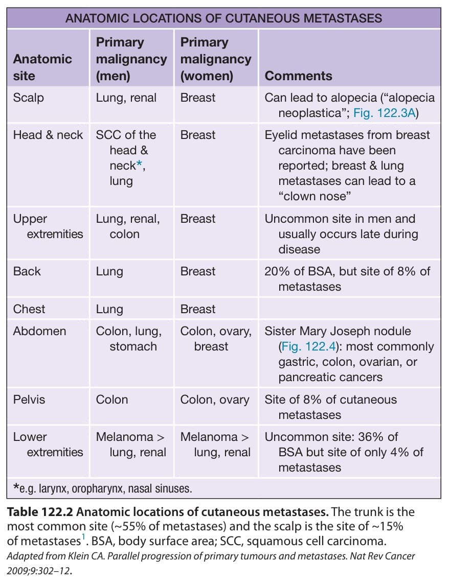
Table 122.2 Anatomic locations of cutaneous metastases. The trunk is the most common site (~55% of metastases) and the scalp is the site of ~15% of metastases. BSA, body surface area; SCC, squamous cell carcinoma. Adapted from Klein CA. Parallel progression of primary tumours and metastases. Nat Rev Cancer 2009;9:302–12.
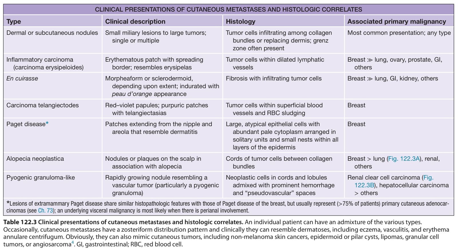
Table 122.3 Clinical presentations of cutaneous metastases and histologic correlates. An individual patient can have an admixture of the various types. Occasionally, cutaneous metastases have a zosteriform distribution pattern and clinically they can resemble dermatoses, including eczema, vasculitis, and erythema annulare centrifugum. Obviously, they can also mimic cutaneous tumors, including non-melanoma skin cancers, epidermoid or pilar cysts, lipomas, granular cell tumors, or angiosarcoma. GI, gastrointestinal; RBC, red blood cell.