臨床特徵(CLINICAL FEATURES)
皮膚轉移的臨床特性可提供重要線索。當一位有已知癌症病史的病人表現為極度堅實且快速生長的紅斑性結節,或在原發腫瘤鄰近處出現多發性皮膚結節的疹發(Table 122.2)時,皮膚轉移的診斷相對直接。然而,皮膚病灶可能生長較慢,且雖然轉移典型上在原發惡性腫瘤診斷後數年內出現,它們也可能於數十年後表現。
皮膚轉移的顏色多變,自膚色至粉紅—紅色至藍—黑色,後者最常見於轉移性黑色素瘤(Fig. 122.3)。一般而言,真皮與皮下轉移在觸診時比脂肪瘤或表皮樣與毛根鞘(毛髮)囊腫更為堅實。皮膚轉移可源自血行性或淋巴性擴散,以及直接延伸。在後兩種情境中,如前所述,
轉移與原發惡性腫瘤的鄰近性可作為診斷線索。淋巴性擴散的一個例子是黑色素瘤的在途轉移(in-transit metastases),而直接延伸則最常見於乳癌與頭頸部鱗狀細胞癌。
還有其他解剖定位的例子,包括腸胃道惡性腫瘤好發轉移至臍周區域,稱為 Sister Mary Joseph nodule(Fig. 122.4)。偶爾,腫瘤的植入發生於手術操作當時,例如因肺癌所致惡性肋膜積液而置放胸管時。雖然在血行性擴散的情況下皮膚轉移的分布通常較為隨機,但頭皮對某些惡性腫瘤(例如腎細胞癌)可能是偏好的部位。
A Pink nodule on the scalp with associated alopecia due to a metastasis from lung carcinoma (alopecia neoplastica). B Red–purple eroded nodule due to a metastasis from renal cell carcinoma; such lesions may be confused with vascular tumors. C Multiple small dark purple to black papules on the left neck and chest due to metastatic melanoma. D Pink papules and plaques on the chest due to metastases from adenocarcinoma of the colon. The patient was referred for treatment of dermatitis. A, Courtesy Lorenzo Cerroni, MD; B, Courtesy Edward Cowen, MD; C, Courtesy Chris Bunick, MD; D, Courtesy Kalman Watsky, MD.
在所有轉移至皮膚的癌症中,乳癌可能是臨床表現範圍最廣者(Table 122.3),從丘疹結節、模擬丹毒的紅斑性斑(發炎性癌 inflammatory carcinoma),到具橘皮(peau d’orange)外觀的木質樣硬化(Fig. 122.5)。後者常被稱為「en cuirasse」,因其臨床上類似士兵(cuirassier)的皮革盔甲。此外,血管內轉移可導致紅斑性丘疹,可能類似血管增生,包括乳房照射後可能發生的血管肉瘤(見 Ch. 114)。
較不常見的表現包括 Paget disease 類似皮膚炎的外觀,乳房型與乳房外型皆然(見 Table 122.3)。腎細胞癌的轉移可能類似化膿性肉芽腫,包括易出血的傾向(見 Fig. 122.3B)。罕見情況下,血管內轉移導致網狀的血管型態,反映一種血管阻塞過程,或皮膚轉移呈現不尋常的分布型態,例如帶狀疱疹樣(zosteriform)。
除了源自實體器官的轉移之外,白血病與全身性淋巴瘤亦可侵犯皮膚(見 Chs. 119–121)。這些病灶常為粉紫色至紅棕色的丘疹結節,因此在臨床上可能與源自實體器官惡性腫瘤的皮膚轉移無法區分。白血病或淋巴瘤的皮膚侵犯在傳統上不稱為「轉移」。
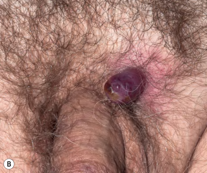
圖 122-3:皮膚轉移
Fig. 122.3 Cutaneous metastases.
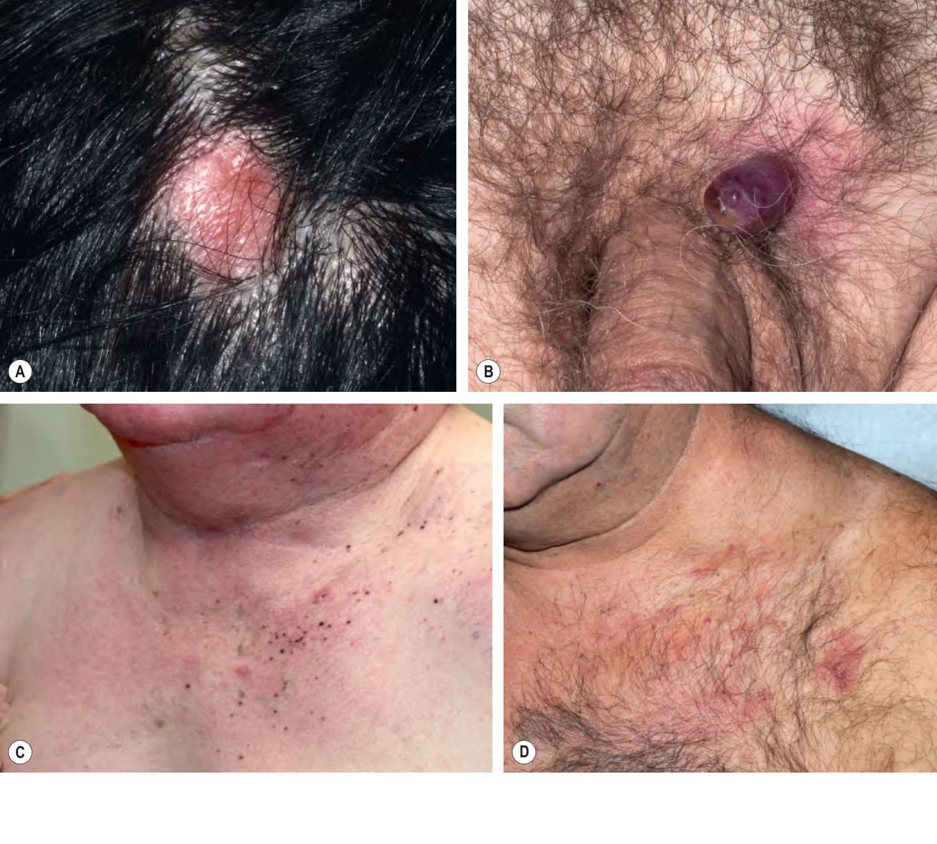
圖 122-4:結腸癌的皮膚轉移。臍部 Sister Mary Joseph nodule,表現為帶鱗屑痂皮的粉紅色斑塊
Fig. 122.4 Cutaneous metastasis of colon carcinoma. An umbilical Sister Mary Joseph nodule presenting as a pink plaque with scalecrust. Courtesy Stuart Lessin, MD.
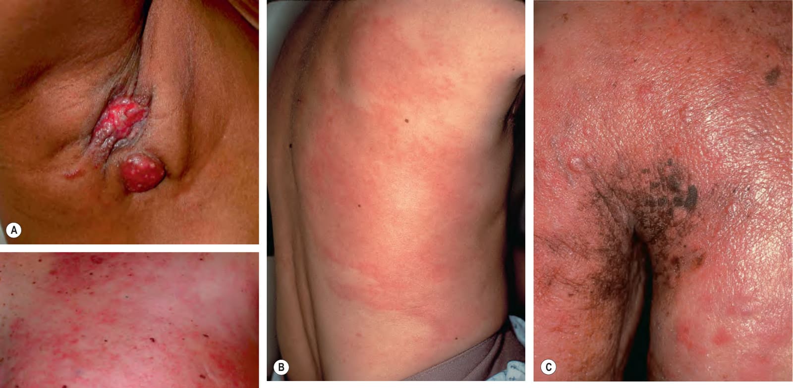
圖 122-5:乳癌皮膚轉移的各種表現。A 腋窩處糜爛的紅斑性結節。B 發炎型(carcinoma erysipeloides),有紅斑性斑,起初可能被誤診為感染性蜂窩性組織炎。C 主要為 en cuirasse 型,除丘疹結節外還有明顯硬化與橘皮外觀。D 混合型態——carcinoma erysipeloides 的網狀紅斑,以及乳暈附近的橘皮外觀
Fig. 122.5 Various presentations of cutaneous metastases of breast carcinoma.A Eroded erythematous nodules in the axilla. B Inflammatory form (carcinoma erysipeloides) with patches of erythema that may initially be misdiagnosed as infectious cellulitis. C Primarily en cuirasse form with obvious induration and peau d’orange appearance in addition to papulonodules. D Mixed pattern – reticulated erythema of carcinoma erysipeloides as well as peau d’orange appearance near the areola. A, D, Courtesy Stuart Lessin, MD.
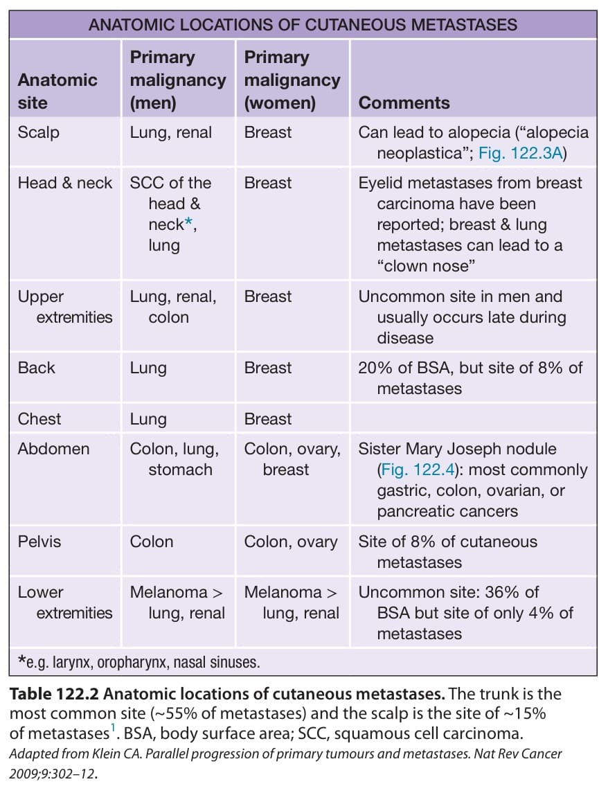
表 122-2:皮膚轉移的解剖部位
Table 122.2 Anatomic locations of cutaneous metastases. The trunk is the most common site (~55% of metastases) and the scalp is the site of ~15% of metastases. BSA, body surface area; SCC, squamous cell carcinoma. Adapted from Klein CA. Parallel progression of primary tumours and metastases. Nat Rev Cancer 2009;9:302–12.
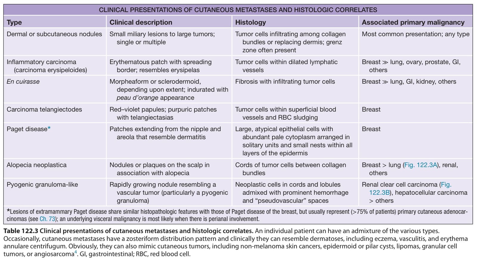
表 122-3:皮膚轉移的臨床表現與組織學對應
Table 122.3 Clinical presentations of cutaneous metastases and histologic correlates. An individual patient can have an admixture of the various types. Occasionally, cutaneous metastases have a zosteriform distribution pattern and clinically they can resemble dermatoses, including eczema, vasculitis, and erythema annulare centrifugum. Obviously, they can also mimic cutaneous tumors, including non-melanoma skin cancers, epidermoid or pilar cysts, lipomas, granular cell tumors, or angiosarcoma. GI, gastrointestinal; RBC, red blood cell.