PATHOLOGY
Adenocarcinoma, squamous cell carcinoma (SCC), and melanoma represent the three major categories of cutaneous metastases (Table 122.4). In broad terms, these metastatic tumors generally lack an epidermal connection or involvement, are situated in the dermis or subcutis, and are composed of nodules, nests, or cords of atypical cells with mitoses (Table 122.5). However, it is important to note that occasionally there are primary “dermal” nodular melanomas, and primary cutaneous spindle cell SCCs often lack a prominent epidermal component. In addition, cutaneous metastases of both melanoma and
SCC (cutaneous, oropharyngeal, or solid organ) may show significant epidermal involvement so that distinction from a synchronous or metachronous primary skin tumor can be difficult. Four primary histopathologic patterns of cutaneous metastases are: (1) nodular; (2) diffuse, including infiltrative; (3) intravascular, including intralymphatic; and (4) epidermotropic (Fig. 122.6).
The following can serve as histologic clues to the diagnosis of cutaneous metastases, in addition to those outlined in Table 122.5: (1) atypical single cells infiltrating through collagen bundles (breast carcinoma; see Fig. 122.6B); (2) clear cells plus abundant hemorrhage (renal cell carcinoma; Fig. 122.7); (3) “dirty” necrosis within glandular structures (colon carcinoma); (4) formation of colloid within
lumina (thyroid carcinoma); (5) psammoma bodies (thyroid or ovarian carcinoma); and (6) signet ring cells (gastric or breast carcinoma; Fig. 122.8). In addition to melanoma and SCC (see above), epidermo tropism can occasionally be seen in cutaneous metastases from other malignancies, in particular breast carcinoma, and less often carcinomas of the prostate, colon, larynx, penis, or vagina. Rarely, epidermotropic metastases, especially from breast carcinoma, may contain pigment and even an intratumoral increase in melanocytes, leading to the misdiagnosis of a melanocytic tumor (see Fig. 122.6D). When there is intra-vascular involvement, this may be confused with benign entities such as intralymphatic histiocytosis (Fig. 122.9).
Complete evaluation of a cutaneous metastasis often requires the use of a panel of histochemical stains (Fig. 122.10; see Table 122.4). While such stains are not a substitute for a detailed history and physical examination plus diagnostic imaging and biopsy of the primary tumor, the resulting information does provide direction and may circumvent the need for a more invasive biopsy of another tissue. In order to distinguish between a primary cutaneous tumor (usually an adnexal carcinoma) and a cutaneous metastasis (especially of adenocarcinoma), immunostains, in particular p40/p63, CK5/6 and D2–40 (podoplanin), can prove helpful. Primary cutaneous tumors are typically positive for some or all these markers and metastases lack staining. Important exceptions are p63-positivity in some metastatic adenocarcinomas (breast, urothelial, and lung) and an absence of p63 staining in some primary cutaneous adnexal carcinomas.
In the case of cutaneous metastases from adenocarcinomas, various combinations of CK7 and CK20 expression can provide useful information (see Fig. 122.10). This panel is also employed to evaluate extramammary Paget disease, in which CK20 positivity points to a secondary form of the disease that is originating from a visceral malignancy (see Fig. 73.16).
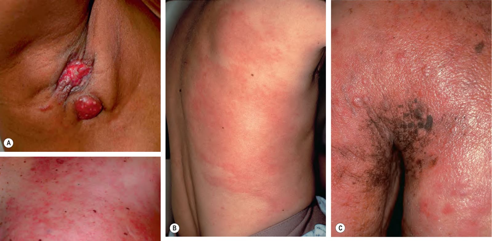
Fig. 122.5 Various presentations of cutaneous metastases of breast carcinoma.A Eroded erythematous nodules in the axilla. B Inflammatory form (carcinoma erysipeloides) with patches of erythema that may initially be misdiagnosed as infectious cellulitis. C Primarily en cuirasse form with obvious induration and peau d’orange appearance in addition to papulonodules. D Mixed pattern – reticulated erythema of carcinoma erysipeloides as well as peau d’orange appearance near the areola. A, D, Courtesy Stuart Lessin, MD.
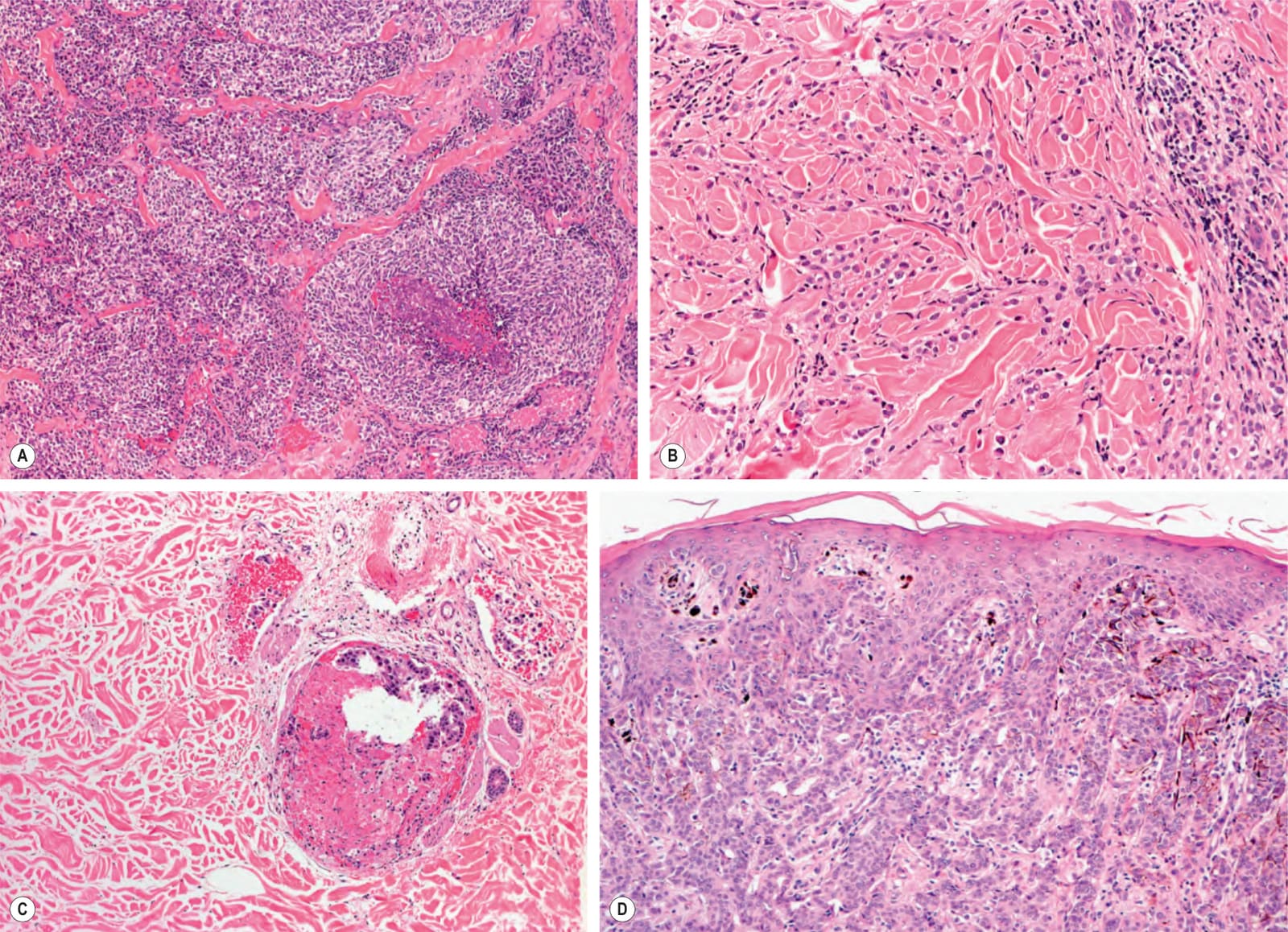
Fig. 122.6 Major patterns of cutaneous metastases.A Nodular pattern with focal, prominent necrosis in a cutaneous metastasis of small cell lung carcinoma. B Diffuse pattern with linear arrays of single cells in a metastasis of breast carcinoma. C Intralymphatic pattern in a metastatic breast carcinoma; tumor cells are confined to dilated lymphatic vessels. D Epidermotropic pattern in a metastasis of breast carcinoma; tumor cells are in both the dermis and the epidermis. The presence of focal pigmentation and melanocyte hyperplasia could lead to the misdiagnosis of a melanocytic tumor. Courtesy Lorenzo Cerroni, MD.
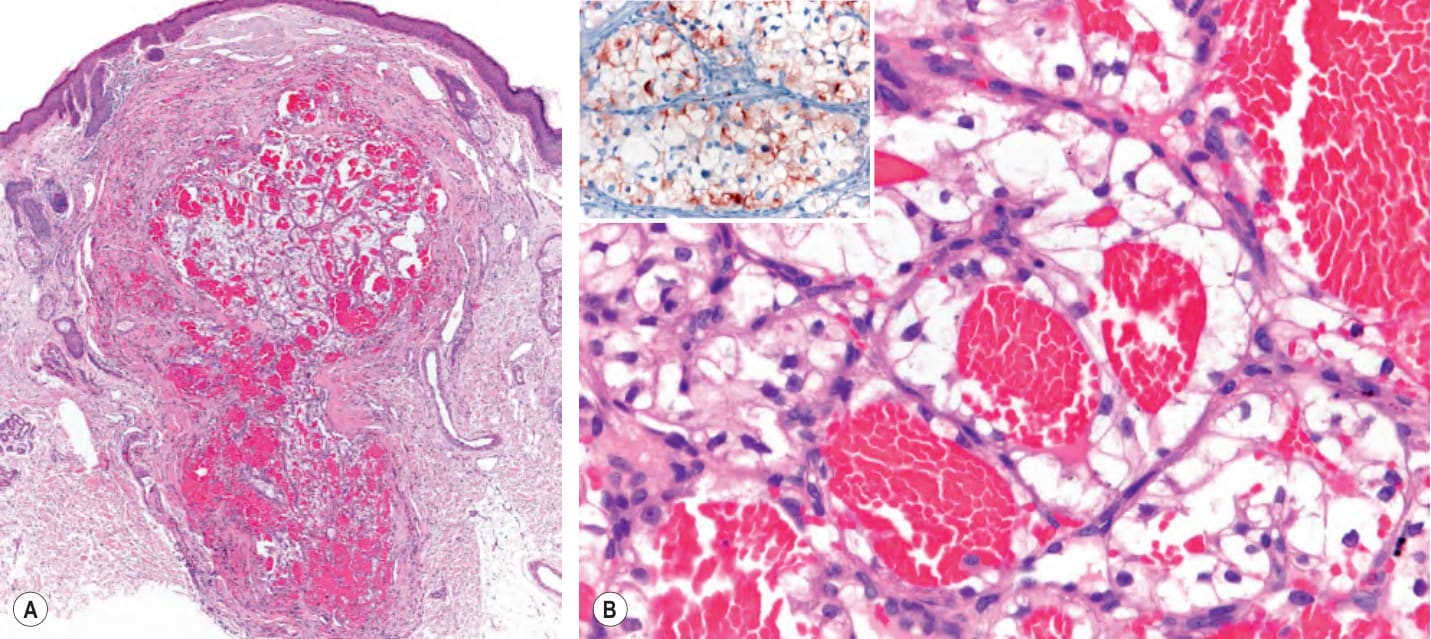
Fig. 122.7 Histopathologic features of cutaneous metastasis from clear cell renal cell carcinoma.A Vertically oriented hemorrhagic tumor within the dermis resembling an angiomatous tumor. B Tumor cells with clear cytoplasm arranged as fine trabecular structures and lobules surrounding hemorrhagic areas. The lack of prominent atypia can be misleading and lead to the erroneous diagnosis of hemangioma. Positive immunohistochemical staining with RCC-Ma (renal cell carcinoma marker), a monoclonal antibody directed against a proximal tubule antigen, labels clear cell renal cell carcinoma (inset). Courtesy Lorenzo Cerroni, MD.
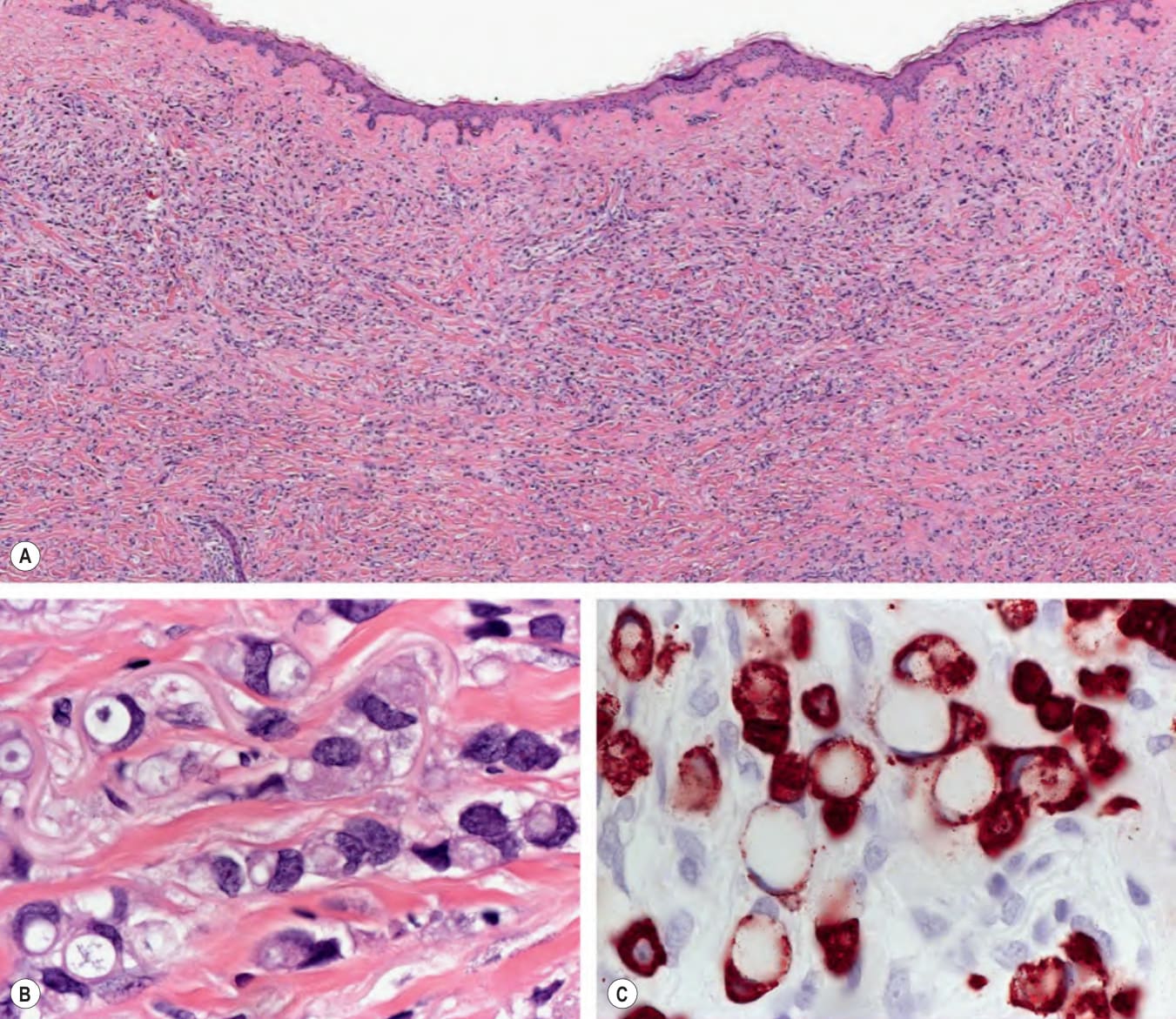
Fig. 122.8 Histopathologic features of cutaneous metastases of signet ring adenocarcinoma of the breast (A, B) and stomach (C).A Diffuse infiltration of neoplastic cells throughout the entire dermis. B Detail of neoplastic cells with signet ring cell morphology due to intracellular mucin accumulation. C Signet ring cells are not unique to breast carcinoma and may be observed in primary cutaneous carcinomas (e.g. histiocytoid carcinoma of the eyelid), non-epithelial neoplasms (e.g. melanoma), and a variety of adenocarcinomas, particularly those from the gastrointestinal tract. A cytokeratin (pan-CK) stain in this metastatic signet ring cell carcinoma of the stomach highlights the peculiar morphology of the cells, with intracellular mucin pressing the nuclei and distorting them, thus conferring the characteristic shape similar to that of a signet ring. Courtesy Lorenzo Cerroni, MD.
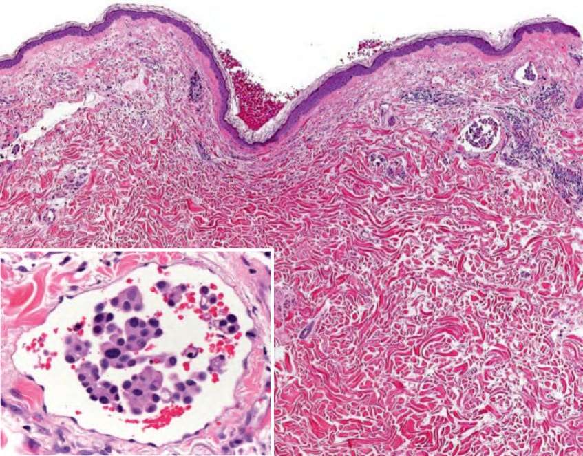
Fig. 122.9 Histopathologic features of cutaneous metastasis of urothelial micropapillary bladder carcinoma. Complexes of neoplastic cells within dilated lymphatic spaces in the dermis. Detail of a lymphatic space with intravascular tumor complexes admixed with erythrocytes (inset). Neoplastic urothelial (transitional) cells exhibit round–ovoid nuclei and abundant eosinophilic cytoplasm, and they form small rosette-like aggregates. These histopathologic features may simulate a benign condition, namely, intralymphatic histiocytosis. Courtesy Lorenzo Cerroni, MD.
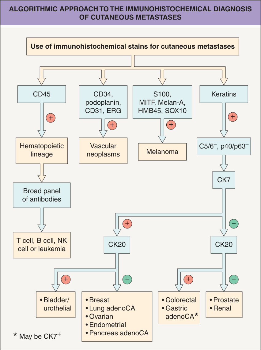
Fig. 122.10 Algorithmic approach to the immunohistochemical diagnosis of cutaneous metastases. The main differential diagnosis of metastatic adenocarcinoma is a primary cutaneous adnexal tumor (generally CK5/6+, p40+/p63+). For tumors not clearly classifiable as a primary skin tumor, clinical history and directed immunohistochemical stains can be key to confirming the diagnosis. adenoCA, adenocarcinoma. Adapted from Handa U, Kundu R, Dimri K. Cutaneous metastasis: a study of 138 cases diagnosed by fine-needle aspiration cytology. Acta Cytol 2017;61:47–54; Saeed S, Keehn CA, Morgan MB. Cutaneous metastasis: a clinical, pathological, and immunohistochemical appraisal. J Cutan Pathol 2004;31:419–30.
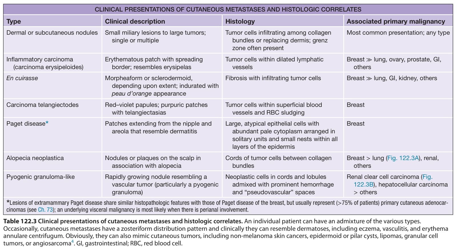
Table 122.3 Clinical presentations of cutaneous metastases and histologic correlates. An individual patient can have an admixture of the various types. Occasionally, cutaneous metastases have a zosteriform distribution pattern and clinically they can resemble dermatoses, including eczema, vasculitis, and erythema annulare centrifugum. Obviously, they can also mimic cutaneous tumors, including non-melanoma skin cancers, epidermoid or pilar cysts, lipomas, granular cell tumors, or angiosarcoma. GI, gastrointestinal; RBC, red blood cell.
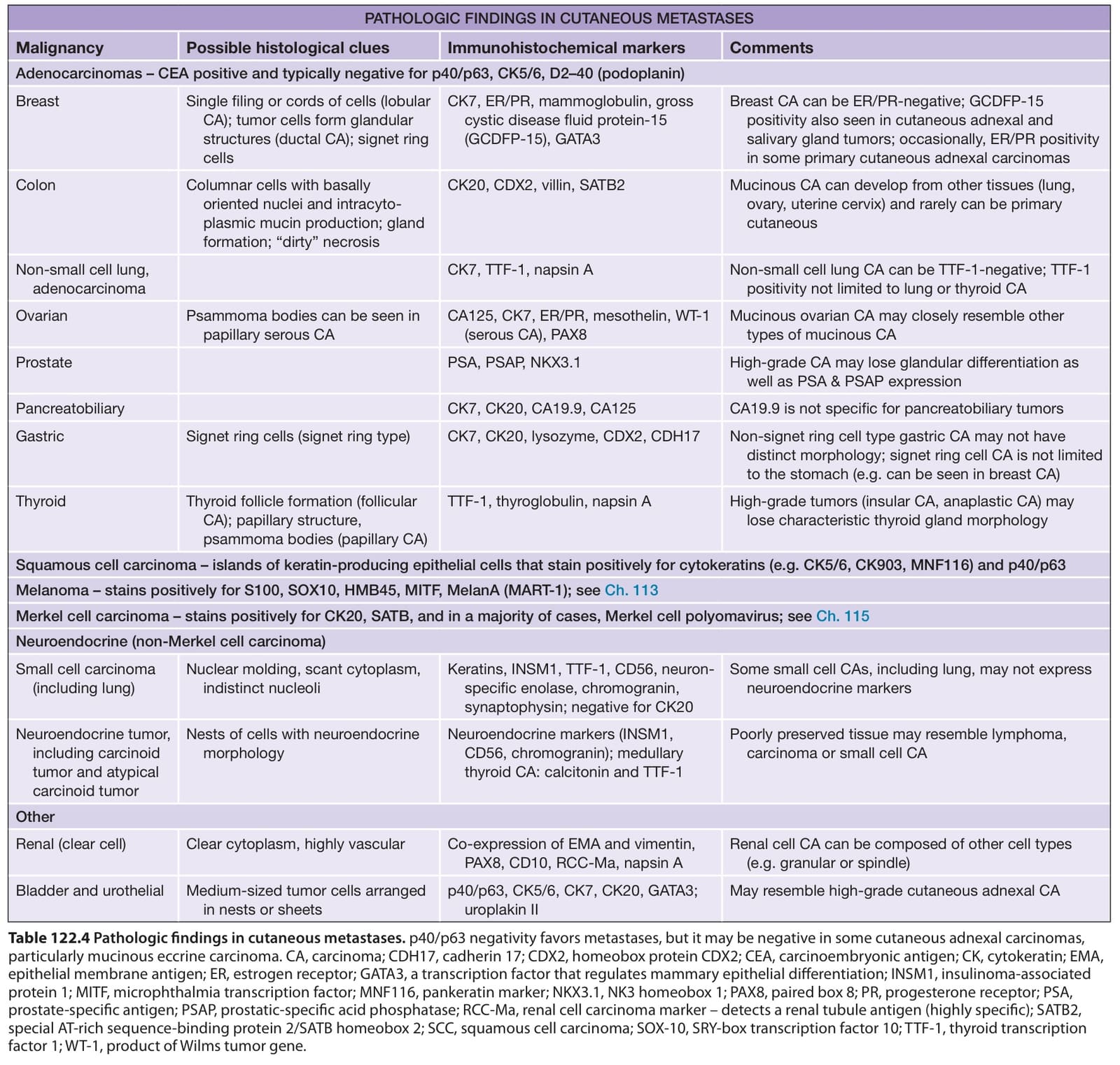
Table 122.4 Pathologic findings in cutaneous metastases. p40/p63 negativity favors metastases, but it may be negative in some cutaneous adnexal carcinomas, particularly mucinous eccrine carcinoma. CA, carcinoma; CDH17, cadherin 17; CDX2, homeobox protein CDX2; CEA, carcinoembryonic antigen; CK, cytokeratin; EMA, epithelial membrane antigen; ER, estrogen receptor; GATA3, a transcription factor that regulates mammary epithelial differentiation; INSM1, insulinoma-associated protein 1; MITF, microphthalmia transcription factor; MNF116, pankeratin marker; NKX3.1, NK3 homeobox 1; PAX8, paired box 8; PR, progesterone receptor; PSA, prostate-specific antigen; PSAP, prostatic-specific acid phosphatase; RCC-Ma, renal cell carcinoma marker – detects a renal tubule antigen (highly specific); SATB2, special AT-rich sequence-binding protein 2/SATB homeobox 2; SCC, squamous cell carcinoma; SOX-10, SRY-box transcription factor 10; TTF-1, thyroid transcription factor 1; WT-1, product of Wilms tumor gene.
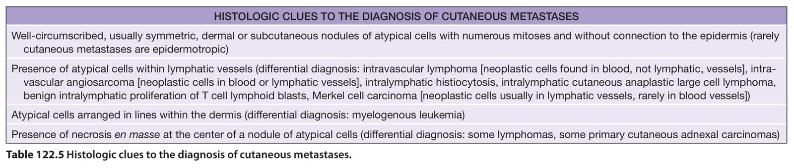
Table 122.5 Histologic clues to the diagnosis of cutaneous metastases.