病理(PATHOLOGY)
腺癌(adenocarcinoma)、鱗狀細胞癌(squamous cell carcinoma, SCC)與黑色素瘤代表皮膚轉移的三大類別(Table 122.4)。廣義而言,這些轉移性腫瘤一般缺乏與表皮的連接或表皮侵犯,位於真皮或皮下,並由具有絲分裂的非典型細胞所構成的結節、細胞巢或索狀構造組成(Table 122.5)。然而,重要的是要注意,偶爾會有原發性的「真皮型(dermal)」結節性黑色素瘤,而原發性皮膚梭形細胞 SCC 也常缺乏明顯的表皮成分。此外,黑色素瘤與
SCC(皮膚、口咽或實體器官)的皮膚轉移可能顯示顯著的表皮侵犯,以致難以與同時性或異時性的原發皮膚腫瘤區分。皮膚轉移的四種主要組織病理型態為:(1) 結節型(nodular);(2) 瀰漫型(diffuse),包括浸潤型(infiltrative);(3) 血管內型(intravascular),包括淋巴管內型(intralymphatic);以及 (4) 表皮親和型(epidermotropic)(Fig. 122.6)。
除了 Table 122.5 所列者之外,下列各項可作為診斷皮膚轉移的組織學線索:(1) 非典型單一細胞穿行浸潤於膠原纖維束之間(乳癌;見 Fig. 122.6B);(2) 透明細胞加上大量出血(腎細胞癌;Fig. 122.7);(3) 腺體結構內的「髒性(dirty)」壞死(結腸癌);(4) 管腔內
膠質(colloid)的形成(甲狀腺癌);(5) 砂粒體(psammoma bodies)(甲狀腺或卵巢癌);以及 (6) 印戒細胞(signet ring cells)(胃癌或乳癌;Fig. 122.8)。除黑色素瘤與 SCC(見上)之外,表皮親和性偶爾也可見於源自其他惡性腫瘤的皮膚轉移,特別是乳癌,較少見於攝護腺、結腸、喉部、陰莖或陰道的癌症。罕見情況下,表皮親和性轉移(尤其源自乳癌)可能含有色素,甚至有腫瘤內黑色素細胞的增加,導致誤診為黑色素細胞腫瘤(見 Fig. 122.6D)。當有血管內侵犯時,可能與良性疾病實體如淋巴管內組織球增生症混淆(Fig. 122.9)。
皮膚轉移的完整評估往往需要使用一組組織化學染色(Fig. 122.10;見 Table 122.4)。雖然此類染色無法取代詳細的病史與理學檢查,加上診斷性影像與原發腫瘤的切片,但所得資訊確實提供方向,並可能避免對另一組織進行更具侵入性切片的必要。為了區分原發性皮膚腫瘤(通常為附屬器癌)與皮膚轉移(尤其是腺癌的轉移),免疫染色,特別是 p40/p63、CK5/6 與 D2–40(podoplanin),可能有幫助。原發性皮膚腫瘤典型上對這些標記中的部分或全部呈陽性,而轉移則缺乏染色。重要的例外是某些轉移性腺癌(乳房、泌尿上皮與肺部)的 p63 陽性,以及某些原發性皮膚附屬器癌缺乏 p63 染色。
在源自腺癌的皮膚轉移中,CK7 與 CK20 表現的各種組合可提供有用資訊(見 Fig. 122.10)。此組合亦用於評估乳房外 Paget disease,其中 CK20 陽性指向源自內臟惡性腫瘤的續發型疾病(見 Fig. 73.16)。
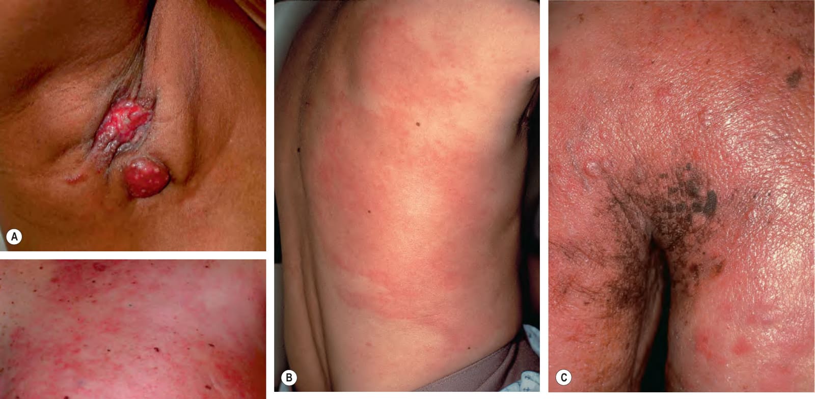
圖 122-5:乳癌皮膚轉移的各種表現。A 腋窩處糜爛的紅斑性結節。B 發炎型(carcinoma erysipeloides),有紅斑性斑,起初可能被誤診為感染性蜂窩性組織炎。C 主要為 en cuirasse 型,除丘疹結節外還有明顯硬化與橘皮外觀。D 混合型態——carcinoma erysipeloides 的網狀紅斑,以及乳暈附近的橘皮外觀
Fig. 122.5 Various presentations of cutaneous metastases of breast carcinoma.A Eroded erythematous nodules in the axilla. B Inflammatory form (carcinoma erysipeloides) with patches of erythema that may initially be misdiagnosed as infectious cellulitis. C Primarily en cuirasse form with obvious induration and peau d’orange appearance in addition to papulonodules. D Mixed pattern – reticulated erythema of carcinoma erysipeloides as well as peau d’orange appearance near the areola. A, D, Courtesy Stuart Lessin, MD.
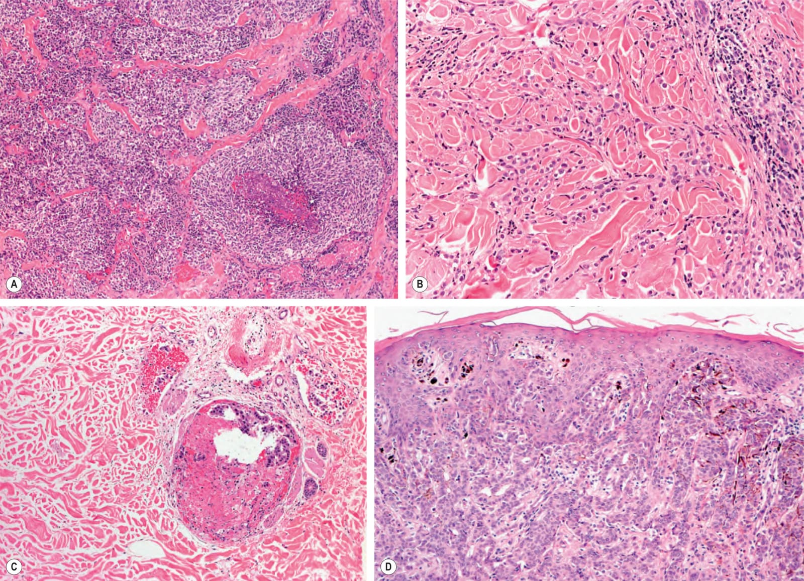
圖 122-6:皮膚轉移的主要型態。A 小細胞肺癌皮膚轉移中的結節型態伴局部明顯壞死。B 乳癌轉移中的瀰漫型態,單一細胞呈線狀排列。C 轉移性乳癌中的淋巴管內型態;腫瘤細胞侷限於擴張的淋巴管內。D 乳癌轉移中的表皮親和型態;腫瘤細胞同時位於真皮與表皮
Fig. 122.6 Major patterns of cutaneous metastases.A Nodular pattern with focal, prominent necrosis in a cutaneous metastasis of small cell lung carcinoma. B Diffuse pattern with linear arrays of single cells in a metastasis of breast carcinoma. C Intralymphatic pattern in a metastatic breast carcinoma; tumor cells are confined to dilated lymphatic vessels. D Epidermotropic pattern in a metastasis of breast carcinoma; tumor cells are in both the dermis and the epidermis. The presence of focal pigmentation and melanocyte hyperplasia could lead to the misdiagnosis of a melanocytic tumor. Courtesy Lorenzo Cerroni, MD.
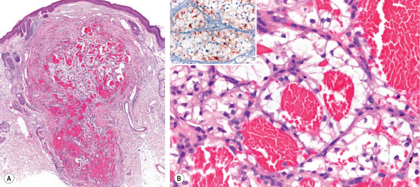
圖 122-7:透明細胞腎細胞癌皮膚轉移的組織病理特徵。A 真皮內垂直走向的出血性腫瘤,類似血管瘤樣腫瘤。B 具透明細胞質的腫瘤細胞排列成細小樑狀結構與小葉,環繞出血區域
Fig. 122.7 Histopathologic features of cutaneous metastasis from clear cell renal cell carcinoma.A Vertically oriented hemorrhagic tumor within the dermis resembling an angiomatous tumor. B Tumor cells with clear cytoplasm arranged as fine trabecular structures and lobules surrounding hemorrhagic areas. The lack of prominent atypia can be misleading and lead to the erroneous diagnosis of hemangioma. Positive immunohistochemical staining with RCC-Ma (renal cell carcinoma marker), a monoclonal antibody directed against a proximal tubule antigen, labels clear cell renal cell carcinoma (inset). Courtesy Lorenzo Cerroni, MD.
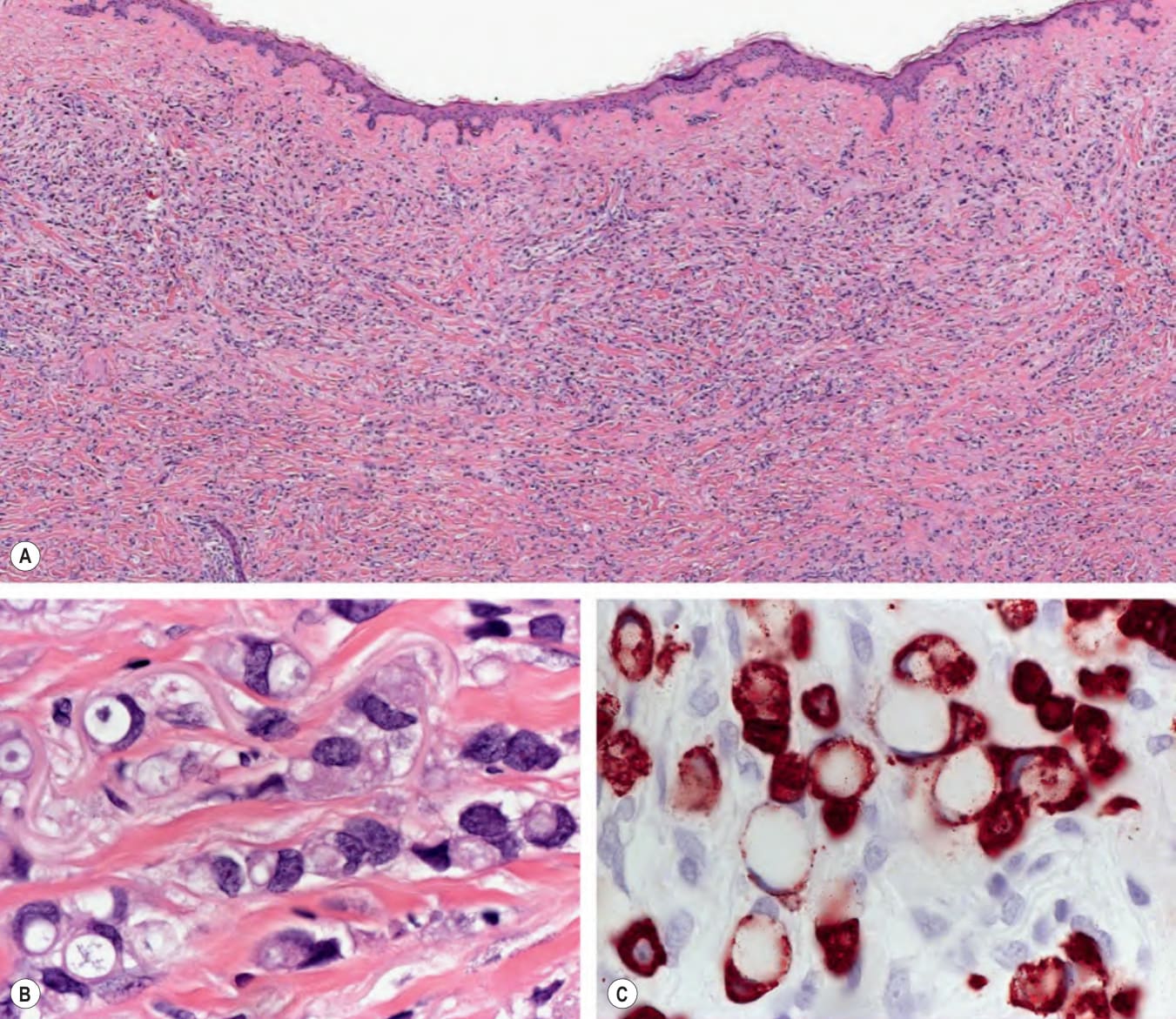
圖 122-8:乳房(A、B)與胃(C)印戒細胞腺癌皮膚轉移的組織病理特徵。A 腫瘤細胞在整層真皮的瀰漫性浸潤。B 因細胞內黏液蓄積而呈印戒細胞形態之腫瘤細胞的細部
Fig. 122.8 Histopathologic features of cutaneous metastases of signet ring adenocarcinoma of the breast (A, B) and stomach (C).A Diffuse infiltration of neoplastic cells throughout the entire dermis. B Detail of neoplastic cells with signet ring cell morphology due to intracellular mucin accumulation. C Signet ring cells are not unique to breast carcinoma and may be observed in primary cutaneous carcinomas (e.g. histiocytoid carcinoma of the eyelid), non-epithelial neoplasms (e.g. melanoma), and a variety of adenocarcinomas, particularly those from the gastrointestinal tract. A cytokeratin (pan-CK) stain in this metastatic signet ring cell carcinoma of the stomach highlights the peculiar morphology of the cells, with intracellular mucin pressing the nuclei and distorting them, thus conferring the characteristic shape similar to that of a signet ring. Courtesy Lorenzo Cerroni, MD.
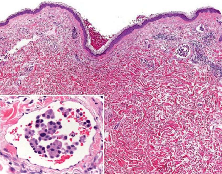
圖 122-9:泌尿上皮微乳頭狀膀胱癌皮膚轉移的組織病理特徵。真皮內擴張淋巴管腔隙中的腫瘤細胞複合體
Fig. 122.9 Histopathologic features of cutaneous metastasis of urothelial micropapillary bladder carcinoma. Complexes of neoplastic cells within dilated lymphatic spaces in the dermis. Detail of a lymphatic space with intravascular tumor complexes admixed with erythrocytes (inset). Neoplastic urothelial (transitional) cells exhibit round–ovoid nuclei and abundant eosinophilic cytoplasm, and they form small rosette-like aggregates. These histopathologic features may simulate a benign condition, namely, intralymphatic histiocytosis. Courtesy Lorenzo Cerroni, MD.
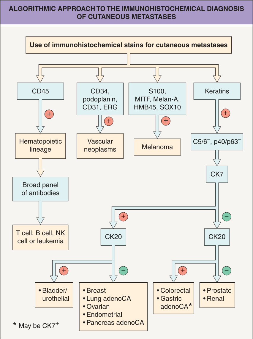
圖 122-10:皮膚轉移免疫組織化學診斷的演算取徑
Fig. 122.10 Algorithmic approach to the immunohistochemical diagnosis of cutaneous metastases. The main differential diagnosis of metastatic adenocarcinoma is a primary cutaneous adnexal tumor (generally CK5/6+, p40+/p63+). For tumors not clearly classifiable as a primary skin tumor, clinical history and directed immunohistochemical stains can be key to confirming the diagnosis. adenoCA, adenocarcinoma. Adapted from Handa U, Kundu R, Dimri K. Cutaneous metastasis: a study of 138 cases diagnosed by fine-needle aspiration cytology. Acta Cytol 2017;61:47–54; Saeed S, Keehn CA, Morgan MB. Cutaneous metastasis: a clinical, pathological, and immunohistochemical appraisal. J Cutan Pathol 2004;31:419–30.
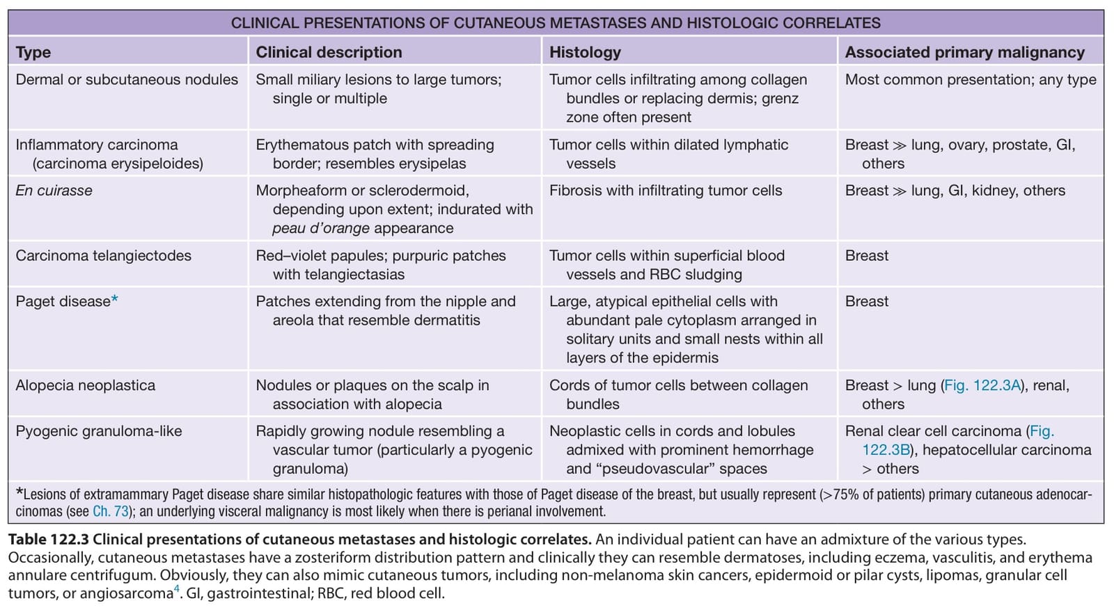
表 122-3:皮膚轉移的臨床表現與組織學對應
Table 122.3 Clinical presentations of cutaneous metastases and histologic correlates. An individual patient can have an admixture of the various types. Occasionally, cutaneous metastases have a zosteriform distribution pattern and clinically they can resemble dermatoses, including eczema, vasculitis, and erythema annulare centrifugum. Obviously, they can also mimic cutaneous tumors, including non-melanoma skin cancers, epidermoid or pilar cysts, lipomas, granular cell tumors, or angiosarcoma. GI, gastrointestinal; RBC, red blood cell.
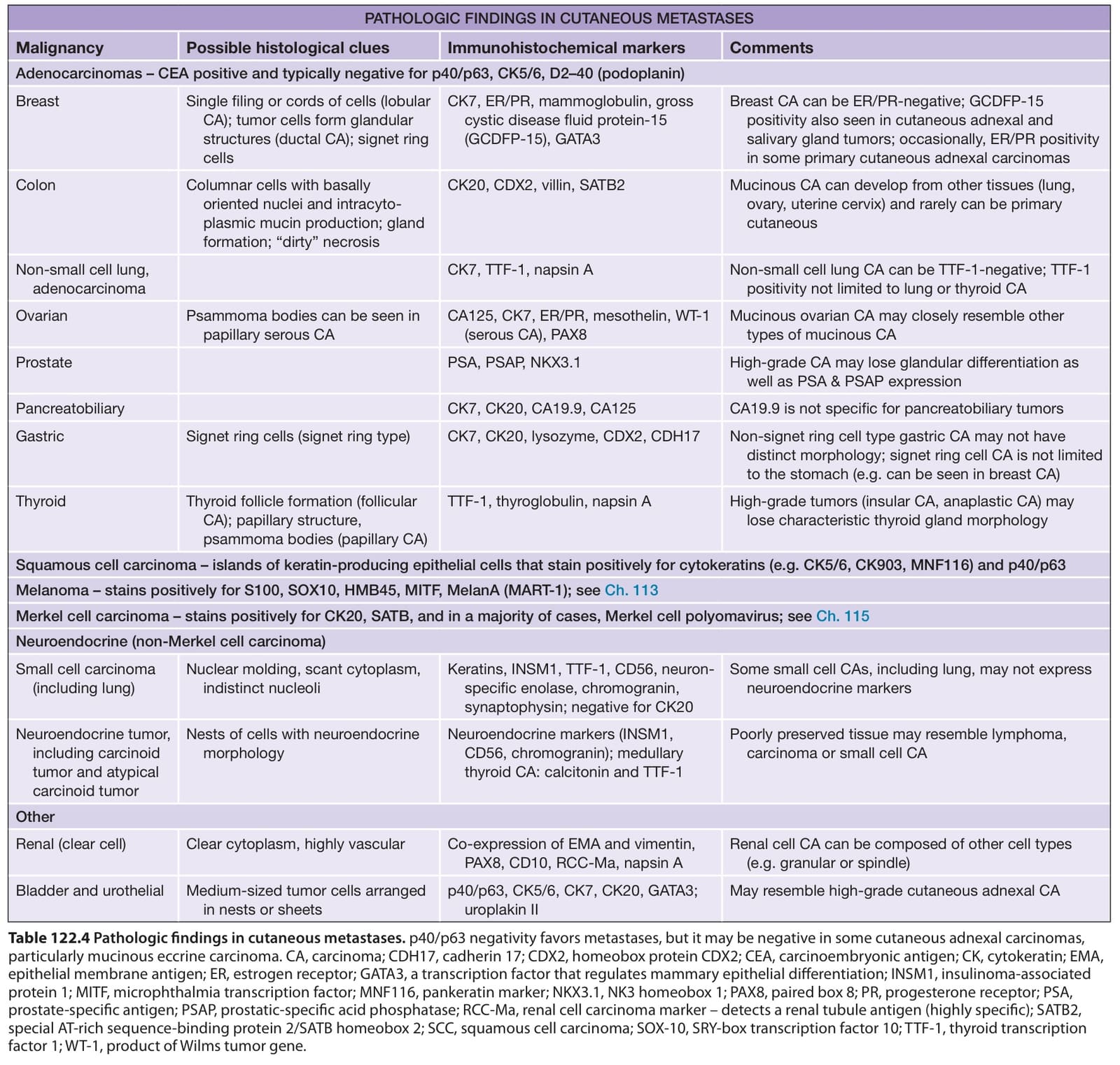
表 122-4:皮膚轉移的病理所見
Table 122.4 Pathologic findings in cutaneous metastases. p40/p63 negativity favors metastases, but it may be negative in some cutaneous adnexal carcinomas, particularly mucinous eccrine carcinoma. CA, carcinoma; CDH17, cadherin 17; CDX2, homeobox protein CDX2; CEA, carcinoembryonic antigen; CK, cytokeratin; EMA, epithelial membrane antigen; ER, estrogen receptor; GATA3, a transcription factor that regulates mammary epithelial differentiation; INSM1, insulinoma-associated protein 1; MITF, microphthalmia transcription factor; MNF116, pankeratin marker; NKX3.1, NK3 homeobox 1; PAX8, paired box 8; PR, progesterone receptor; PSA, prostate-specific antigen; PSAP, prostatic-specific acid phosphatase; RCC-Ma, renal cell carcinoma marker – detects a renal tubule antigen (highly specific); SATB2, special AT-rich sequence-binding protein 2/SATB homeobox 2; SCC, squamous cell carcinoma; SOX-10, SRY-box transcription factor 10; TTF-1, thyroid transcription factor 1; WT-1, product of Wilms tumor gene.
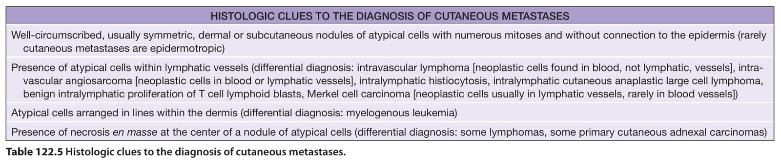
表 122-5:診斷皮膚轉移的組織學線索
Table 122.5 Histologic clues to the diagnosis of cutaneous metastases.