TREATMENT AND PROGNOSIS
Typically, cutaneous metastases are a harbinger of late-stage or progressive disease and as such portend a poor prognosis. The average survival time following the initial diagnosis of cutaneous metastasis is 7.5 months and half of patients expire within the first 6 months. Whether the patient has a known or unknown primary malignancy, the development of cutaneous metastases calls for a multidisciplinary approach, including medical and surgical oncologists, radiation oncologists, and mental health care providers when appropriate. Localized treatment of cutaneous metastases can be undertaken for functional, palliative, or cosmetic indications. When feasible, surgical excision is performed; however, radiation and focal chemotherapeutics or immunotherapies can be considered on a case-by-case basis. Systemic therapies are indicated for immuno- or chemotherapy-responsive tumors. When malodorous, topical metronidazole solution (supplied for intravenous use) can be applied via cotton gauze or pump spray once to twice daily or crushed metronidazole tablets can be sprinkled onto the tumor twice daily.
Additional figures available in our eBook (see inside front cover for access code).
-
Klein CA. Parallel progression of primary tumours and tases in patients with metastatic carcinoma: a retrospective study of 4020 patients. J Am Acad Dermatol. 1993;29:228–236.2. Nashan D, Lucas Muller M, Braun-Falco M, et al.
-
Lee JJ, Mochel MC, Piris A, et al. p40 exhibits better metastases. Nat Rev Cancer. 2009;9:302–312.9. Swetter SM, Ecker PM, Johnson DL, Harvell JD. Primary specificity than p63 in distinguishing primary skin adnexal carcinomas from cutaneous metastases. Hum Pathol. 2014;45:1078–1083.15. Chu P, Wu E, Weiss LM. Cytokeratin 7 and cytokeratin dermal melanoma. A distinct subtype of melanoma. Arch Dermatol. 2004;140:99–103.10. McKee PH. Cutaneous metastases. J Cutan Pathol.
Cutaneous metastases of visceral tumors: a review. J Cancer Res Clin Oncol. 2009;135:1–14.3. Schwartz RA. Cutaneous metastatic disease. J Am Acad
20 expression in epithelial neoplasms: a survey of 435 cases. Mod Pathol. 2000;13:962–972.16. Handa U, Kundu R, Dimri K. Cutaneous metastasis: a
1985;12:239–250.11. Schwartz RA. Histopathologic aspects of
Dermatol. 1995;33:161–182.4. Sariya D, Ruth K, Adams-McDonnell R, et al.
cutaneous metastatic disease. J Am Acad Dermatol. 1995;33:649–657.12. Bellizzi AM. An algorithmic immunohistochemical
Clinicopathologic correlation of cutaneous metastases: experience from a cancer center. Arch Dermatol. 2008;144:1155–1162.5. Brodland DG, Zitelli JA. Mechanisms of metastasis. J Am study of 138 cases diagnosed by fine-needle aspiration cytology. Acta Cytol. 2017;61:47–54.17. Saeed S, Keehn CA, Morgan MB. Cutaneous metastasis:
approach to define tumor type and assign site of origin. Adv Anat Pathol. 2020;27:114–163.13. Mahalingam M, Nguyen LP, Richards JE, et al. The
Acad Dermatol. 1992;27:1–10.6. Brodland DG. The life of a skin cancer. Mayo Clin Proc.
a clinical, pathological, and immunohistochemical appraisal. J Cutan Pathol. 2004;31:419–430.18. Schoenlaub P, Sarraux A, Grosshans E, et al. Survival diagnostic utility of immunohistochemistry in distinguishing primary skin adnexal carcinomas from metastatic adenocarcinom to skin: an immunohistochemical reappraisal using cytokeratin 15, nestin, p63, D2-40, and calretinin. Mod Pathol. 2010;23:713–719.
1997;72:475–478.7. Morgan-Parkes JH. Metastases: mechanisms, pathways, and cascades. Adv Clin Med. 1995;164:1075–1082.
after cutaneous metastasis: a study of 200 cases. Ann Dermatol Venereol. 2001;128:1310–1315.
Certain organ-restricted markers may also be useful including: CD10, RCC, and PAX8 (positive in renal and ovarian tumors); thyroid-specific transcription factor-1 (TTF-1) (thyroid, lung; see Fig. 122.2); thyroglobulin (thyroid); prostate specific antigen (PSA) and NKX3.1 (prostate); CDX2, CDH17, and SATB2 (gastrointestinal); Hep Par1 and arginase-1 (hepatocellular carcinoma); and CD56 and insulinoma-associated protein 1 (INSM1) (neuroendocrine) (see Table 122.4). In cutaneous metastases of SCC, staining for pankeratins and EMA may be positive, but this information is not helpful in determining the site of the primary SCC.
In addition to the histologic examination of conventional skin biopsies, cutaneous metastases can be sampled via fine-needle aspiration cytology.
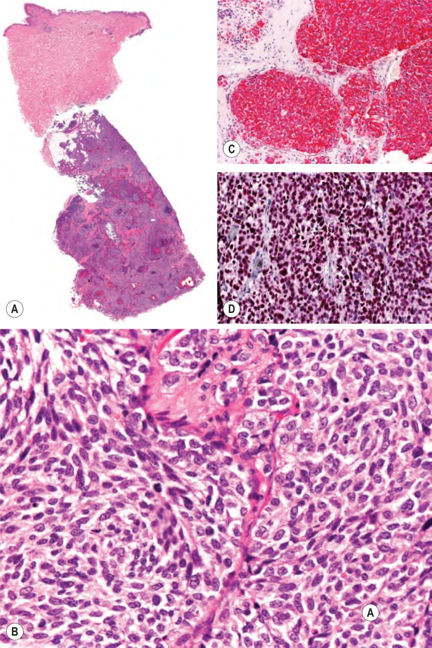
Fig. 122.2 Subcutaneous metastasis of small cell lung carcinoma.A Note sparing of the dermis. B Higher magnification demonstrating clusters of basaloid cells. C The tumor cells stain positively for CK7. D The tumor cells also stain positively for TTF-1 (thyroid transcription factor-1). Courtesy Lorenzo Cerroni, MD.
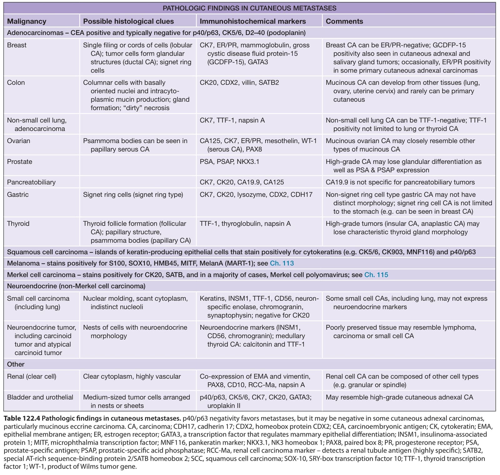
Table 122.4 Pathologic findings in cutaneous metastases. p40/p63 negativity favors metastases, but it may be negative in some cutaneous adnexal carcinomas, particularly mucinous eccrine carcinoma. CA, carcinoma; CDH17, cadherin 17; CDX2, homeobox protein CDX2; CEA, carcinoembryonic antigen; CK, cytokeratin; EMA, epithelial membrane antigen; ER, estrogen receptor; GATA3, a transcription factor that regulates mammary epithelial differentiation; INSM1, insulinoma-associated protein 1; MITF, microphthalmia transcription factor; MNF116, pankeratin marker; NKX3.1, NK3 homeobox 1; PAX8, paired box 8; PR, progesterone receptor; PSA, prostate-specific antigen; PSAP, prostatic-specific acid phosphatase; RCC-Ma, renal cell carcinoma marker – detects a renal tubule antigen (highly specific); SATB2, special AT-rich sequence-binding protein 2/SATB homeobox 2; SCC, squamous cell carcinoma; SOX-10, SRY-box transcription factor 10; TTF-1, thyroid transcription factor 1; WT-1, product of Wilms tumor gene.