治療與預後(TREATMENT AND PROGNOSIS)
典型上,皮膚轉移是晚期或進行性疾病的先兆,因而預示不良預後。皮膚轉移初次診斷後的平均存活時間為 7.5 個月,且半數病人於最初 6 個月內死亡。無論病人的原發惡性腫瘤是已知或未知,皮膚轉移的發生都需要多專科取徑,包括內科與外科腫瘤醫師、放射腫瘤醫師,以及在適當時的心理健康照護提供者。皮膚轉移的局部治療可基於功能性、緩和性或美觀上的適應症而施行。在可行時執行手術切除;然而,放射治療與局部化學治療藥物或免疫治療可依個案考量。全身性療法適用於對免疫治療或化學治療有反應的腫瘤。當有惡臭時,可將外用 metronidazole 溶液(以靜脈注射用製劑提供)以棉紗布或噴瓶每日塗抹一至二次,或將壓碎的 metronidazole 錠劑每日兩次灑於腫瘤上。
Additional figures available in our eBook (see inside front cover for access code).
-
Klein CA. Parallel progression of primary tumours and tases in patients with metastatic carcinoma: a retrospective study of 4020 patients. J Am Acad Dermatol. 1993;29:228–236.2. Nashan D, Lucas Muller M, Braun-Falco M, et al.
-
Lee JJ, Mochel MC, Piris A, et al. p40 exhibits better metastases. Nat Rev Cancer. 2009;9:302–312.9. Swetter SM, Ecker PM, Johnson DL, Harvell JD. Primary specificity than p63 in distinguishing primary skin adnexal carcinomas from cutaneous metastases. Hum Pathol. 2014;45:1078–1083.15. Chu P, Wu E, Weiss LM. Cytokeratin 7 and cytokeratin dermal melanoma. A distinct subtype of melanoma. Arch Dermatol. 2004;140:99–103.10. McKee PH. Cutaneous metastases. J Cutan Pathol.
Cutaneous metastases of visceral tumors: a review. J Cancer Res Clin Oncol. 2009;135:1–14.3. Schwartz RA. Cutaneous metastatic disease. J Am Acad
20 expression in epithelial neoplasms: a survey of 435 cases. Mod Pathol. 2000;13:962–972.16. Handa U, Kundu R, Dimri K. Cutaneous metastasis: a
1985;12:239–250.11. Schwartz RA. Histopathologic aspects of
Dermatol. 1995;33:161–182.4. Sariya D, Ruth K, Adams-McDonnell R, et al.
cutaneous metastatic disease. J Am Acad Dermatol. 1995;33:649–657.12. Bellizzi AM. An algorithmic immunohistochemical
Clinicopathologic correlation of cutaneous metastases: experience from a cancer center. Arch Dermatol. 2008;144:1155–1162.5. Brodland DG, Zitelli JA. Mechanisms of metastasis. J Am study of 138 cases diagnosed by fine-needle aspiration cytology. Acta Cytol. 2017;61:47–54.17. Saeed S, Keehn CA, Morgan MB. Cutaneous metastasis:
approach to define tumor type and assign site of origin. Adv Anat Pathol. 2020;27:114–163.13. Mahalingam M, Nguyen LP, Richards JE, et al. The
Acad Dermatol. 1992;27:1–10.6. Brodland DG. The life of a skin cancer. Mayo Clin Proc.
a clinical, pathological, and immunohistochemical appraisal. J Cutan Pathol. 2004;31:419–430.18. Schoenlaub P, Sarraux A, Grosshans E, et al. Survival diagnostic utility of immunohistochemistry in distinguishing primary skin adnexal carcinomas from metastatic adenocarcinom to skin: an immunohistochemical reappraisal using cytokeratin 15, nestin, p63, D2-40, and calretinin. Mod Pathol. 2010;23:713–719.
1997;72:475–478.7. Morgan-Parkes JH. Metastases: mechanisms, pathways, and cascades. Adv Clin Med. 1995;164:1075–1082.
after cutaneous metastasis: a study of 200 cases. Ann Dermatol Venereol. 2001;128:1310–1315.
某些器官限定性的標記也可能有用,包括:CD10、RCC 與 PAX8(在腎臟與卵巢腫瘤中呈陽性);thyroid-specific transcription factor-1(TTF-1)(甲狀腺、肺;見 Fig. 122.2);thyroglobulin(甲狀腺);prostate specific antigen(PSA)與 NKX3.1(攝護腺);CDX2、CDH17 與 SATB2(腸胃道);Hep Par1 與 arginase-1(肝細胞癌);以及 CD56 與 insulinoma-associated protein 1(INSM1)(神經內分泌)(見 Table 122.4)。在 SCC 的皮膚轉移中,pankeratins 與 EMA 的染色可能呈陽性,但此資訊對判定原發 SCC 的部位並無幫助。
除了對傳統皮膚切片進行組織學檢查之外,皮膚轉移亦可經由細針抽吸細胞學(fine-needle aspiration cytology)取樣。
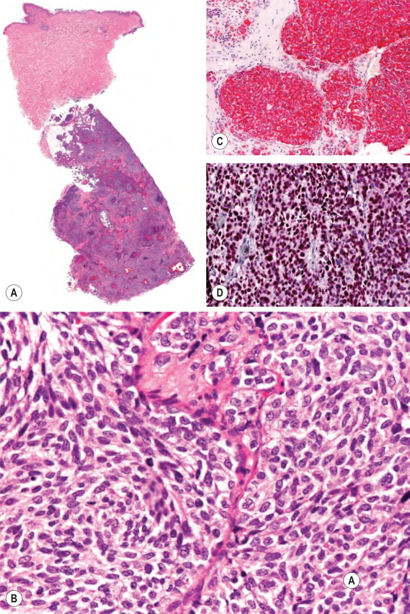
圖 122-2:小細胞肺癌的皮下轉移。A 注意真皮的倖免。B 高倍放大顯示基底樣細胞的群集。C 腫瘤細胞對 CK7 染色呈陽性。D 腫瘤細胞亦對 TTF-1(thyroid transcription factor-1)染色呈陽性
Fig. 122.2 Subcutaneous metastasis of small cell lung carcinoma.A Note sparing of the dermis. B Higher magnification demonstrating clusters of basaloid cells. C The tumor cells stain positively for CK7. D The tumor cells also stain positively for TTF-1 (thyroid transcription factor-1). Courtesy Lorenzo Cerroni, MD.
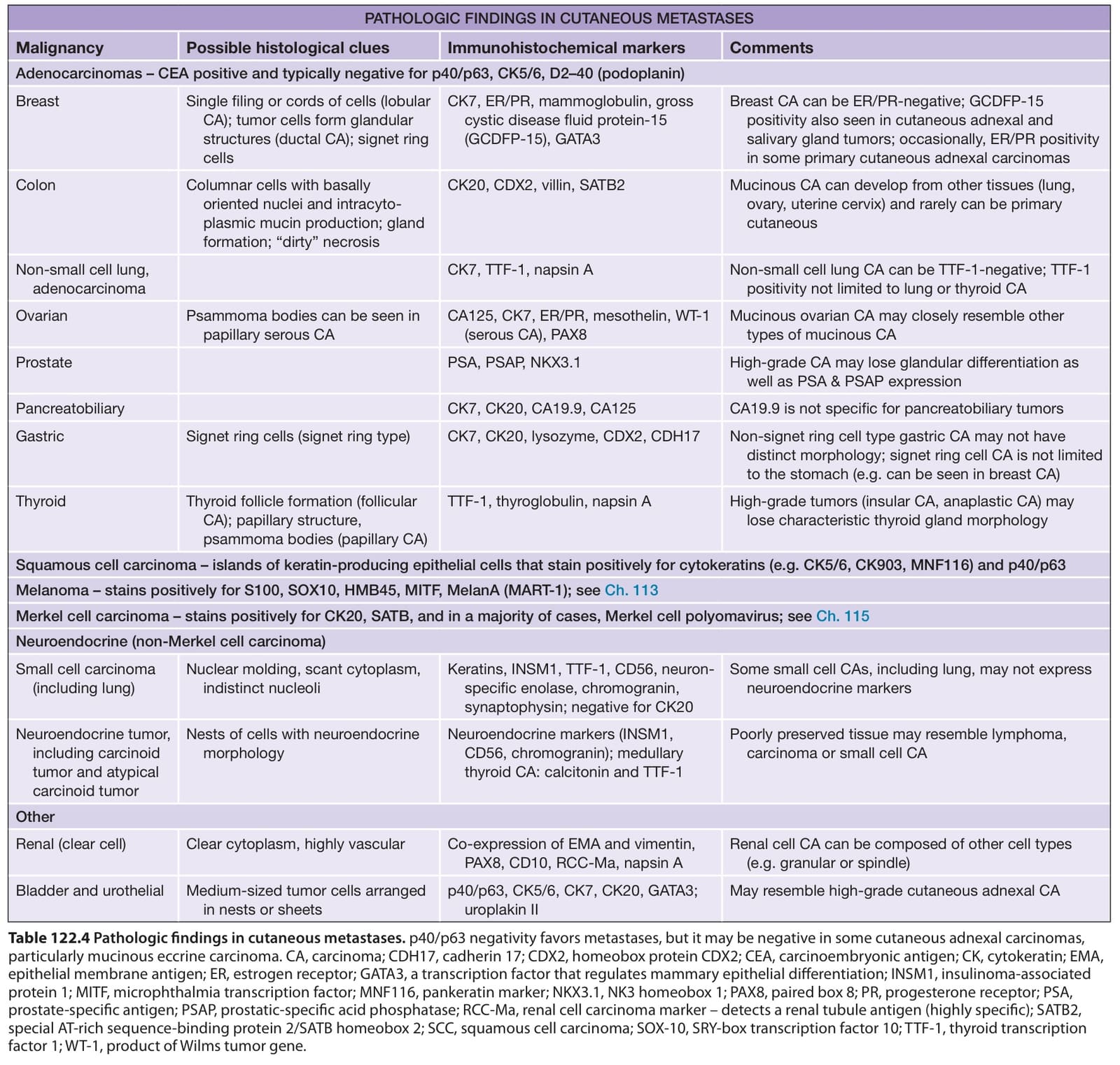
表 122-4:皮膚轉移的病理所見
Table 122.4 Pathologic findings in cutaneous metastases. p40/p63 negativity favors metastases, but it may be negative in some cutaneous adnexal carcinomas, particularly mucinous eccrine carcinoma. CA, carcinoma; CDH17, cadherin 17; CDX2, homeobox protein CDX2; CEA, carcinoembryonic antigen; CK, cytokeratin; EMA, epithelial membrane antigen; ER, estrogen receptor; GATA3, a transcription factor that regulates mammary epithelial differentiation; INSM1, insulinoma-associated protein 1; MITF, microphthalmia transcription factor; MNF116, pankeratin marker; NKX3.1, NK3 homeobox 1; PAX8, paired box 8; PR, progesterone receptor; PSA, prostate-specific antigen; PSAP, prostatic-specific acid phosphatase; RCC-Ma, renal cell carcinoma marker – detects a renal tubule antigen (highly specific); SATB2, special AT-rich sequence-binding protein 2/SATB homeobox 2; SCC, squamous cell carcinoma; SOX-10, SRY-box transcription factor 10; TTF-1, thyroid transcription factor 1; WT-1, product of Wilms tumor gene.