CUTANEOUS DISEASES AND BARRIER DYSFUNCTION
Abnormalities of the skin barrier are observed in both common and rare skin diseases. Causes of barrier dysfunction include (but are not limited to): mechanical trauma, exposure to detergents or solvents, seborrhea, prolonged water exposure, ultraviolet radiation, infection, inflammatory dermatoses, certain drugs, and genetic mutations and polymorphisms. Low humidity downregulates barrier competence while cold temperatures impair permeability barrier homeostasis. Many disorders of cornification, including the ichthyoses, are due to pathogenic variants in genes that encode key components of the skin barrier, such as keratin and enzymes involved in extracellular lipid synthesis, among others.
Mutations and genetic polymorphisms in barrier-related genes also contribute to the pathogenesis of more common skin diseases, e.g. filaggrin in atopic dermatitis (see Ch. 12) or cornified envelope proteins LCE3B and LCE3C in psoriasis. Figure 124.5 depicts the impact of reduced filaggrin production on various functions of the stratum corneum. Aging skin also displays barrier abnormalities and increased susceptibility to xerotic eczema and pruritus due to age-associated declines in CE proteins and stratum corneum lipid content as well as elevated skin surface pH.
Barrier dysfunction induces a wide range of cellular responses in epidermal keratinocytes. Beyond controlling barrier recovery, they also affect epidermal proliferation (important in wound healing) and inflammation (outside-in link between barrier and inflammation). The latter is characterized by the production and release of epidermal cytokines involved in both innate immunity (e.g. TNF, IL-1α) and leukocyte recruitment and activation (e.g. IL-17, IL-22). When epidermal inflammation occurs on a large scale, it can have a local as well as a systemic impact via circulating cytokines. This may explain in part the association between skin diseases with barrier dysfunction and disorders such as cardiovascular disease.
Inflammation can also initiate barrier dysfunction (inside-out link between inflammation and barrier) via two major mechanisms: (1) disruption of epidermal integrity (e.g. spongiotic, vesiculobullous, or ulcerative diseases), with defects in or loss of the stratum corneum; and (2) permeabilization of the stratum corneum interstices via changes in the lipid membranes. The first mechanism is relatively well characterized, with structural defects in the stratum corneum a rather straightforward explanation for impaired barrier function. The second mechanism typically involves a pronounced disorganization of stratum corneum lipid lamellae, as for example in psoriasis. Psoriatic scale contains immature CEs and CLEs (as assessed by covalently bound lipid composition) that make them fragile and less hydrophobic than normal corneocytes, which could explain the failure to properly organize extracellular lipids into barrier-providing mature lipid lamellae. There are also changes in the composition of the extracellular lipids in some inflammatory skin diseases, particularly in the chain length of ceramides and fatty acids, which correlate with alterations in the lateral packing of lipid membranes and increased permeability. How inflammation induces these changes is an area of active investigation since the role of several inflammatory cytokines (e.g. IL-1, TNF) on epidermal homeostasis and barrier function remains unclear. Finally, psychological stress disrupts permeability barrier homeostasis, impairs stratum corneum cohesion, and compromises epidermal innate immunity by increasing endogenous glucocorticoids.
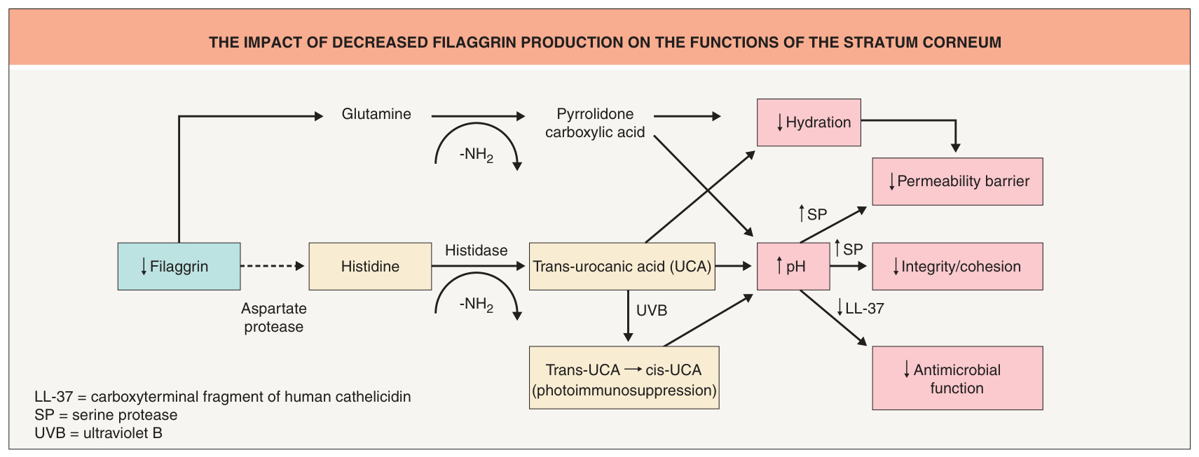
Fig. 124.5 The impact of decreased filaggrin production on the functions of the stratum corneum.