MECHANISM OF ACTION
Retinoids are involved in the regulation of diverse biologic functions (Table 126.1). They can: (1) affect cellular growth, differentiation and morphogenesis; (2) inhibit tumor promotion and malignant cell growth; (3) exert immunomodulatory actions; and (4) alter cellular cohesiveness.
Vitamin A Metabolism
Vitamin A (retinol) must be acquired through the diet as retinyl esters and provitamin A carotenoids; of the latter, β-carotene is particularly efficient in its ability to be converted to vitamin A. Within the intestinal lumen, retinyl esters are hydrolyzed to retinol, which is then absorbed and stored in the liver in the ester form (especially retinyl palmitate; Fig. 126.2). Retinol and its esters represent the main dietary source, as well as transport and storage form, of vitamin A. Once inside the bloodstream, after release of the retinyl ester (storage form) from the liver, retinol is transported bound to a complex of retinol-binding protein (RBP) and transthyretin. The uptake of retinol from the RBP–transthyretin complex to target cells is mediated by STRA6 (stimulated by retinoic acid 6), a dimeric transmembrane protein that acts as an RBP– retinol receptor (see Fig. 126.2).
The cellular pathway leading to conversion of retinol to at-RA, the biologically active ligand that binds to nuclear RARs, consists of a two-step process. Retinol (vitamin A alcohol) is first reversibly oxidized to retinaldehyde (vitamin A aldehyde), which is then irreversibly converted to retinoic acid (vitamin A acid). Cellular RBP (CRBP-I) facilitates these enzymatic reactions by delivering retinol to appropriate enzymes (see Fig. 126.2).
Two additional intracellular carriers, cellular retinoic acid-binding proteins (CRABP-I and CRABP-II), are thought to transport retinoic acid to the nucleus and buffer the level of free at-RA in the cell. The latter is accomplished by sequestering at-RA and by promoting metabolism of at-RA by cytochrome P450 enzymes belonging to the CYP26 family (e.g. CYP26A1). CRABP-I regulates the metabolic fate of its ligands by directly affecting the activities of RA-metabolizing cytochrome P450 enzymes, while CRABP-II markedly stimulates the RA-induced transcriptional activity of RAR through protein–protein interactions.
Intracellularly, retinoic acid is found in both all-trans and 9-cis configurations (see Fig. 126.2). Physiologically, at-RA (tretinoin) is the predominant retinoic acid form, and only a small fraction is isomerized (enzymatically or non-enzymatically) into 13-cis-RA (isotretinoin) or converted to polar metabolites with unknown biologic activity. at-RA is the primary active ligand that binds to the three known nuclear RARs,
which mediate, at least in part, the molecular and cellular effects of retinoic acid. Although the biosynthesis of 9-cis-RA (alitretinoin) is not well-defined, it might be produced from all-trans-retinol via 9-cis-retinol and 9-cis-retinaldehyde.
Retinoid Receptors
Retinoids exert most of their physiologic effects on DNA transcription by binding to two distinct families of nuclear receptors, RARs and RXRs (see Fig. 126.2). These receptor families belong to a superfamily of nuclear receptors that act as ligand-activated transcription factors and include the steroid, vitamin D, and thyroid hormone receptors as well as peroxisome proliferator-activated receptors (PPARs). The RAR and RXR receptor families each contain three receptor isotypes (α, β and γ) encoded by different genes. at-RA (tretinoin) binds only to RARs, whereas 9-cis-RA (alitretinoin) binds both to RARs and RXRs (Table 126.2).
RARs function as heterodimers with RXRs, whereas RXRs may also act as homodimers or participate in the formation of heterodimers with a variety of other nuclear receptors, including vitamin D, thyroid hormone, and PPARs. Such heterodimers provide a mechanism for cross-talk between nuclear hormone signaling pathways.
Dimers of retinoid receptors (RAR/RXR or RXR/RXR) are localized to the nucleus and bind, even in the unliganded state, to specific DNA regulatory sequences called retinoid hormone response elements (RAREs) in the promoter regions of retinoid-responsive genes (see Fig. 126.2). Unliganded receptors bind to co-repressor molecules and repress transcription. However, when the receptor binds its ligand, it undergoes a conformational change resulting in the release of co-repressors and recruitment of co-activators. These molecules include histone acetylases that change the conformation of chromatin and allow access to DNA by transcriptional machinery. The retinoid– receptor complex thereby modulates the transcription of specific sets of genes, and it is likely that many of the differentiation-inducing actions of retinoids are mediated by this mechanism.
In addition to direct upregulation of gene transcription via binding of retinoid–receptor complexes to RAREs, retinoids can have indirect effects on gene transcription that result from the downregulation of genes that do not contain RAREs in their promoter region. The retinoid–receptor complex is thought to antagonize transcription factors such as activator protein-1 (AP1) and nuclear factor-interleukin-6 (NF-IL6) by competing for the same set of required co-activator proteins, thereby leading to decreased expression of AP1 and NF-IL6-responsive genes; expression of the latter genes promotes cellular proliferation and inflammation. The antiproliferative and anti-inflammatory actions of retinoids are believed to be mediated by this type of indirect negative gene regulation. In particular, retinoic acid has important roles in inhibiting IL-6-driven induction of proinflammatory Th17 cells and in promoting differentiation of anti-inflammatory regulatory T cells (see Ch. 4). The anti-inflammatory effects of at-RA (tretinoin) have also been associated with its ability to regulate the expression and activation of Toll-like receptors (TLRs), especially TLR2. The biologic relevance of non-genomic effects of retinoids (e.g. cross-talk with extracellular protein/peptide signals acting via membrane receptors with downstream phosphorylation) remains under investigation.
The existence of several types and varied distributions of receptors, dimers, hormone response elements, and regulatory proteins shows that retinoid action is mediated via multiple pathways and results in a complex process of activation or inhibition of a large set of coordinately regulated genes. However, the exact mechanism of retinoid action in many dermatologic conditions is still unknown, and, so far, the nuclear receptor concept does not fully explain the biologic diversity of retinoid effects.
Retinoids in the Skin
Vitamin A and its bioactive metabolites play a major role in promoting the keratinocyte differentiation that occurs from the epidermal basal layer to the stratum corneum. Retinyl esters and retinol are the major retinoids found in the skin, with the former reaching a concentration of >1000 pmol/g in the epidermis. Epidermal retinoids can absorb UVB radiation and have ~2%–3% of the UVB-filtering capacity of the melanin in phototype II/III skin. The UVB absorption process results in dramatic depletion of epidermal retinoids, which can be prevented by application of topical retinoids. Nutritional deficiency, aging, and oxidative stress can also lead to lowered retinoid levels in the skin. There has been ongoing debate regarding possible generation of reactive oxygen species upon the interaction of UVA radiation with retinyl esters (e.g. retinyl palmitate) present in cosmetics and sunscreens, but the theoretical potential for photocarcinogenicity has not been shown to be clinically relevant.
Synthetic Retinoids
There are now four generations of retinoids (see Fig. 126.1). The firstgeneration non-aromatic retinoids (tretinoin, isotretinoin, alitretinoin) are synthesized by chemically modifying the polar end group and the polyene side chain of vitamin A. Second-generation mono-aromatic retinoids (etretinate, acitretin, motretinide) are formed by replacing the cyclic moiety of vitamin A with different substituted and unsubstituted ring systems. Third-generation polyaromatic retinoids (adapalene, bexarotene, tazarotene), called arotinoids, are produced by cyclization of the polyene side chain. Fourth-generation polyaromatic retinoids (trifarotene, seletinoid G, tamibarotene) were designed to have increased selectivity of binding.
Commercially available retinoids differ not only in their spectrum of clinical efficacy but also in their observed toxicity and pharmacokinetics. Each retinoid should be investigated as a unique drug, and the lack of disease response to one retinoid does not necessarily signify unresponsiveness to other retinoids.
The oral bioavailability of retinoids is considerably enhanced when administrated with food, especially with a fatty meal, due to their lipophilic properties. More recently, a lidose–isotretinoin formulation with enhanced absorption when taken without food was developed. Retinoid metabolism is predominantly hepatic; it involves oxidation and chain shortening to biologically inactive and polar metabolites, facilitating biliary and/or renal elimination. The oxidative metabolism is induced by the retinoids themselves and possibly also by other agents known to stimulate hepatic cytochrome P450 isoforms. Table 126.2 summarizes the key pharmacologic features and nuclear binding profiles of retinoids used in dermatology.
First-generation retinoids
Tretinoin, the predominant physiologic form of retinoic acid, was the first topical retinoid to be utilized for the treatment of acne and photoaging. It binds to cellular retinoic acid-binding proteins (CRABPs) and non-selectively to nuclear RARs (see above), resulting in modulation of keratinocyte proliferation and differentiation, inflammation, and extracellular matrix production. Because the standard tretinoin formulation is photolabile, night-time application is recommended to prevent degradation.
Isotretinoin is a naturally occurring retinoid resulting from the metabolism of vitamin A (see Fig. 126.2). 13-cis-RA and at-RA are two physiologically interconvertible isomers that differ in their elimination half-lives: approximately 20 hours and 1 hour, respectively. Isotretinoin undergoes first-pass metabolism in the liver and subsequent enterohepatic recycling. In plasma, more than 99% of isotretinoin is proteinbound, mainly to albumin. There is neither liver nor adipose tissue storage, in contrast to vitamin A. The major metabolite of isotretinoin, 4-oxo-isotretinoin, is produced by oxidation. Isotretinoin and its metabolites are excreted in the urine and feces. After discontinuation of isotretinoin, physiologic concentrations of the drug and its major metabolites are reached within 2 weeks, ranging from 2 days for at-RA to 10 days for 4-oxo-isotretinoin. Therefore, 1 month of post-therapy contraception provides an adequate safety margin to avoid teratogenicity.
Among natural and synthetic retinoids, only oral isotretinoin and its 4-oxo-isotretinoin metabolite significantly suppress sebum production, which explains their dramatic effects on acne. Isotretinoin-induced seboatrophy is unlikely to be RAR/RXR-mediated since isotretinoin has no clear affinity for any known retinoid receptor, and potent RAR/RXR ligands such as tretinoin and alitretinoin do not lead to seboatrophy. The mechanism of isotretinoin-induced seboatrophy is thought to involve sebocyte apoptosis, but inhibition of either sebocyte progenitor differentiation or the genes encoding sebogenic enzymes represent two other possible pathomechanisms.
Alitretinoin is a naturally occurring pan-agonist retinoid that binds to both RARs and RXRs, representing the endogenous ligand for the latter. Topical alitretinoin 0.1% gel is FDA-approved for the treatment of cutaneous Kaposi sarcoma, and oral alitretinoin therapy is currently utilized outside the US for refractory chronic hand eczema. The absorption of oral alitretinoin from the gastrointestinal tract is variable and dose-proportional over the therapeutic range of 10–30 mg daily; the absolute bioavailability of alitretinoin has not been determined. Alitretinoin is metabolized via oxidation by CYP3A4 enzymes of the liver into 4-oxo-alitretinoin, and both the parent drug and its metabolite also undergo isomerization into at-RA and 4-oxo-at-RA, respectively. Elimination is primarily renal, with the half-life of unchanged alitretinoin ranging between 2 and 10 hours. Alitretinoin concentrations return to normal within 1–3 days after treatment cessation.
The mechanism of action of alitretinoin in chronic hand eczema is unknown, although downregulated expression of RARs and RXRs in lesional skin was recently described. Alitretinoin has immunomodulatory and anti-inflammatory effects that include suppressing the expansion of cytokine-activated leukocyte subsets and antigenpresenting cells.
Second-generation retinoids
Acitretin is the major metabolite and the pharmacologically active form of etretinate. Although acitretin and etretinate are equally effective, acitretin has much more rapid elimination. Etretinate is approximately 50 times more lipophilic than acitretin and binds strongly to plasma proteins, particularly lipoproteins and albumin. Etretinate is stored in adipose tissue (including subcutaneous fat), from which it is released slowly, with a long terminal half-life of up to 120 days. Under identical conditions, acitretin carries a negatively charged group that makes it much less lipophilic than etretinate. As a result, acitretin does not accumulate in adipose tissue and is eliminated from the body more rapidly, with a half-life of 2 days.
The major serious adverse effect of oral retinoids is teratogenicity, and, therefore, the length of time that these drugs are present in the body is of great importance. Re-esterification of acitretin to etretinate may occur when acitretin is taken simultaneously with alcohol. This finding prompted the manufacturer to extend the time of contraception for female patients with childbearing potential taking acitretin to 2 years after its discontinuation, as it is for etretinate. The FDA advises a contraceptive period of at least 3 years for acitretin, based on the pharmacokinetics of acitretin and etretinate observed in clinical trials and on previous safety experience with etretinate. However, acitretin retains its shorter half-life in women who strictly avoid alcohol during treatment and for 2 months thereafter. Because of its improved risk–benefit ratio, acitretin was approved by the FDA in 1996 as a substitute for etretinate, which is no longer commercially available.
Acitretin’s mechanism of action is still not clearly understood. Paradoxically, it activates all three RAR subtypes despite binding poorly to them. Evidence supports a normalization of differentiation and proliferation as well as a modification of inflammatory responses and neutrophil function.
Third-generation retinoids
Adapalene is a photostable, rigid, and highly lipophilic synthetic retinoid with higher affinity for RAR-β/γ than for RAR-α. Since RAR-β is not expressed in keratinocytes, RAR-γ is the primary target receptor for adapalene in the epidermis. Adapalene does not bind to cellular retinoic acid-binding proteins (CRABPs) but does induce expression of CRABP-II. Its lipophilic properties may contribute to better pilosebaceous uptake and anti-acne activity. Given its negligible transdermal absorption, the teratogenic risks of topical adapalene appear to be minimal. Adapalene affects the cellular differentiation, keratinization, and inflammatory processes that are abnormal in acne. Although adapalene has not been shown to be sebostatic in vivo in humans, it was found to inhibit triacylglycerol biosynthesis and lipid droplet accumulation in vitro in hamster sebocytes.
Tazarotene is a prodrug that is rapidly converted by cutaneous esterases to its free carboxylic acid (tazarotenic acid), which is the active metabolite. It has a higher affinity for RAR-β/γ than RAR-α and no affinity for RXR. Because of its rapid metabolism, systemic exposure is low. Tazarotene appears to modulate the pathogenesis of psoriasis by regulating expression of retinoid responsive genes, including those involved in cell proliferation, cell differentiation, and inflammation.
Bexarotene, an RXR-selective retinoid (rexinoid), is FDA-approved in oral (1999) and topical (2000) forms for treatment of CTCL. This compound is about 100-fold more potent at binding to RXRs than to RARs. In plasma, bexarotene is highly bound (>99%) to various proteins that have yet to be characterized. Bexarotene has a clearance profile similar to that of isotretinoin, with a terminal half-life of between 7 and 9 hours. Bexarotene is metabolized by CYP3A4 and generates its own oxidative metabolites via hepatic CYP3A4 induction. Neither bexarotene nor its metabolites are excreted in the urine; their elimination is thought to occur primarily via the hepatobiliary system. The exact mechanism of action of bexarotene in CTCL is still unknown, but it probably acts through regulation of cellular differentiation and proliferation, as well as via induction of apoptosis.
Fourth-generation retinoids
Trifarotene, a fourth-generation retinoid with 20-fold higher affinity for RAR-γ than RAR-α/β, was FDA-approved in 2019 as a topical acne treatment for individuals ≥9 years of age. This agent was designed to be stable in keratinocytes but rapidly metabolized in hepatic microsomes, thereby minimizing systemic drug levels. In addition to effects on known retinoid-mediated pathways, gene expression profiling has shown that trifarotene modulates proteolysis, reduces intercellular adhesion, and activates transporters involved in skin hydration.
Retinoic acid metabolism blocking agents
Retinoic acid metabolism blocking agents (RAMBAs) inhibit catabolism of at-RA by P450 enzymes in the CYP26 family, thereby raising the intracellular levels of endogenous at-RA in the skin and other targeted tissues. This limits systemic exposure and the potential for toxicity, and at-RA levels return to the physiologic range within 24 hours of discontinuing these medications. Liarozole, an imidazole derivative without antifungal properties, is a RAMBA with low isoenzyme specificity that also inhibits P450-mediated pathways of steroid biosynthesis whereas talarozole, a triazole derivative, is a more selective and highly active CYP26 enzyme inhibitor. DX314, a CYP26B1-selective RAMBA that may protect epidermal barrier integrity, is also under investigation. Liarozole has been granted orphan drug status for congenital ichthyosis by the European Commission and the FDA. In a randomized, placebo-controlled phase II/III trial in 64 patients with moderate-to-severe lamellar ichthyosis, a 41%–50% response rate was observed in patients receiving liarozole, compared to 11% with placebo, with borderline statistical significance (p = 0.056).
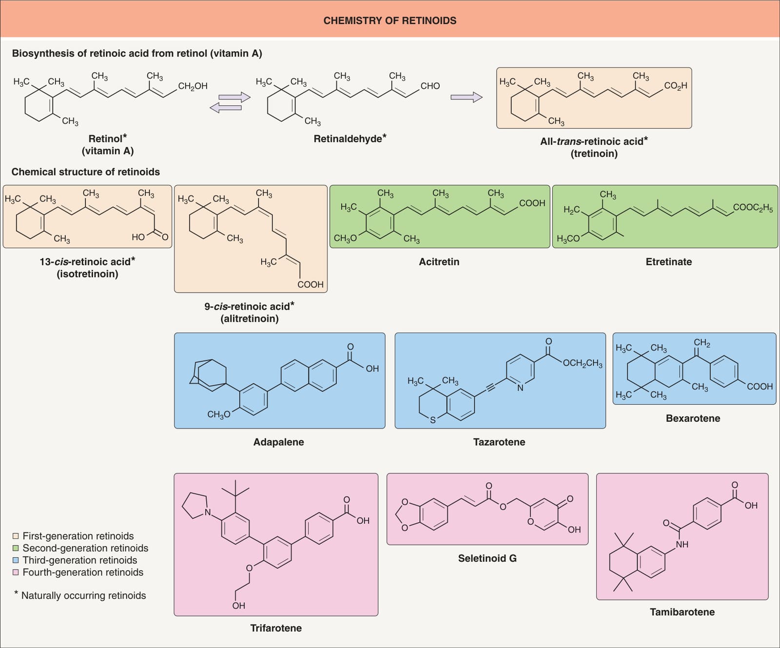
Fig. 126.1 Chemical structure of natural and synthetic retinoids. Motretinide is an additional second-generation retinoid.
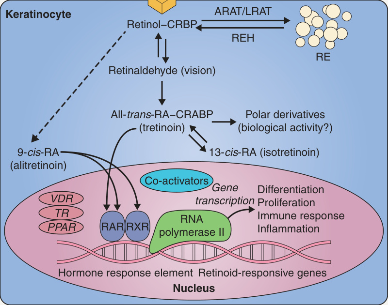
Fig. 126.2 Metabolism and mechanism of action of natural retinoids. ARAT, acyl-CoA:retinol acyl transferase; CRABP, cellular retinoic acid-binding protein; CRBP, cellular retinol-binding protein; LRAT, lecithin:retinol acyl transferase; PPAR, peroxisome proliferator-activated receptor; RA, retinoic acid; RAR, retinoic acid receptor; RBP, retinol-binding protein; RE, retinyl esters; REH, retinyl ester hydrolase; RXR, retinoid X receptor; STRA6, stimulated by retinoic acid 6; TR, thyroid receptor; VDR, vitamin D receptor.
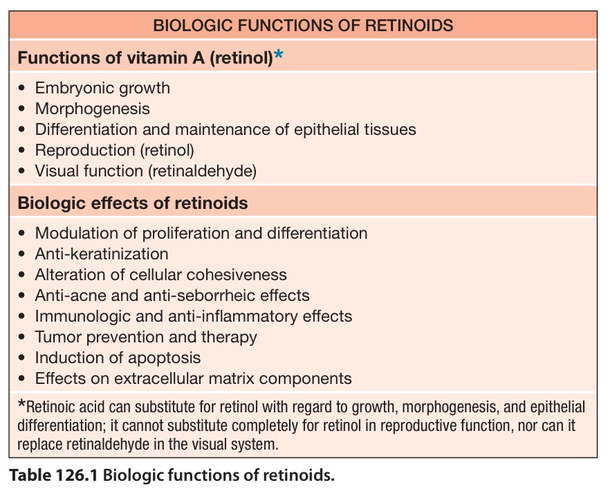
Table 126.1 Biologic functions of retinoids.
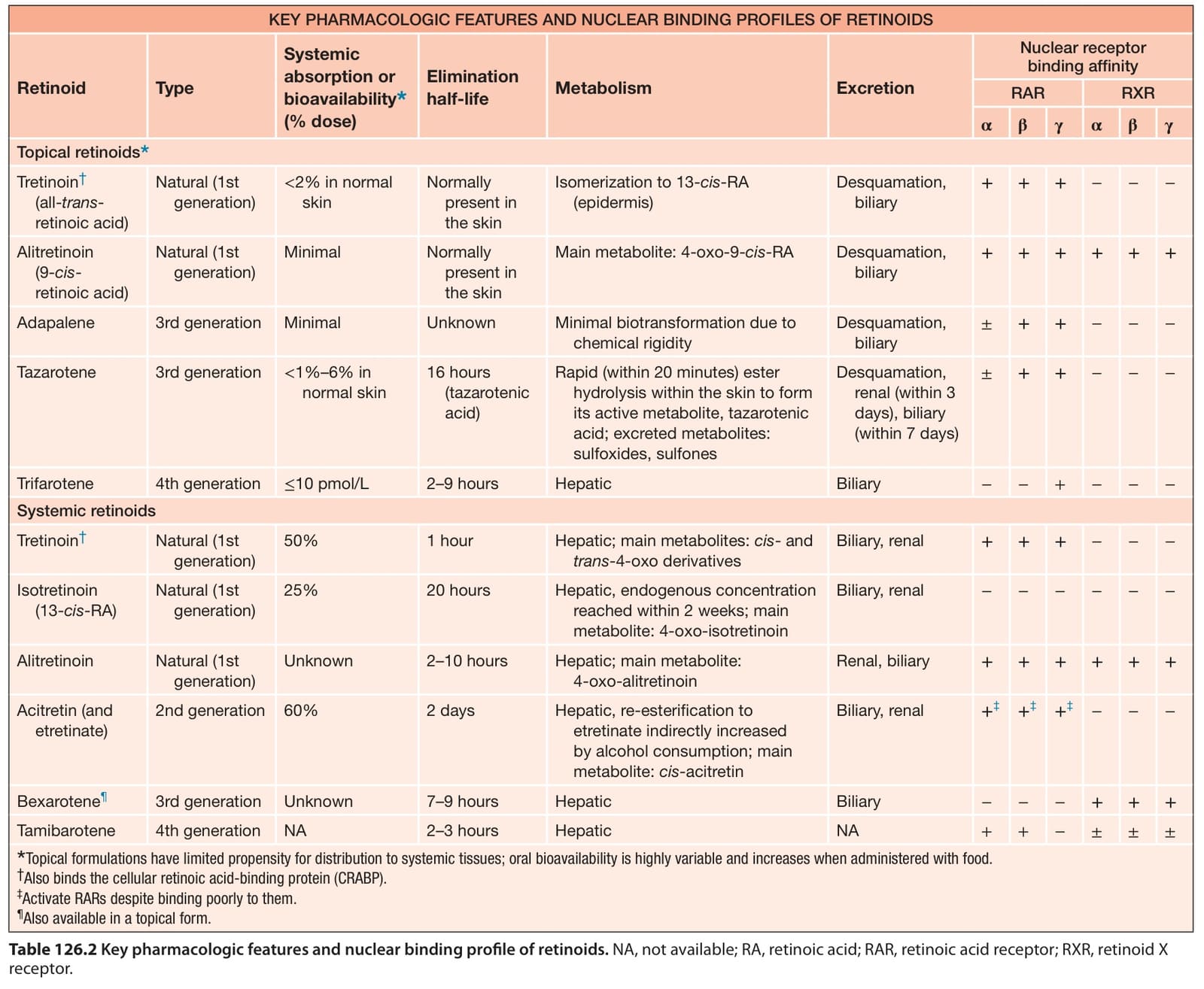
Table 126.2 Key pharmacologic features and nuclear binding profile of retinoids. NA, not available; RA, retinoic acid; RAR, retinoic acid receptor; RXR, retinoid X receptor.