PATHOGENESIS
The pathogenesis of AD can be divided into three major categories: (1) epidermal barrier dysfunction; (2) immune dysregulation; and (3) alteration of the microbiome. These mechanisms interact with each other and are modulated by both genetic and environmental factors (Fig. 12.2).
Genetic Factors
Genetic factors account for a large proportion of susceptibility to earlyonset AD, with a significantly higher concordance rate in monozygotic twins (77%) compared to dizygotic twins (15%). Although the entities in the atopic triad cluster together in families, a parental history of AD is a stronger risk factor for the development of AD than either asthma or allergic rhinitis, supporting the existence of genes specific to AD susceptibility. Genes that encode proteins important to the epidermal barrier and immunologic functions have been implicated in AD pathogenesis (Table 12.2). AD is a complex genetic disease, and both gene–gene and gene–environment interactions have important roles.
Epidermal Barrier Dysfunction
A defective epidermal permeability barrier represents a consistent feature of AD and is evident in non-lesional as well as lesional skin of affected individuals. Manifestations of epidermal barrier dysfunction include increases in transepidermal water loss (TEWL), pH and permeability, as well as altered lipid composition. The level of TEWL in non-lesional skin of children with AD correlates with disease severity. Epidermal barrier dysfunction permits an easier entry for irritants, allergens, and microbes, which trigger immune responses that include the release of proinflammatory cytokines. In infants, greater TEWL is associated with an increased likelihood of epicutaneous sensitization to aeroallergens, which could potentially play a role in the development of asthma and allergic rhinoconjunctivitis. Factors that contribute to the impaired cutaneous barrier in AD are discussed next.
Filaggrin and other structural proteins
Filaggrin is a keratin filament-aggregating protein that serves as a major structural component of the stratum corneum. FLG loss-of-function (LOF) variants represent the strongest known genetic risk factor for AD and are also responsible for ichthyosis vulgaris (see Ch. 57), with carrier frequencies of up to 10% in European and ~3% in East Asian populations. Approximately 20%–50% of European and Asian children with moderate-to-severe AD have at least one FLG LOF variant; the penetrance of AD is ~40% for one and ~90% for two LOF alleles. This implicates epidermal barrier dysfunction in the initiation of AD, with subsequent development of Th2-biased immune responses. Although FLG LOF variants also play a role in the pathogenesis and persistence of AD in children of African descent, they appear to be rare in individuals from sub-Saharan Africa; however, technical limitations may have reduced detection of uncommon FLG variants in earlier studies. Of note, filaggrin expression is also affected by intragenic copy number variation and reduced by increased local pH, protease activity, and Th2 cytokine levels.
FLG mutations are associated with early-onset AD, greater disease severity, and persistence into adulthood as well as enhanced epicutaneous sensitization and an increased risk of irritant contact dermatitis, hand eczema, herpes simplex virus (HSV) infections, and food allergy. FLG mutations have also been linked to an increased risk for the development of asthma and greater asthma severity; however, these effects are only seen in patients with pre-existing AD. Since filaggrin is not found in the gastrointestinal or bronchial mucosa, the association of FLG mutations with food allergy and asthma strongly suggests that epicutaneous sensitization and/or cutaneous inflammation can contribute to the development of systemic atopic disease.
Filaggrin breakdown products such as histidine contribute to epidermal hydration, acid mantle formation, lipid processing, and barrier function. Gene expression profiling and immunohistochemical analysis of lesional and non-lesional skin from AD patients have shown broad defects in terminal differentiation, with downregulation of other epidermal barrier proteins such as loricrin, corneodesmosin, involucrin, small prolene rich proteins 3/4 (SPRR3/4), claudin-1, and late cornified envelope protein 2B.
Stratum corneum lipids
The composition, organization, and biochemical processing of stratum corneum lipids are critical determinants of epidermal permeability barrier function (see Ch. 124). In AD, a filaggrin-deficient cytoskeletal scaffold contributes to abnormal loading and secretion of lamellar bodies, with subsequent defects in post-secretory lipid organization and processing. Disruption of the skin’s acidic mantle leads to reduced activity of lipid-processing enzymes such as β-glucoscerebrosidase and acid sphingomyelinase. Th2 cytokines also negatively affect generation of stratum corneum lipid components. Lastly, Staphylococcus aureus colonization of the skin affects lipid composition and contributes to epidermal barrier impairment.
Proteases and protease inhibitors
Lesional AD skin demonstrates elevated levels of endogenous serine proteases, e.g. kallikrein 5 and 7 (KLK5/7), due to an imbalance in the activities of these proteolytic enzymes and protease inhibitors, such as the lymphoepithelial Kazal-type trypsin inhibitor (LEKTI) encoded by SPINK5. Biallelic loss-of-function SPINK5 mutations underlie Netherton syndrome, which features profoundly compromised barrier function and atopy (see Ch. 57), while SPINK5 polymorphisms have been linked to increased risk of AD in some populations. Other factors that enhance proteolysis include increased skin surface pH and exogenous proteases from allergens (e.g. house dust mites, pollens), S. aureus, and Malassezia.
LEKTI deficiency results in excessive degradation of the corneodesmosomal component desmoglein-1 (Dsg1), causing abnormal stratum corneum detachment and thereby disrupting the epidermal barrier. The S. aureus extracellular V8 protease, which has a sequence similar to those of S. aureus exfoliative toxins, is also thought to degrade Dsg1. In addition, unrestrained protease activity leads to degradation of lipidprocessing enzymes and antimicrobial peptides as well as activation of proinflammatory cytokines.
Immune Dysregulation
Both innate and adaptive immune systems play dynamic interrelated roles in the pathogenesis of AD. The inflammatory profile is diverse, with activation of skin-resident inflammatory dendritic cells, group 2 innate lymphoid cells, and Langerhans cells. Acute AD lesions have a predominance of Th2 cytokines, but there is subsequent evolution to a chronic phase characterized by Th1 and Th22 cytokine profiles, as well as variable levels of Th17 cytokines in both acute and chronic AD. The acute phase features IL-4, IL-5, and IL-13; activation of eosinophils and mast cells; and production of allergen-specific IgE. Keratinocyte-derived cytokines, including IL-1, thymic stromal lymphopoietin (TSLP), IL-25 (IL-17E) and IL-33, promote a Th2 immune response. Th2 cytokines inhibit expression of major terminal differentiation proteins such as loricrin, filaggrin, and involucrin as well as β-defensin-2/3 antimicrobial peptides.
Thymic stromal lymphopoietin (TSLP)
TSLP is an IL-7-like cytokine that is known as the “master-switch of allergic inflammation” due to its central role in evoking a Th2 response via dendritic cell activation. Exposure to allergens, viral infections, trauma, and other cytokines (e.g. IL-1β, TNF) can trigger TSLP production by keratinocytes, fibroblasts, and mast cells. TSLP is highly expressed in acute and chronic lesions of AD, but not in the non-lesional skin of patients with AD or in unaffected individuals.
IL-4 and IL-13
IL-4 has a key role in driving Th2 cell differentiation, IgE production, and eosinophil recruitment. Transgenic mice overexpressing IL-4 in their epidermis develop atopic dermatitis-like lesions, pruritus, an altered microbiome, and elevated IgE levels. The heterodimeric receptors for IL-4 and IL-13 both contain the IL-4 receptor α subunit (IL-4Rα; see Fig. 128.9 C) and activate signal transducer and activator of transcription 6 (STAT6), which promotes the differentiation of naive T cells into Th2 effector cells. Although IL-4 and IL-13 share 25% sequence homology and effector functions, studies in human subjects, and human keratinocyte cell lines, support an independent role for IL-13 in AD pathogenesis. Monoclonal antibodies that target IL-4/-13 represent effective treatments for AD (see Ch. 128 and below).
Other cytokines
Th17 cells are important in the regulation of innate immunity, in particular neutrophil recruitment, and have also been implicated in allergic disorders. Th17 cells are found in acute as well as chronic AD lesions, and production of IL-17 and IL-19 is especially characteristic of new-onset pediatric AD. IL-26, which is also produced by Th17 cells, induces production of Th2- and Th17-associated cytokines, thereby potentially acting as a bridge between Th2 and Th17 responses.
IL-31 is a Th2 cytokine that is highly expressed in lesional skin and serum of patients with AD as well as in other pruritic skin disorders such as prurigo nodularis. Cutaneous exposure to staphylococcal superantigen rapidly induces IL-31 expression in atopic individuals, establishing a link between staphylococcal colonization of the skin and pruritus. The heterodimeric receptor for IL-31 is expressed by keratinocytes, eosinophils, activated macrophages, cutaneous C nerve fibers, and dorsal root ganglia (see Ch. 5). Randomized, placebo-controlled trials (RCTs) have shown that nemolizumab, a humanized monoclonal antibody against the IL-31 receptor A subunit, can significantly reduce pruritus in patients with moderate to severe AD, although improvements in disease severity scores were modest.
IL-33, a member of the IL-1 cytokine family, protects against helminth infection by promoting a Th2-type immune response. IL-33 expression is increased in AD lesions compared to the skin of unaffected individuals, and AD therapies targeting IL-33 are under development.
Innate lymphoid cells
The innate lymphoid cell (ILC) family includes natural killer cells and three groups of non-cytotoxic ILCs that orchestrate immunity, inflammation, and homeostasis in multiple tissues. The group 2 ILC (ILC2) population is expanded in AD lesions and stimulated by TSLP, IL-25 (IL-17E), and IL-33. ILC2s interact with other immune cells (e.g. mast cells, eosinophils) in the skin to promote Th2-type inflammation in a T cell independent manner.
The Cutaneous Microbiome
The cutaneous microbiome represents a complex and highly diverse community of pathogenic and commensal bacteria, fungi, and viruses that play a critical role in epidermal homeostasis, with microbial dysbiosis contributing to the pathogenesis of AD. More than 90% of patients with AD have skin colonized with S. aureus, compared to about 5% of unaffected individuals, presumably reflecting the disrupted acid mantle, decreased antimicrobial peptides (e.g. cathelicidins, defensins), and altered cytokine milieu of AD skin. During AD flares, bacterial diversity decreases and the proportion of the microbiome accounted for by Staphylococcus spp. increases from ~35% to ~90%. Conversely, normalization of the microbial population correlates with clinical improvement in AD.
Superantigens can promote the development of a Th2 immune response, and exotoxins with superantigenic properties are produced by up to 65% of the S. aureus strains that colonize AD patients. Compared to unaffected controls, an IgE response to the S. aureus superantigens enterotoxin A and enterotoxin B occurs more frequently in patients with AD. The S. aureusδ-toxin also stimulates mast cell degranulation and Th2 inflammation. In addition, filaggrin deficiency increases the susceptibility of keratinocytes to S. aureusα-toxininduced cytotoxicity. Lastly, Malassezia spp. may also contribute to inflammation in AD, and adults with severe head and neck disease often display IgE reactivity to Malassezia antigens.
Alterations in the skin microbiome of AD patients related to the use of cleansers and topical immunomodulatory or antimicrobial agents may have potential effects on cutaneous inflammation and barrier function. Topical administration of coagulase-negative Staphylococcus strains with antimicrobial activity or the Gram-negative commensal Roseomonas mucosa has been shown to markedly reduce S. aureus colonization in AD patients. R. mucosa application was also associated with decreased AD severity and topical corticosteroid requirement, providing the basis for bacteriotherapy as a potential AD treatment.
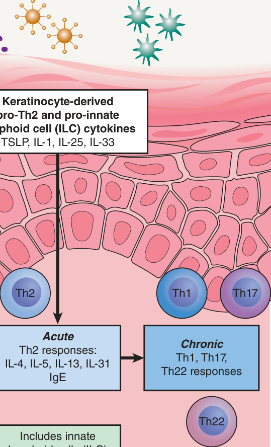
Fig. 12.2 Atopic dermatitis results from defects in epidermal barrier function, immune dysregulation, and environmental influences. SC, stratum corneum; KLK, kallikrein; TEWL, transepidermal water loss; TSLP, thymic stromal lymphopoietin. Courtesy Harvey Lui, MD.
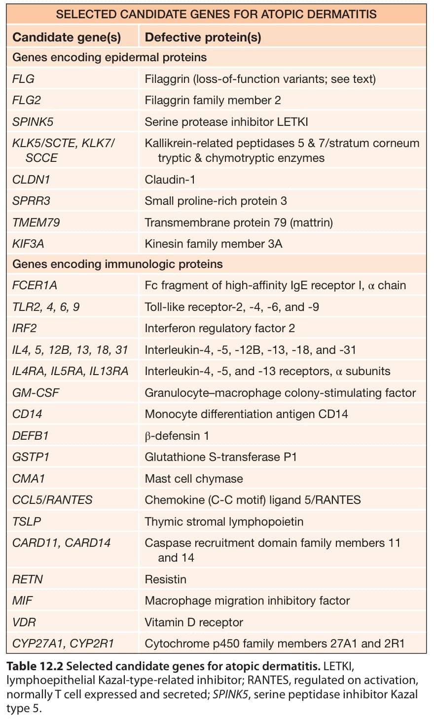
Table 12.2 Selected candidate genes for atopic dermatitis. LETKI, lymphoepithelial Kazal-type-related inhibitor; RANTES, regulated on activation, normally T cell expressed and secreted; SPINK5, serine peptidase inhibitor Kazal type 5.