CLINICAL FEATURES
Age-Related Cutaneous Findings
AD has a broad clinical spectrum that varies depending upon the age of the patient. It is divided into infantile, childhood, and adolescent/adult stages (Fig. 12.3). In each stage, patients may develop acute, subacute, and chronic eczematous lesions, all of which are intensely pruritic and often excoriated. Acute lesions predominate in infantile AD and are characterized by edematous, erythematous papules and plaques that may exhibit vesiculation, oozing, and serous crusting. Subacute eczematous lesions display erythema, scaling, and variable crusting. Chronic lesions, which typify adolescent/adult AD, present as thickened plaques with lichenification as well as scale; prurigo nodule-like lesions can also develop (see below). Perifollicular accentuation and small, flattopped papules (papular eczema) are particularly common in Black or Asian patients (Fig. 12.4). In any stage of AD, a generalized exfoliative erythroderma may develop in the most severely affected patients (see Ch. 10). All types of AD lesions can leave postinflammatory hyper-, hypo-, or occasionally depigmentation upon resolution (Fig. 12.5).
Infantile AD (age <2 years) typically develops after the second month of life, often initially appearing as edematous papules and papulovesicles on the cheeks, with sparing of the central face; the lesions may evolve to form large plaques with oozing and crusting (Fig. 12.6). The scalp, neck, extensor aspects of the extremities, and trunk may also be involved, usually with sparing of the diaper area (Fig. 12.7). In the first
Courtesy Thomas Bieber, MD and Caroline Bussmann, MD.
6 months of life, the face is affected in >90% of patients with AD. Young infants may attempt to relieve itch through rubbing movements against their bedding, whereas older infants are better able to directly scratch affected areas.
In childhood AD (age 2 to 12 years), the lesions tend to be less exudative and often become lichenified. The classic sites of predilection are the antecubital and popliteal fossae (flexural eczema) (Fig. 12.8). Other common locations include the wrists, hands, ankles, feet, neck, and eyelids, although any area can be involved (Fig. 12.9). Xerosis typically becomes pronounced and widespread.
Adult/adolescent AD (age >12 years) also features subacute to chronic, lichenified lesions, and involvement of the flexural folds typically continues (Fig. 12.10). However, the clinical picture may also change. Adults with AD frequently present with chronic hand dermatitis that has both endogenous and exogenous components (Fig. 12.11), while others have primarily facial dermatitis (Fig. 12.12), often with severe eyelid involvement (see below). Patients who have suffered from continuous AD since childhood are more likely to have extensive disease that is resistant to treatment. Such individuals may also have severe excoriations and chronic papular skin lesions because of habitual scratching and rubbing (Fig. 12.13A).
AD in the elderly (age >60 years) is characterized by marked xerosis. Most of these patients do not have the lichenified flexural lesions typical of AD in children and younger adults.
Regional Variants of Atopic Dermatitis
Several regional variants of AD can occur in isolation or together with the classic age-related patterns of involvement described above (see Fig. 12.3). The face is a frequent location for site-specific manifestations. Eczema of the lips, referred to as cheilitis sicca, is common in AD patients, especially during the winter (Fig. 12.14A). It is characterized by dryness (“chapping”) of the vermilion lips, sometimes with peeling and fissuring, and may be associated with angular cheilitis. Patients try to moisten their lips by licking, which in turn may irritate the skin around the mouth, resulting in so-called lip-licker’s eczema. Another common feature of childhood AD is ear eczema, presenting as erythema, scaling, and fissures under the earlobe and in the retroauricular area, sometimes in association with bacterial superinfection. Eyelid eczema can represent the only manifestation of AD, especially in adults. In contrast to eyelid eczema due to other causes, it is characterized by lichenification of the periorbital skin.
“Head and neck dermatitis” represents a variant of AD that typically occurs after puberty and primarily involves the face, scalp, and neck. When older children and teenagers present with this form of AD, it usually persists into adulthood. Malassezia yeasts, which are members of the skin microbiome in the head and neck area, may be an aggravating factor for this presentation, and systemic antifungal treatment with itraconazole or fluconazole may be of benefit.
Eczema variants also occur in acral sites. Juvenile plantar dermatosis presents with “glazed” erythema, scale, and fissuring on the balls of the feet and plantar aspect of the toes in children with AD (see Ch. 13). Atopic hand eczema (see Fig. 12.11) affects ~60% of adults with AD and may be the only manifestation of the condition. FLG mutations are associated with increased likelihood of hand eczema in children
A Chronic diffuse involvement of the extensor arms with scaling and hyperpigmentation. Note the follicular prominence on the trunk. B Nummular lesions on the extensor aspect of the arm and nipple eczema. B, Courtesy Julie V. Schaffer, MD.
and adults, and frequent exposure to water and other irritants in household or occupational settings represents another risk factor. Atopic hand eczema typically involves the volar wrists and dorsum of the hands. The palms and sides of the fingers may develop the deepseated vesicles of dyshidrotic eczema (see Ch. 13).
The prurigo form of AD favors the extensor aspects of the extremities and is characterized by firm, dome-shaped papules and nodules with central scale-crust, similar to prurigo nodularis lesions in non-atopic patients (Fig. 12.13B). Nummular (discoid) lesions also tend to develop on the extremities in children and adults with AD, appearing as coinshaped eczematous plaques, usually 1 to 3 cm in diameter and often with prominent oozing and crusting (Fig. 12.13C; see Fig. 12.7B). They are similar in appearance to nummular dermatitis occurring outside the setting of atopy (see Ch. 13). Frictional lichenoid eruption has a predilection for atopic children and presents as multiple small, flat-topped, pink to skin-colored papules on the elbows and (less often) knees and dorsal hands. Lastly, chronic nipple eczema can develop in children and adults with AD (Fig. 12.14B).
Associated Cutaneous Features
Pruritus
Intense pruritus is a hallmark of AD. The itch is often worse in the evening and may be exacerbated by exogenous factors such as sweating or wool clothing. Rubbing and scratching in response to pruritus can initiate flares or exacerbate existing dermatitis, explaining why AD is known as the “itch that rashes”. Excoriations (linear or punctate) are frequently present, providing evidence of scratching (Fig. 12.15; see Figs. 12.8–12.10). With repeated rubbing and scratching, the skin becomes thickened and leathery with exaggerated skin markings, referred to as lichenification (see Fig. 12.10 & 12.11).
Atopic stigmata
Physical findings other than dermatitis that are frequently observed in patients with AD are presented in Table 12.3 and Figs. 12.16–12.18.
Pityriasis alba
Pityriasis alba frequently affects children and adolescents with AD. It is characterized by multiple, ill-defined, hypopigmented macules and patches, usually 0.5 to 2 cm in diameter, with fine scaling. The lesions are typically located on the face, especially the cheeks (see Fig. 12.18), but occasionally appear on the shoulders and arms. Pityriasis alba is most obvious in individuals with darkly pigmented skin and/or following sun exposure. It is thought to result from a low-grade eczematous dermatitis that disrupts the transfer of melanosomes from melanocytes to keratinocytes.
Similar hypopigmented lesions can appear upon resolution of more overtly inflamed, erythematous lesions of AD. The differential diagnosis of pityriasis alba also includes postinflammatory hypopigmentation secondary to other dermatoses such as seborrheic dermatitis or pityriasis lichenoides chronica. Tinea versicolor is typically more sharply demarcated, with small lesions that may coalesce centrally in involved areas, and vitiligo is also sharply demarcated and depigmented rather than hypopigmented. Hypopigmented mycosis fungoides may occasionally represent a diagnostic consideration if there is extrafacial involvement. Regular use of sunscreens and other forms of photoprotection may minimize the appearance of pityriasis alba.
Infectious Complications
Bacterial and viral infections represent the most common complications of AD. Considering that S. aureus colonizes the skin of the vast majority of patients with AD, it is not surprising that impetiginization, which can also be caused by Streptococcus pyogenes, occurs frequently (Fig. 12.19). Bacterial infections may exacerbate AD by stimulating the inflammatory cascade, e.g. via S. aureus exotoxins that act as superantigens (see above).
Eczema herpeticum represents rapid dissemination of a herpes simplex viral infection over the eczematous skin of AD patients. It initially develops as an eruption of vesicles, but affected individuals more often present with numerous monomorphic, punched-out erosions with hemorrhagic crusting (Fig. 12.20). Eczema herpeticum is frequently widespread and may occur at any site, with a predilection for the head, neck, and trunk. It is often associated with fever, malaise, and lymphadenopathy, and complications may include superinfection with S. aureus or S. pyogenes as well as herpetic keratoconjunctivitis and meningoencephalitis. Patients with mutations in the filaggrin gene and those who have both severe AD and asthma have an increased risk for eczema herpeticum, and decreased production of antimicrobial peptides may have a pathogenic role. Patients with AD are also predisposed to the development of widespread molluscum contagiosum (see Ch. 81).
Impact on Quality of Life
AD has profound and multifaceted adverse impacts on the quality of life of affected children and adults as well as their families and caregivers. Intense pruritus and stigmatization often result in sleep disturbances, psychological distress, social isolation, disrupted family dynamics, and impaired functioning at school or work. Children with AD experience greater impairment in their quality of life than those with diabetes mellitus or epilepsy, and detrimental effects on productivity have also been documented in adults.
Associated Extracutaneous Disorders
Ocular manifestations
In addition to allergic rhinoconjunctivitis, the spectrum of atopic eye disease includes chronic manifestations such as atopic keratoconjunctivitis, which typically affects adults, and vernal keratoconjunctivitis that favors children living in warm climates. Symptoms include ocular itching, burning, tearing, and mucus discharge, often in association with conjunctival injection and blepharitis manifesting as swelling and scaling of the eyelids. Vernal keratoconjunctivitis features large, cobblestone-like papillae on the upper palpebral conjunctiva, and atopic keratoconjunctivitis is more prone to scarring. Additional infrequent ocular complications of AD include keratoconus and subcapsular cataracts, with anterior cataracts more specifically related to AD and posterior cataracts occurring more commonly; rarely there is retinal detachment.
Psychiatric disease and psychosocial burden
AD is associated with an increased risk of depression, anxiety, suicidal ideation, sleep disorders, and (in children) attention deficit hyperactivity disorder. Sleep deprivation, pruritus, visible disease, stress, and low self-esteem contribute the psychosocial burden of AD.
Cardiovascular complications
A recent meta-analysis of cohort studies found that AD is associated with an increased risk of cardiovascular complications such as angina and myocardial infarction, cerebrovascular accidents, and heart failure during adulthood.
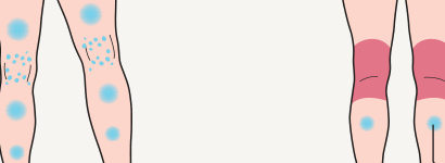
*Fig. 12.3 Distribution patterns of atopic dermatitis (AD) and regional variants. May be the only manifestation of AD in adults. †Not to be confused with nummular eczema occurring outside the setting of AD.
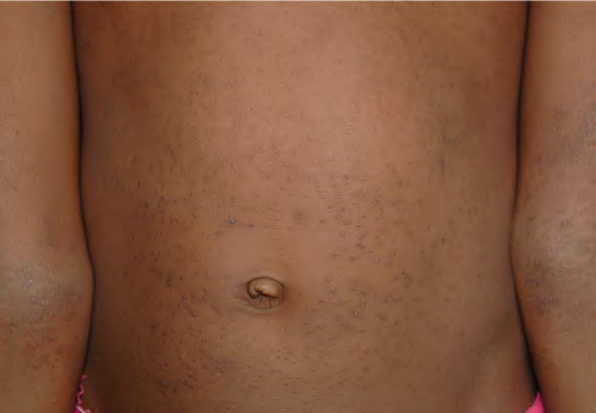
Fig. 12.4 Atopic dermatitis – papular pattern. This clinical variant is more commonly observed in patients with more darkly pigmented skin. There is also lichenification in the antecubital fossae. Courtesy Gail Todd, MD.
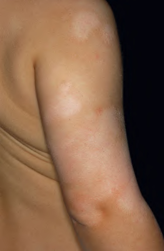
Fig. 12.5 Postinflammatory hypopigmentation in atopic dermatitis.
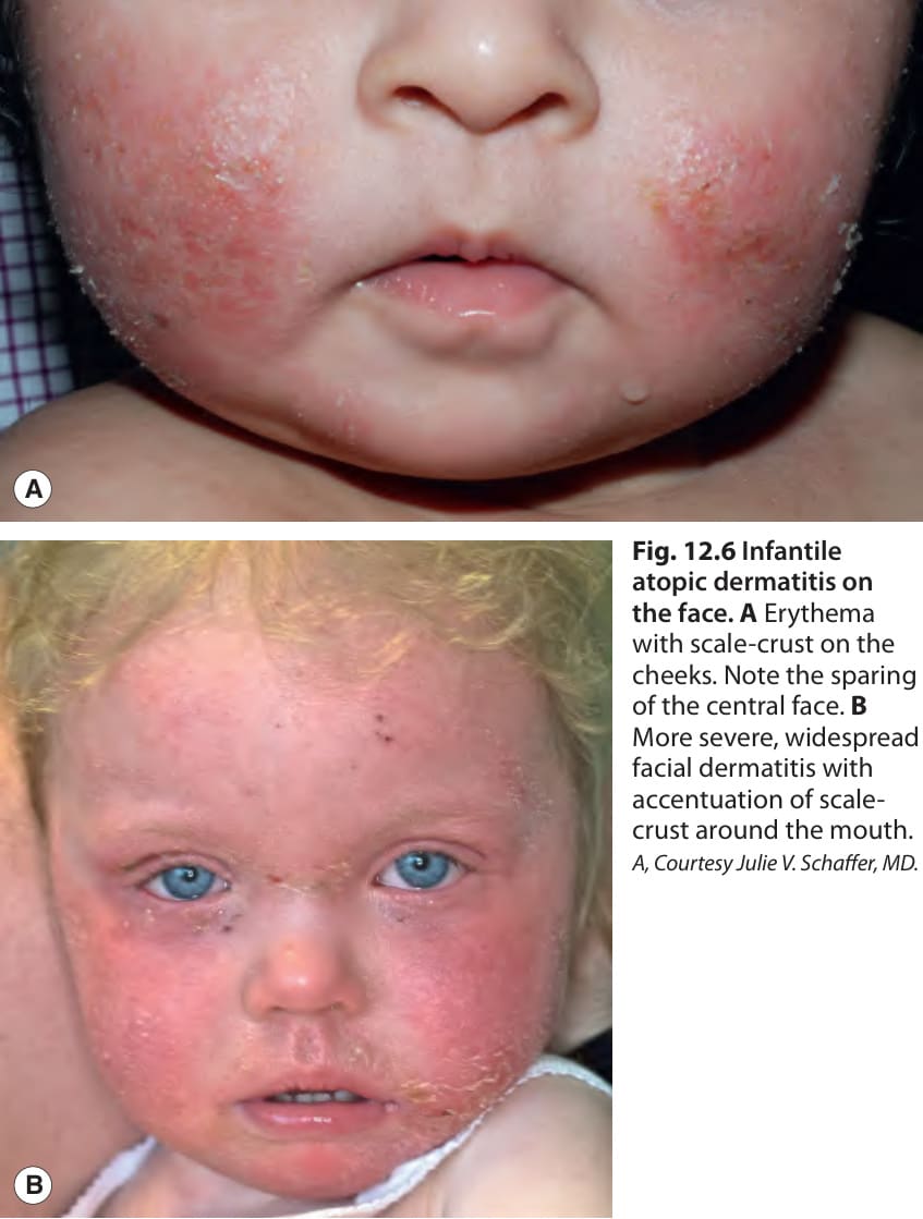
Fig. 12.6 Infantile atopic dermatitis on the face.A Erythema with scale-crust on the cheeks. Note the sparing of the central face. B More severe, widespread facial dermatitis with accentuation of scalecrust around the mouth. A, Courtesy Julie V. Schaffer, MD.
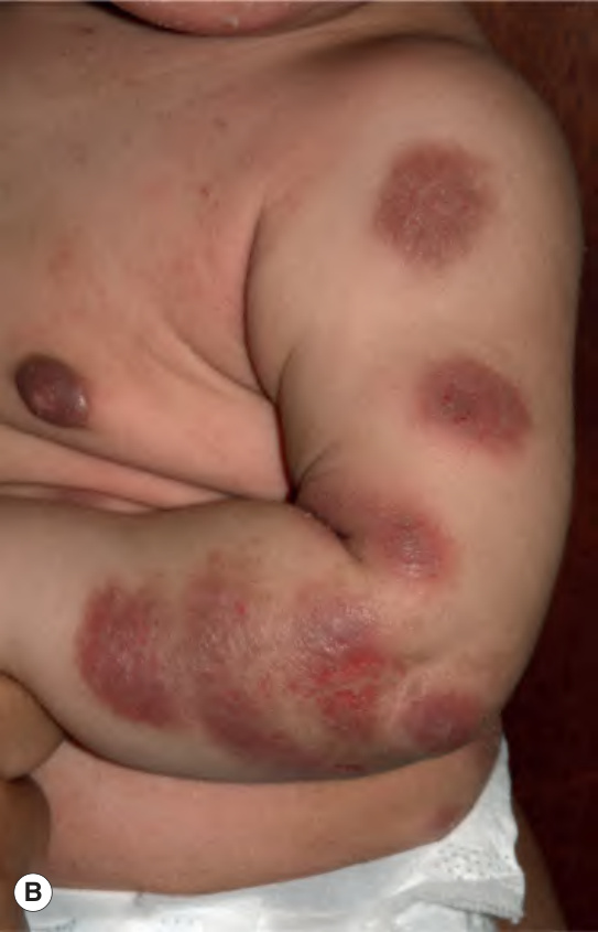
Fig. 12.7 Infantile atopic dermatitis on the extensor arms.
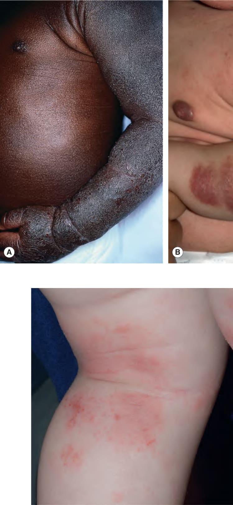
Fig. 12.8 Flexural atopic dermatitis in a child. The popliteal fossa is a typical location. Note the excoriations. Courtesy Julie V. Schaffer, MD.
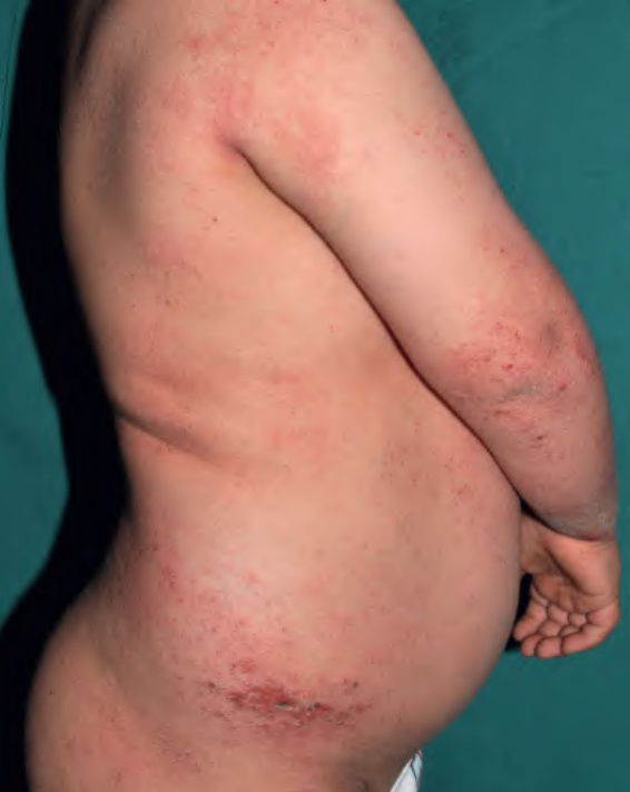
Fig. 12.9 Extensive atopic dermatitis in a child. Excoriations, crusting, and lichenification are evident.
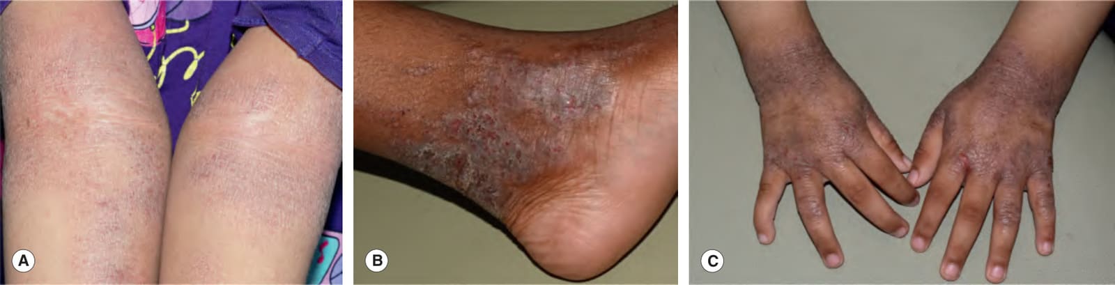
Fig. 12.10 Chronic atopic dermatitis.A Lichenification, scale, and punctate excoriations in the antecubital fossae. B Coalescing papules and lichenification on the ankle due to chronic scratching and rubbing. C Thick eczematous plaques with lichenification and excoriation on the dorsal hands and wrists. Courtesy Julie V. Schaffer, MD.
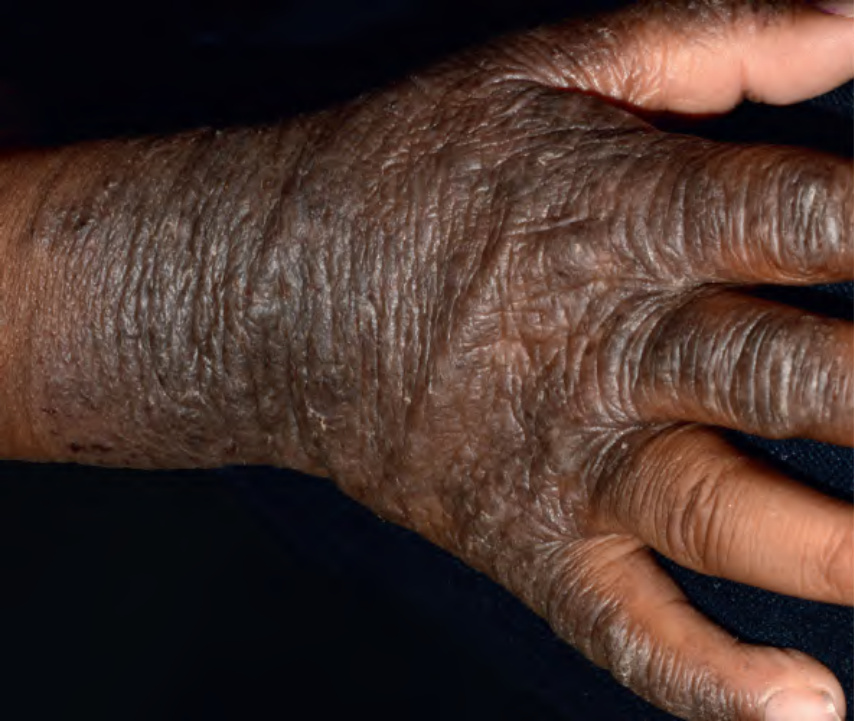
Fig. 12.11 Atopic dermatitis with severe chronic hand involvement. Note the marked lichenification. Courtesy Julie V. Schaffer, MD.
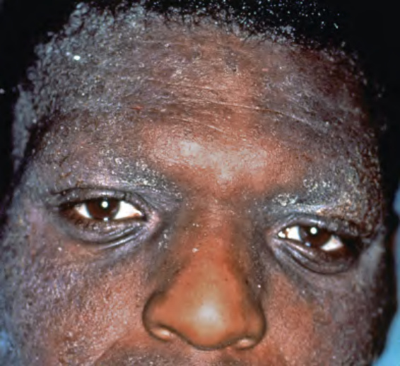
Fig. 12.12 Severe atopic dermatitis with facial involvement in an adult.
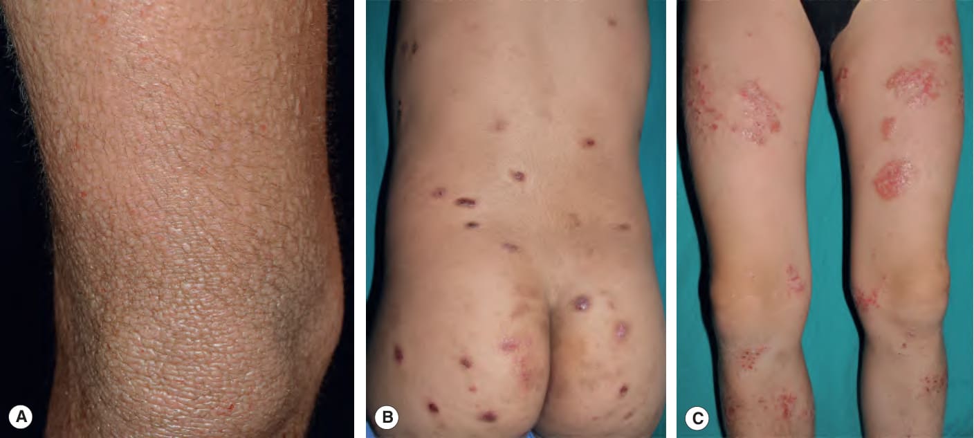
Fig. 12.13 Atopic dermatitis variants.A Chronic papular lesions resulting from habitual rubbing and scratching in the setting of longstanding disease. B Prurigo lesions presenting as firm, dome-shaped papules and nodules with central hemorrhagic crust. C Nummular plaques with oozing and crusting on the legs. A, Courtesy Thomas Bieber, MD and Caroline Bussmann, MD; B, C, Courtesy Antonio Torrelo, MD.
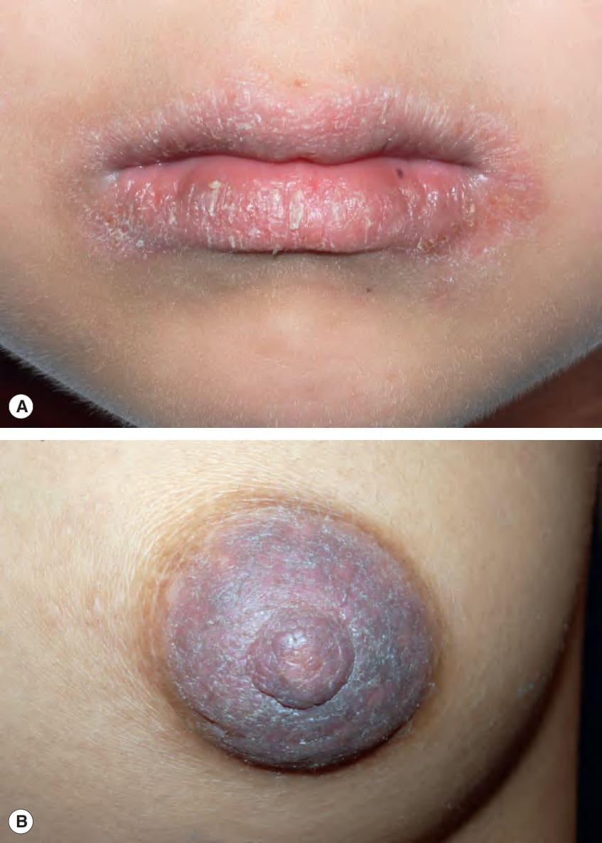
Fig. 12.14 Regional variants of atopic dermatitis.A Atopic cheilitis involving both the vermilion lip and surrounding skin (lip-licker’s eczema). B Nipple eczema in an adolescent. Courtesy Julie V. Schaffer, MD.
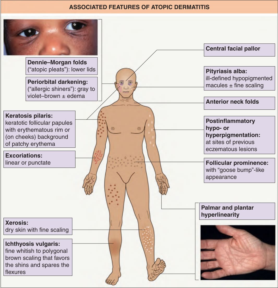
Fig. 12.15 Associated features of atopic dermatitis. See Table 12.3. Inset of hand: Courtesy Jean L. Bolognia, MD.
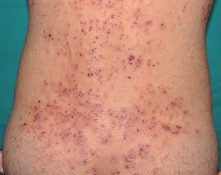
Fig. 12.16 Excoriations. Numerous punctate and a few linear excoriations in an area of papular eczema on the lower back. Courtesy Antonio Torrelo, MD.
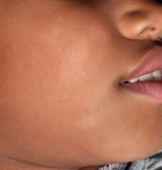
Fig. 12.18 Pityriasis alba. Note the slight scale associated with the hypopigmented macules and patches on the cheeks. Courtesy Antonio Torrelo, MD.
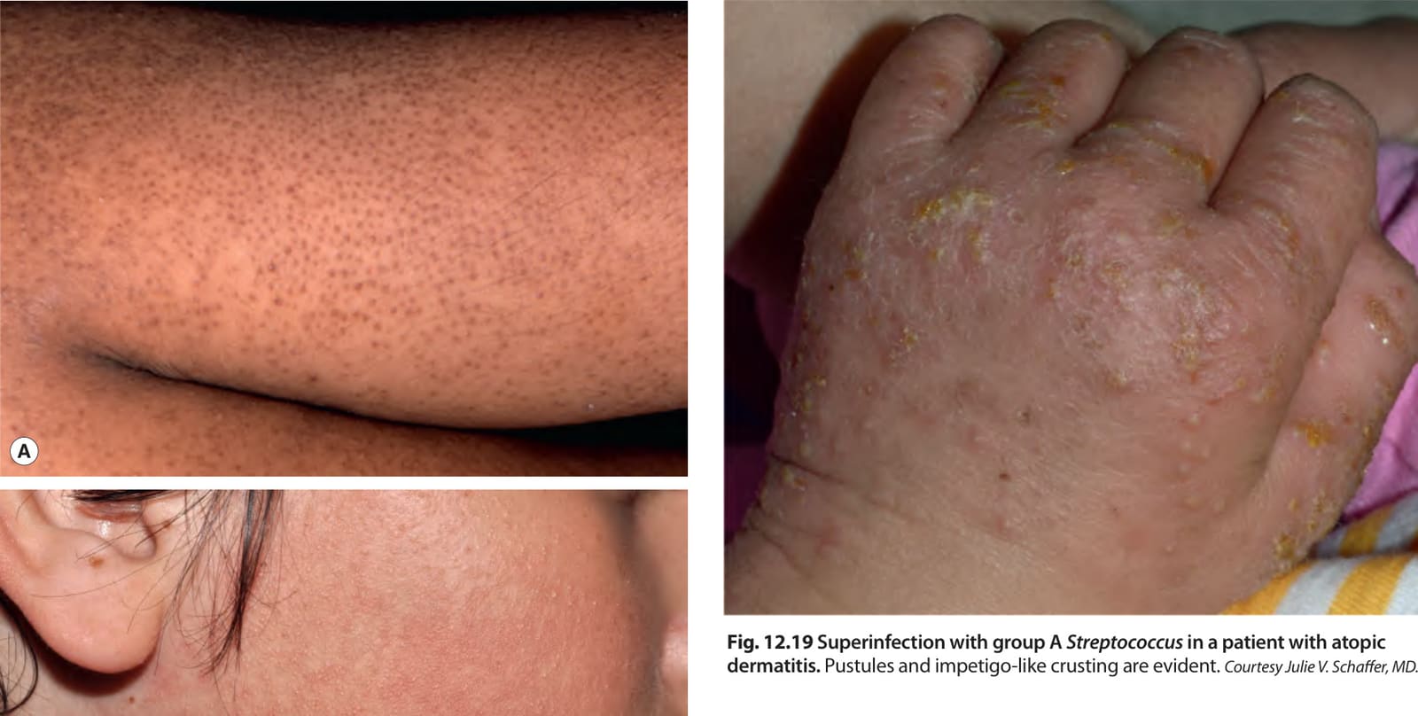
Fig. 12.19 Superinfection with group A Streptococcus in a patient with atopic dermatitis. Pustules and impetigo-like crusting are evident. Courtesy Julie V. Schaffer, MD.
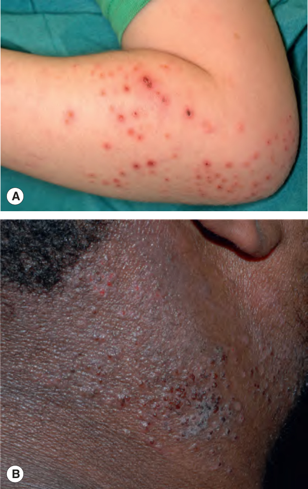
Fig. 12.20 Eczema herpeticum.

Table 12.3 Associated features of atopic dermatitis (“atopic stigmata”). AD, atopic dermatitis. See Fig. 12.15.