DIFFERENTIAL DIAGNOSIS
The differential diagnosis of AD is broad and includes other chronic dermatoses, infections, infestations, and malignancies as well as metabolic, genetic (e.g. primary immunodeficiencies), and autoimmune disorders (Table 12.4).
In infants, AD is often preceded and/or accompanied by seborrheic dermatitis, which commonly presents during the first month of life as yellowish-white, adherent scale-crusts on the scalp. In contrast to the typical distribution of infantile AD on the extensor surfaces of the extremities and cheeks as well as the scalp, infantile seborrheic dermatitis has a predilection for the skin folds, where lesions tend to lack scale, and the forehead. Scabies in infants often has generalized involvement and can mimic AD; in addition to the presence of burrows or identification of the mite or eggs via dermoscopy or skin scrapings, scabies can usually be distinguished by the predominance of discrete small crusted papules, involvement of the axillae and diaper area, and the presence of acral vesiculopustules. Other less common conditions that occasionally represent a diagnostic consideration in infants, such as primary immunodeficiencies, are listed in Table 12.4.
Adolescents and adults without a history of atopy who present with an eczematous eruption should have a thorough history and consideration of patch testing to assess for allergic contact dermatitis. This diagnosis should also be considered in children and adults with established AD who fail to respond as expected to treatment or who develop lesions in an atypical distribution pattern. Components of emollients or topical corticosteroid preparations represent potential allergens in these individuals. Protein contact dermatitis has a predilection for atopic individuals and can also present as a chronic eczematous dermatitis. Causes include a variety of foods and animal products (Table 12.5; see Ch. 16), and it is diagnosed by prick testing or observation of an urticarial reaction within 30 minutes of patch testing on previously affected skin.
Mycosis fungoides (MF) should be considered in adolescents and adults with chronic dermatitis poorly responsive to topical corticosteroid treatment. Because the histologic findings of early MF may be difficult to distinguish from those of AD, multiple biopsies are recommended, preferably from untreated areas of skin since corticosteroids
can eliminate the epidermotropic T cells that point to the diagnosis of MF. Longitudinal evaluation of such individuals is required, especially when the clinical and/or histologic features are not classic for AD, with additional biopsies as indicated.
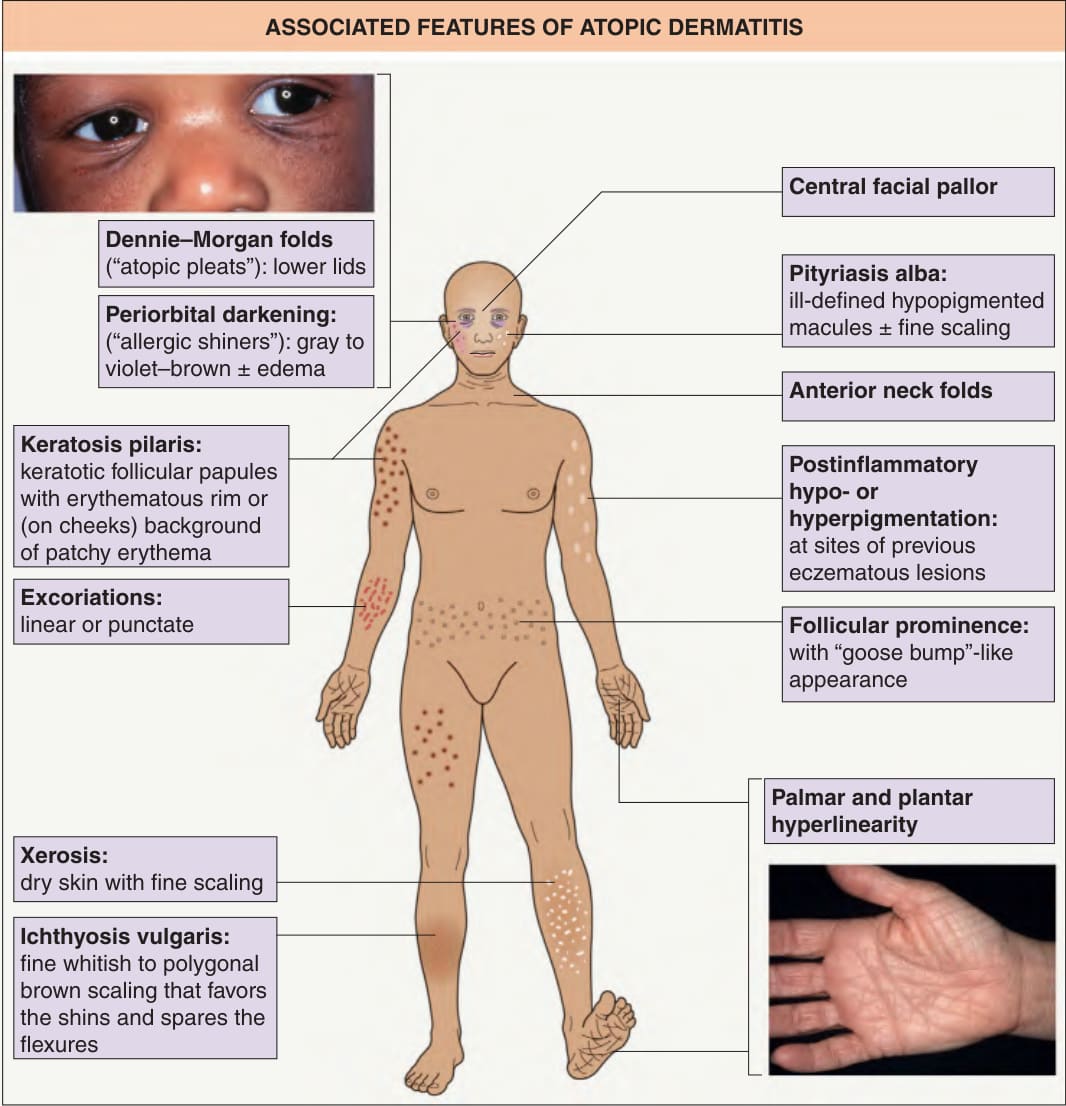
Fig. 12.15 Associated features of atopic dermatitis. See Table 12.3. Inset of hand: Courtesy Jean L. Bolognia, MD.
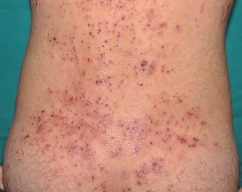
Fig. 12.16 Excoriations. Numerous punctate and a few linear excoriations in an area of papular eczema on the lower back. Courtesy Antonio Torrelo, MD.

Table 12.3 Associated features of atopic dermatitis (“atopic stigmata”). AD, atopic dermatitis. See Fig. 12.15.
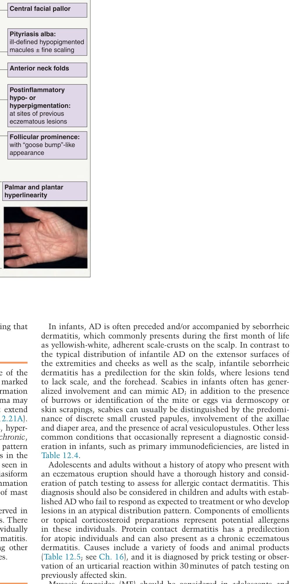
Table 12.4 Differential diagnosis of atopic dermatitis. A, adults; B, both; C, children/infants.
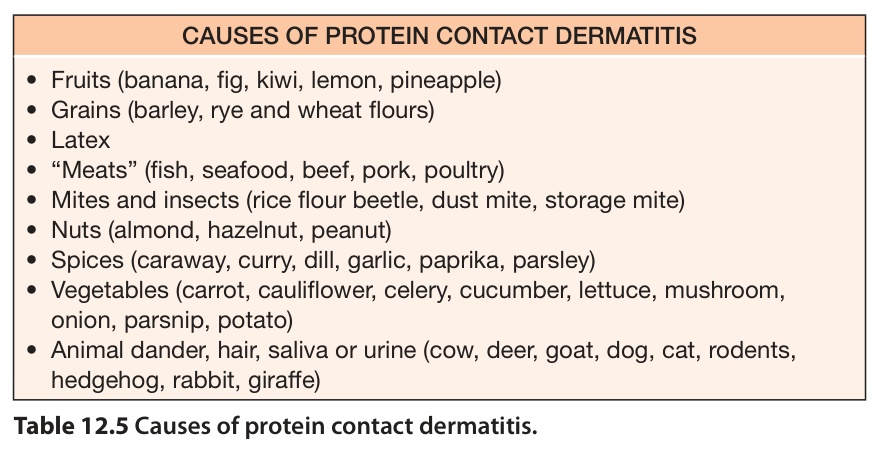
Table 12.5 Causes of protein contact dermatitis.