TREATMENT
General Approach
Because AD is a chronic, relapsing disease, the traditional approach was reactive management targeting acute flares with short-term treatment regimens. Based on insights into the underlying skin barrier defect and its relationship to inflammatory processes in the skin and other organs, a proactive approach that includes long-term maintenance therapy is now recommended (Fig. 12.22). This treatment strategy may modify the overall disease course and possibly prevent the development of atopic comorbidities. Management of AD includes education of patients/parents, gentle skin care, moisturizer use, and anti-inflammatory therapy to control subclinical inflammation as well as overt flares. Topical agents represent the mainstay of treatment. Severe disease may require phototherapy or systemic medications, usually in conjunction with continued topical therapy (Table 12.6). Factors that can potentially exacerbate AD should be identified and, if possible, avoided (see Table 12.1).
Educational interventions
Education of patients and families to promote AD self-management has been shown to improve outcomes. Knowledge of disease mechanisms and course, potential triggers, appropriate use of therapies, and the goals of management can increase treatment adherence and reduce fear and misconceptions. Parents often seek an eradicable cause for their child’s AD and have difficulty accepting “control” rather than a “cure” for the condition. Addressing their specific concerns and acknowledging the stresses associated with this chronic disease, as well
as noting the improvement in pruritus and sleep disturbances likely to result from treatment, can lead to more effective care. Educational approaches include counseling clinic visits with a team approach/ multidisciplinary input, online resources (e.g. nationaleczema.org), and structured teaching programs (“eczema schools”). Written action
Grouped punchedout monomorphic erosions with hemorrhagic crusts on the arm (A) and posterior neck (B). Vesicles are rarely evident. B, Courtesy Julie V. Schaffer, MD.
plans have also been shown to be beneficial in AD management. Psychological interventions to aid in coping with AD may prove useful, with reported approaches including biofeedback, cognitive-behavioral therapy, and stress management.
Bathing
Bathing can hydrate the skin and remove scale, crust, irritants, and allergens. Although there is a paucity of objective data on optimal bathing practices for AD, it is generally recommended that patients bathe or shower once daily for 5–10 minutes in warm (not hot) water, with use
Acute lesion showing prominent spongiosis, epidermal hyperplasia, and a mild inflammatory infiltrate in the upper dermis. B Subacute lesion with parakeratosis and less spongiosis. A, Courtesy Lorenzo Cerroni, MD.
of a fragrance-free non-soap cleanser with a neutral to low pH as needed (e.g. syndets; see Ch. 153). Application of a moisturizer shortly after bathing is essential to maintain skin hydration. If treatment with a topical corticosteroid or other anti-inflammatory agent is needed, it should be applied immediately after bathing, prior to the moisturizer; this has been referred to as the “soak and smear” technique. With the exception of bleach (see below), there are no RCTs to support the use of bath oils, other bath additives, acidic spring water, or water-softening devices for AD.
Moisturizers
Daily use of moisturizers to counteract dry skin and reduce transepidermal water loss is a cornerstone of AD management that helps to prevent disease flares. Moisturizer application can reduce xerosis, pruritus, erythema, fissuring, and lichenification, thereby decreasing the amount of anti-inflammatory medication required for disease control. Standard moisturizers contain varying amounts of emollient agents that lubricate the skin, occlusive agents that prevent water loss, and humectants that attract water. Preparations should be free of dyes, fragrances, food-derived allergens such as peanut protein, and other potentially sensitizing ingredients. Factors in selecting the formulation of the moisturizer include the degree of xerosis, the sites of application, acceptance by the patient, and the season. Ointments (e.g. petrolatum) contain higher concentrations of lipids, have occlusive properties, and are typically preservative-free; although they tend to cause less stinging when applied to inflamed skin, the greasiness of ointments is bothersome to some patients. Creams may be a more acceptable option for such individuals, whereas lotions contain a higher water content and are not ideal for the xerosis of AD. Products with higher concentrations of urea or α-/β-hydroxy acids, which can decrease scaling, may also sting when used in children and on acutely inflamed or excoriated skin.
There are limited data on the optimal amount and frequency of moisturizer application or comparing the effectiveness of different moisturizing products, and no particular preparation has been shown to be superior. Weekly use of 150–200 g of moisturizer in young children and 250–500 g in older children/adults has been recommended, with liberal and frequent application. Prescription emollient devices (PEDs) that aim to improve the defective skin barrier of AD include preparations that contain specific ratios of lipids (e.g. cholesterol, fatty acids, ceramides), palmitoylethanolamide, glycyrrhetinic acid, and other hydrolipids. There is currently no evidence that these agents are superior to over-the-counter preparations.
Topical Anti-Inflammatory Therapy
Topical corticosteroids
Topical corticosteroids represent first-line pharmacologic therapy for AD. These agents have anti-inflammatory, antiproliferative, immunosuppressive, and vasoconstrictive actions, with effects on cutaneous T cells, macrophages, and dendritic cells (see Ch. 125). The efficacy of topical corticosteroids in AD has been verified in >100 RCTs, and they have been shown to decrease the acute and chronic inflammation of AD as well as associated pruritus. Topical corticosteroids are used to treat acute flares of AD and as maintenance therapy to prevent relapse.
Factors in selecting the potency and vehicle of the topical corticosteroid include the location, type (e.g. acute versus chronic), thickness, and extent of the AD lesions; patient age and preference as well as the cost and availability of different preparations represent additional considerations. The corticosteroid should have an appropriate potency to quickly gain control of the flare, and continuation of daily therapy until the active dermatitis is completely clear can minimize the likelihood of a rebound. Long-term daily use of an inadequately potent topical corticosteroid can result in a greater risk of side effects as well as less control of the eczema than relatively brief use of a more potent agent. RCTs in children and adults with AD have demonstrated that the risk of relapse can be significantly reduced by proactive maintenance with twice-weekly application of a mid-potency topical corticosteroid to the usual areas of involvement when clear, with no evidence of cutaneous atrophy after up to 40 weeks of treatment.
For the face and body folds, high potency corticosteroids (especially long-term use) should be avoided if possible due to risk of cutaneous atrophy and (for the face) acneiform eruptions. However, short-term use of a potent agent (e.g. mometasone furoate ointment) may be required to clear thick, exuberant lesions on the cheeks of infants. More potent corticosteroids (e.g. class 1–2) are often needed for lichenified plaques, nummular or prurigo-like lesions, and involvement of the palms and soles. Corticosteroid ointments (which minimize stinging) and creams are generally preferred considering the dryness of the skin in AD patients and the moisturizing effects of these vehicles. Application immediately after bathing improves cutaneous penetration and also decreases stinging. Corticosteroid solutions, foams, and oils represent options for AD on the scalp.
Systematic reviews have concluded that topical corticosteroids have a favorable safety profile with short-term (up to several weeks) daily use and long-term intermittent use (see Ch. 125). However, aversion or hesitancy regarding use of topical corticosteroids (“steroid phobia”) is very common among AD patients and their parents, and it often leads to delayed and inadequate treatment. It is essential that these fears and incorrect beliefs about topical corticosteroids are addressed to ensure adherence to the treatment plan.
When AD does not respond as expected to topical corticosteroid therapy, adherence should be assessed, including the amount (grams/ tubes) and duration (consecutive days) of use. If possible, in-patient therapy for patients with severe AD can allow direct observation and
intensive education. Potential complicating factors should be investigated, such as disease exacerbation by superinfection, irritants, or allergens. The latter can include immediate hypersensitivity reactions to foods and aeroallergens as well as delayed hypersensitivity to contact allergens, including components of moisturizers and topical mediations.
Topical calcineurin inhibitors
Two topical calcineurin inhibitors (TCIs) have been FDA-approved for the treatment of AD: (1) tacrolimus 0.03% and 0.1% ointment for moderate to severe disease; and (2) pimecrolimus 1% cream for mild to moderate disease (see Ch. 129). These agents suppress T cell activation and modulate the secretion of cytokines and other proinflammatory mediators; they also decrease mast cell and dendritic cell activity. The efficacy of TCIs in the treatment of AD has been proven in large clinical trials in adults and children ≥2 years of age and, for pimecrolimus, infants ages 3–23 months, although it is not currently approved for the latter group.
TCIs are particularly useful for AD affecting the face and inter-triginous areas, sites where corticosteroid-induced skin atrophy is of increased concern and TCI therapy is especially effective. TCIs are also beneficial in patients with frequently flaring or persistent AD that would otherwise require almost continual use of topical corticosteroids. Randomized controlled studies have shown that the proactive
application of tacrolimus ointment 2–3 times weekly as maintenance can prevent flares of AD without increasing the overall amount of medication used.
The most common adverse effects with TCIs are local stinging and burning. These symptoms lessen after several applications, or if TCI use is preceded by a short topical corticosteroid course. For both topical tacrolimus and pimecrolimus, there has been no short-term or inter-mediate-term (>15 years) evidence of systemic immunosuppression in either clinical studies or post-marketing surveillance, and long-term data collection is ongoing. In 2006 the FDA introduced black box warnings for both drugs concerning a theoretical cancer risk; this was based on the occurrence of lymphomas in mice exposed to systemic levels 30- to 50-fold greater than the highest recorded blood levels in human patients. Pharmacokinetic studies in children and adults with AD have demonstrated minimal systemic absorption of TCIs, with transient detectable but low blood levels occasionally observed in patients with severe AD involving a large portion of the body surface area. Of note, recent meta-analyses found no association between TCI use and overall cancer or skin cancer (keratinocyte carcinoma, melanoma) risk, with a slight increase in lymphoma risk in adults that may potentially be explained by early cutaneous T cell lymphoma being misdiagnosed as AD and treated with TCIs.
Crisaborole
Crisaborole 2% ointment is a phosphodiesterase-4 (PDE-4) inhibitor that is FDA-approved for the treatment of mild to moderate AD in patients ≥3 months of age. In large RCTs, ~30% of such individuals achieved clear/almost clear skin with a ≥2-grade improvement after 4 weeks of twice daily treatment. PDE-4 degrades cAMP, which leads to increased production of cytokines including IL-10 and IL-4 (see Fig. 130.3). The most common side effect is stinging or burning in the area of application.
Topical JAK inhibitors
Ruxolitinib 1.5% cream is a Janus kinase 1/2 (JAK1/2)-selective inhibitor that was recently FDA-approved for short-term or non-continuous chronic treatment (using ≤60 g/week) of mild to moderate AD in patients ≥12 years of age. In RCTs (total n=1249), 50%–55% of individuals treated with ruxolitinib 1.5% cream twice daily for 8 weeks had clear/almost clear skin with a ≥2-grade improvement, compared to 7%–15% for the vehicle control. Mean plasma concentrations were
<15% of the whole blood IC for JAK2 inhibition. However, the JAK inhibitor class-wide black box warning notes potential increased risks of serious infection, death, cancer, cardiovascular events, and thrombosis (see Chs. 128 & 129). Delgocitinib 0.5% ointment (pan-JAK inhibitor; approved in Japan for AD in adults) has shown greater efficacy than vehicle in phase 2 and 3 RCTs in AD patients ≥2 years of age.
Wet wrap therapy
Wet wrap therapy may be helpful in severe recalcitrant AD or during an acute flare. These moist, occlusive dressings increase skin hydration, act as a barrier to scratching, and enhance the penetration of topical corticosteroids. However, because of the latter effect, care should be exercised when using moderate and potent topical corticosteroids with wet wraps, and a possible increased risk of bacterial skin infections has been noted. The wraps consist of application of a topical corticosteroid (sometimes diluted), followed by an inner wet layer and outer dry layer of cotton gauze or garments; they are left in place 8–24 hours per day, and the treatment duration should not exceed 2 weeks.
Phototherapy
Narrowband UVB, UVA1, and UVA combined with UVB have each been shown to improve atopic dermatitis and associated pruritus. The immunomodulatory effects of phototherapy occur via induction of T cell apoptosis, reduction of dendritic cells, and decreased expression of Th2 cytokines such as IL-5, IL-13, and IL-31 (see Ch. 134). In addition, treatment with UVB has been shown to reduce S. aureus colonization of the skin in AD patients. Narrowband UVB and UVA1 can both be helpful for chronic AD lesions, and UVA1 may also be useful in the treatment of acute flares. Phototherapy can be combined with topical corticosteroids, especially in the initial phase of treatment. The side effect profile of phototherapy is favorable compared to systemic immunosuppressive agents, with potential “sunburn” and, with long-term treatment, photoaging and possibly an increased risk of skin cancer. The time required to travel several times a week to a phototherapy center may disrupt school or work for some patients, and a home UV unit may be an option for those receiving chronic treatment. In young children, phototherapy may be difficult for practical reasons, e.g. lack of cooperation.
Systemic Anti-Inflammatory Therapy
Systemic anti-inflammatory medications may be employed for children and adults with moderate to severe AD that has failed to respond adequately to optimized topical treatment. The risk–benefit profile should be carefully considered before starting an immunosuppressive agent, and patients receiving these medications require close monitoring for side effects (see Chs. 128 & 130). Combination of systemic treatment with topical corticosteroid therapy is frequently required to maximize benefit. In addition to the medications discussed below, a number of promising targeted immunomodulatory therapies are under investigation.
Interleukin-4/-13 receptor inhibitors
Dupilumab is a human monoclonal antibody that targets the IL-4Rα subunit of heterodimeric IL-4 and IL-13 receptors (Fig. 128.9 C). It blocks signaling by these cytokines and the resulting Th2-mediated inflammation. Dupilumab is FDA-approved for the treatment of patients ≥6 months of age with moderate to severe atopic dermatitis that is not adequately controlled with topical therapy. Large RCTs have demonstrated a significant benefit in this patient population, with ~40%–50% of adolescents or adults who received dupilumab alone and ~55%–70% of infants, children or adults who received dupilumab plus topical corticosteroids achieving a 75% improvement in their EASI score after 16 weeks of treatment, with sustained efficacy through 52 weeks77,165–168a. Dupilumab is administered via subcutaneous injection. Adults receive a 600 mg loading dose followed by 300 mg every 2 weeks. Pediatric patients have weight-tiered dosing: the adult regimen if ≥60 kg; a 400 mg loading dose then 200 mg every 2 weeks if 30–59 kg; a 600 mg loading dose then 300 mg every 4 weeks if 15–29 kg (with no loading dose if <6 years of age); and 200 mg every 4 weeks if 5–14 kg. Dupilumab has a favorable side effect profile, with injection site reactions occurring in ~10% of patients and conjunctivitis in ~10% and ~25% of patients in clinical trials and real-world settings, respectively. Ocular surface disease appears to be an AD-specific drug–disease interaction and can often be managed with topical therapies while continuing dupilumab. Some patients develop facial erythema distinct from their usual AD, with possible etiologies including rosacea, allergic contact dermatitis, and Malassezia-related dermatitis. Lastly, dupilumab therapy for AD is associated with a decreased risk of skin infections including eczema herpeticum.
Interleukin-13 inhibitors
Tralokinumab is a human monoclonal antibody that binds to free IL-13, blocking its interaction with IL-13Rα1 and IL-13Rα2 (see Fig. 128.9 C). It was recently FDA-approved for the treatment of adults with moderate to severe atopic dermatitis that is not adequately controlled with topical therapy. In large RCTs, tralokinumab monotherapy and combination therapy with topical corticosteroids were superior to placebo at 16 weeks, with ~30% and 55% of patients achieving a 75% improvement in their EASI score, respectively. The majority of responders maintained their response at 52 weeks, and side effects included conjunctivitis. Lebrikizumab is a human monoclonal antibody that also binds to soluble IL-13 and blocks its interaction with IL-4Rα, preventing heterodimerization with IL-13Rα1 and resultant IL-13 signaling (see Fig. 128.9 C). In RCTs, ~55% of adolescents and adults with moderate to severe AD treated with lebrikizumab achieved a 75% improvement in their EASI score at 16 weeks176a, and ~80% of these individuals maintained this response at 52 weeks.
The oral JAK1-selective inhibitors upadacitinib and abrocitinib were recently FDA-approved for the treatment of recalcitrant, moderate to severe AD that is not adequately controlled with other systemic medications in patients ≥12 years of age (with weight ≥40 kg) and ≥18 years of age, respectively. In large RCTs involving adolescents and adults with moderate to severe AD, these medications were superior to placebo, with a 75% improvement in the EASI score in 65% (15 mg) and 75% (30 mg) of those who received upadacitinib daily for 16 weeks, and 40% (100 mg) and 60% (200 mg) of those who received abrocitinib daily for 12 weeks; efficacy was sustained through 52 weeks of treatment. The 15 mg and 100 mg daily starting doses for upadacitinib and abrocitinib, respectively, can be doubled if the response is not adequate. Baricitinib is a JAK1/2-selective inhibitor approved in Europe for treatment of adults with moderate to severe AD, with RCTs demonstrating a 75% improvement in the EASI score in 18% (2 mg) and 23% (4 mg) of such individuals.
Oral JAK inhibitor therapy for AD has a rapid onset of action, typically with reduction in pruritus within 1–2 weeks and improvement of clinical signs within 2–4 weeks. Laboratory monitoring is required and possible adverse effects include acne, serious infections (e.g. bacterial pneumonia, herpes zoster, tuberculosis), cytopenias, thrombosis, hyperlipidemia, major cardiovascular events, and for upadacitinib and baricitinib, gastrointestinal perforation (see Ch. 128). Long-term studies are needed to better establish the risk–benefit ratios of oral JAK inhibitors with different selectivity profiles.
Cyclosporine typically leads to rapid improvement of AD in adults and children, and its efficacy has been established in RCTs. However, because of potential side effects such as nephrotoxicity and hypertension, it is mainly used as a short-term treatment for AD, serving as a bridge between other therapies. Doses utilized for AD range from 3 to 6 mg/kg/day; treatment is often initiated at 5 mg/kg/day with subsequent tapering.
Azathioprine can be an effective treatment for moderate to severe AD in children and adults, with modest benefit documented in RCTs. When dosages are based upon thiopurine methyltransferase (TPMT) activity and/or genotyping for TPMT polymorphisms, the risk of myelotoxicity is reduced (see Table 130.6). It has a slow onset of action, with clinical improvement after 1–2 months and full benefit requiring 2–3 months of treatment.
Methotrexate can lead to improvement of refractory AD in adults and children at a weekly dose of 7.5–25 mg or 0.3–0.5 mg/kg, respectively, together with folic acid supplementation. This regimen is well tolerated, with a clinical effect typically seen after 2–3 months of therapy. Mycophenolate mofetil (MMF) may also be of benefit for recalcitrant AD in adults and children, with 2–3 months of treatment typically required for maximum effect. Dosing generally ranges from 1 to 3 g/day in adults and 30–50 mg/kg/day in children.
Continuous or chronic intermittent use of systemic corticosteroids for AD is not recommended due to a propensity for significant rebound flares upon their discontinuation and the unacceptable side effects of long-term administration (see Fig. 12.22 and Ch. 125). However, a short course of systemic corticosteroids may occasionally be considered for a severe, debilitating acute flare of AD while phototherapy or immunomodulatory treatment is being initiated.
Adjunctive Therapy
Antimicrobials and antiseptics
Although skin colonization and infection with S. aureus can play a role in triggering AD flares, there is no evidence to support the use of topical antibiotics or antiseptic agents to treat AD, with the possible exception of “bleach baths”. In an initial RCT, bathing in 0.005% sodium hypochlorite (0.5 cup of household bleach [6% sodium hypochlorite] in a full 40-gallon bathtub) twice weekly together with a monthly 5-day course of intranasal topical mupirocin for 3 months led to greater improvement of moderate to severe, superinfected AD than placebo; both groups initially received a 2-week course of oral cephalexin and continued their topical anti-inflammatory regimen. Subsequent controlled studies evaluating “bleach baths” in AD patients without a recent superinfection have had inconsistent results regarding efficacy for eczema; no decreases in S. aureus colonization or effects on skin barrier function have been observed.
The routine use of systemic antibiotics for AD is not recommended. However, systemic antibiotics can be utilized when AD patients display clinical evidence of bacterial infection, such as pustules, a purulent exudate, or furuncles. Similarly, systemic antiviral agents should be used to treat eczema herpeticum.
Antihistamines
The role of histamine in the itch of AD is unclear. Topical antihistamines are not effective for AD and are associated with risks of allergic contact dermatitis and systemic side effects. Routine use of oral antihistamines to treat AD is not recommended. Non-sedating antihistamines are not useful in the absence of additional conditions such as urticaria, dermographism, or allergic rhinoconjunctivitis. Short-term use of sedating antihistamines may be employed during an acute AD flare associated with significant sleep disturbance.
Melatonin
Reduced nocturnal melatonin is associated with sleep disturbance and disease severity in pediatric AD. RCTs have demonstrated that melatonin supplementation can improve sleep and decrease AD activity in children.
Omalizumab
The anti-IgE monoclonal antibody omalizumab is FDA-approved for chronic idiopathic urticaria in patients ≥12 years of age and for asthma in patients ≥6 years of age (see Ch. 128). It is administered every 2–4 weeks via subcutaneous injection, and potential side effects include a risk of anaphylaxis. Although observational studies have reported positive effects of omalizumab in AD, small RCTs found that omalizumab was no more effective than placebo.
Systemic immunotherapy
Allergen-specific immunotherapy to abrogate allergic sensitizations has been employed to treat asthma and allergic rhinoconjunctivitis. The benefit of sublingual or subcutaneous immunotherapy for AD with allergens such as dust mites has also been investigated, but heterogeneous studies with poor methodologies make the results difficult to interpret. A Cochrane review concluded that there was no evidence of a benefit for specific allergen immunotherapy in AD, and this treatment approach is currently not recommended.
Dietary manipulation and supplements
In unselected children with AD, there is no high-quality evidence to support dietary manipulation. However, controlled trials have demonstrated significant improvement in AD following exclusion diets in individuals with confirmed egg and cow’s milk allergies. There is also no substantial evidence that oral supplementation with probiotics
(e.g. Lactobacilli), prebiotics (oligosaccharides that promote growth of desirable bacteria), or synbiotics (probiotics plus prebiotics) leads to improvement of AD. Meta-analyses have demonstrated significantly lower vitamin D levels in children with AD compared to controls and vitamin D supplementation is recommended for AD patients with vitamin D insufficiency or deficiency. Nonetheless, a systematic review concluded the evidence was weak for vitamin D supplementation decreasing AD severity in children.
Complementary Therapies
In systemic and meta-analyses as well as two RCTs, there was no evidence of a benefit for Chinese herbal medicines in AD. In one report, analysis of “herbal creams” noted by parents in the UK to improve their children’s AD revealed that 80% contained a corticosteroid, more than half of which represented clobetasol propionate. Currently there also is no evidence that homeopathic treatment is of benefit for AD.
Management of Coexisting Disease
Food allergies
Food hypersensitivity affects up to 30% of infants and young children with AD, and ~90% of reactions in this population are caused by five allergens: eggs (most often linked to AD exacerbations), milk, peanuts, soy, and wheat. Reactivity to peanuts as well as tree nuts, fish, and shellfish tends to persist, but children usually outgrow sensitivities to other foods. In infants with AD, introduction of age-appropriate peanut-containing food as early as 4–6 months of age is recommended to reduce the risk of peanut allergy, with prior peanut-specific IgE and/ or skin prick testing advised in those with severe AD. Exposure to food allergens may exacerbate eczema in ~10%–30% of infants and young children with AD, especially those with severe, recalcitrant disease. However, food allergens more often produce an immediate/ IgE-mediated reaction with urticaria, flushing, or itch within 1–2 hours of exposure.
The National Institute of Allergy and Infectious Diseases (NIAID) recommends consideration of limited food allergy testing in children <5 years of age with moderate to severe AD if they also have: (1) persistent AD activity despite optimized management; or (2) a reliable history of an immediate allergic reaction after ingestion of a specific food. Allergen-specific IgE assays and skin prick tests have high negative predictive values (>95%) but low specificities and positive predictive values (40%–60%). The clinical history and (in selected instances) provocation tests should be used to determine the relevance of positive laboratory and skin prick tests, since these allergens may not necessarily be exacerbating the patient’s AD. When relevant food allergens are identified and avoided, skin-directed AD therapy is still crucial. It is important that parents and/or patients understand that coexistent food allergies are not the “cause” of AD. Even in patients with a clinically relevant allergy, elimination diets can prevent immediate hypersensitivity but are less likely to affect the course of the AD. The potential benefits must be balanced with the possible adverse sequelae from unnecessarily restrictive diets, and modified diets should be supervised by a pediatric dietician to assure that they are nutritionally adequate.
Aeroallergen reactivity
Aeroallergen reactivity increases with age and is more prevalent in those with moderate to severe AD. Common aeroallergens include dust mites, pollens, animal dander, and fungi. Exacerbation by aeroallergens should be considered when AD is more severe in exposed areas, and direct skin contact with aeroallergens may trigger the development of eczematous lesions in some patients. Evaluation includes assessment of specific IgE antibodies and skin prick testing.
Allergic contact dermatitis (ACD)
Patch testing to assess for contact sensitivity should be considered in AD patients with findings suggestive of ACD, a distribution pattern atypical of AD, sudden worsening, or recalcitrance to treatment. Common contact allergens in AD patients include components of topical medications and skin care products, such as fragrance, preservatives, lanolin, propylene glycol, cocamidopropyl betaine, bacitracin, neomycin, and sometimes corticosteroids (see Ch. 14).
Prevention
Although the therapeutic armamentarium available for AD can successfully control the disease in most patients, primary prevention of AD represents a highly desirable goal. There is no evidence that maternal food allergen avoidance during pregnancy or lactation protects against the development of AD in the child. For infants with a family history of atopy, exclusive breastfeeding during the first 3–4 months of life may potentially decrease the risk of AD. However, exclusive breastfeeding for 6 months versus 3–4 months does not confer additional protection against the development of AD. A Cochrane review concluded that there was no evidence for short- or long-term feeding with hydrolyzed formula compared with cow’s milk formula for the prevention of AD in infants. Timing of introduction of allergenic foods during infancy also appears to have no effect on AD risk.
While high intake of fish rich in anti-inflammatory long-chain polyunsaturated fatty acids (LC-PUFAs) during infancy may have a protective effect on the development of AD, meta-analyses found no significant association between either fish intake during pregnancy or maternal (during pregnancy or lactation) or infant PUFA supplementation and AD risk. Low maternal prenatal vitamin D levels may be associated with an increased risk of early-onset AD during infancy. However, there is no evidence that vitamin D supplementation during pregnancy, breastfeeding, or early infancy prevents allergic diseases.
Probiotics and prebiotics
Overall, studies investigating the effects of prebiotics, probiotics, and synbiotics on AD risk lack high-quality evidence due to methodological heterogeneity, differing definitions, and the wide variety of supplementations used. A systematic review and meta-analysis found no significant effect of prebiotic supplementation in pregnant women, breastfeeding mothers, or infants on the incidence of allergic disease. Further investigation is required to determine whether pro-/prebiotic agents are beneficial for AD prevention and, if so, which agents as well as the optimal time of administration.
Emollients
Two large RCTs found that daily emollient application ± adding oil to the bath water during the first 8 to 12 months of life did not reduce the incidence of AD; in one of the studies, emollient use was associated with a mildly increased rate of skin infections. Further investigations are needed to determine whether skin care interventions during infancy promote or prevent AD and whether they influence the risk of food allergy.
Additional figures available in our eBook (see inside front cover for access code).
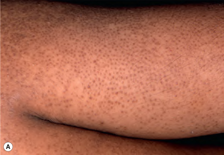
Fig. 12.17 Keratosis pilaris.A Discrete perifollicular papules with central keratotic cores on the extensor surface of the upper arm. Each papule has a rim of erythema. B Keratosis pilaris rubra on the lateral face. This variant is characterized by tiny, “grain-like” follicular papules superimposed on confluent erythema. A, Courtesy Luis Requena, MD; B, Courtesy Angela Hernández-Martín, MD.
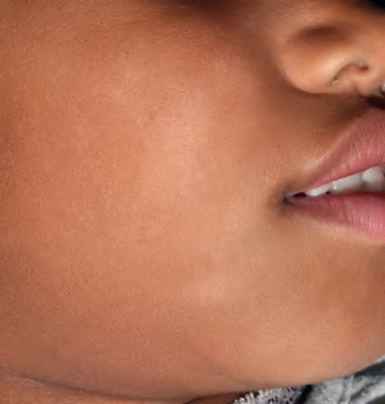
Fig. 12.18 Pityriasis alba. Note the slight scale associated with the hypopigmented macules and patches on the cheeks. Courtesy Antonio Torrelo, MD.
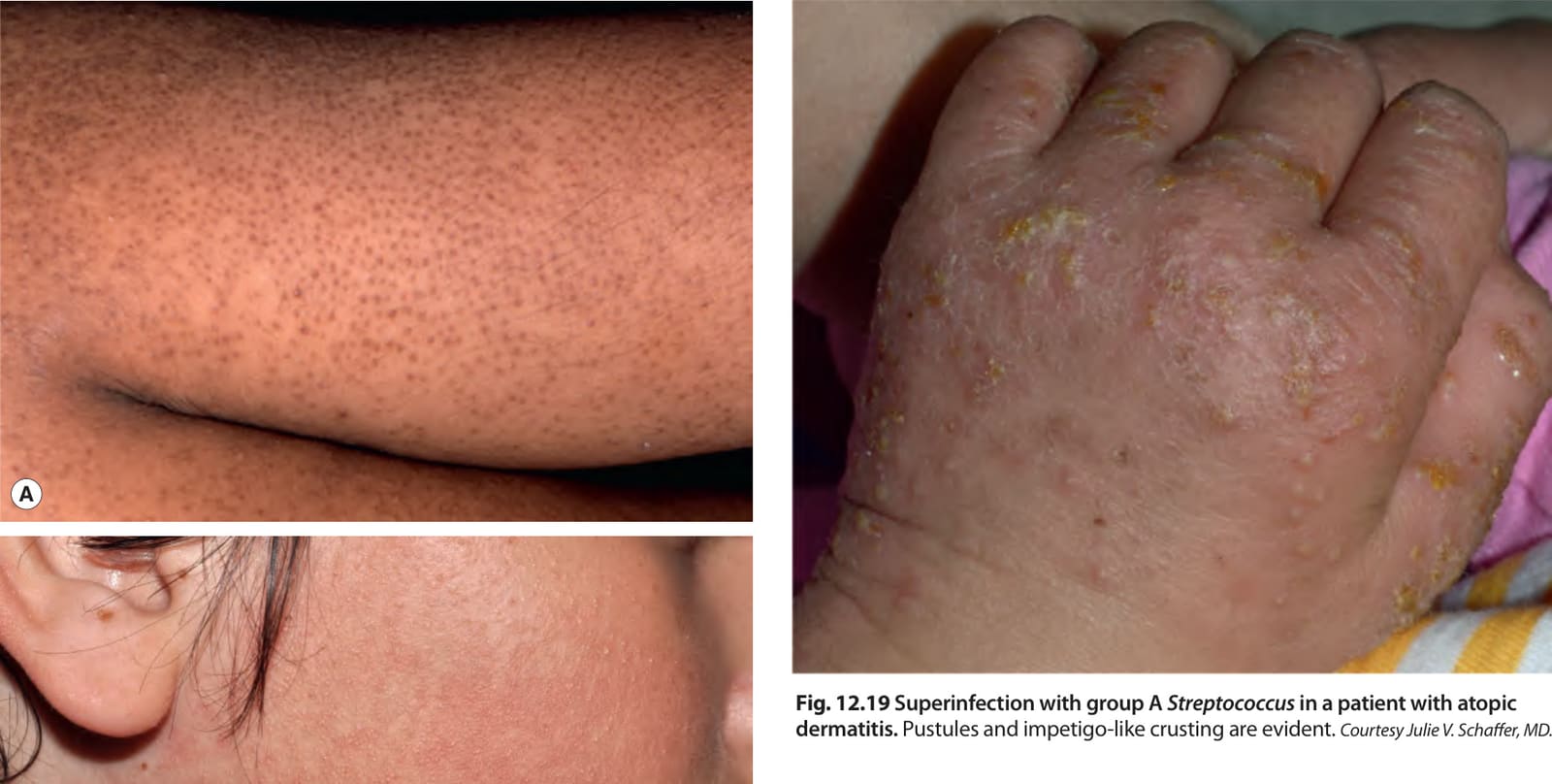
Fig. 12.19 Superinfection with group A Streptococcus in a patient with atopic dermatitis. Pustules and impetigo-like crusting are evident. Courtesy Julie V. Schaffer, MD.
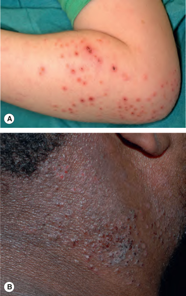
Fig. 12.20 Eczema herpeticum.
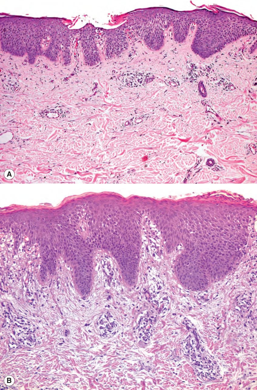
Fig. 12.21 Histologic features of acute and subacute atopic dermatitis.A

Fig. 12.22 Management plan for atopic dermatitis.A The therapeutic regimen should include both treatment of active eczema and maintenance that includes the low-level in all and the high-level in some patients. B Intermittent courses of a systemic corticosteroid result in rebound flares and worsening of disease over time. In contrast, a proactive regimen utilizing topical corticosteroid leads to longer clear periods and milder disease over time.
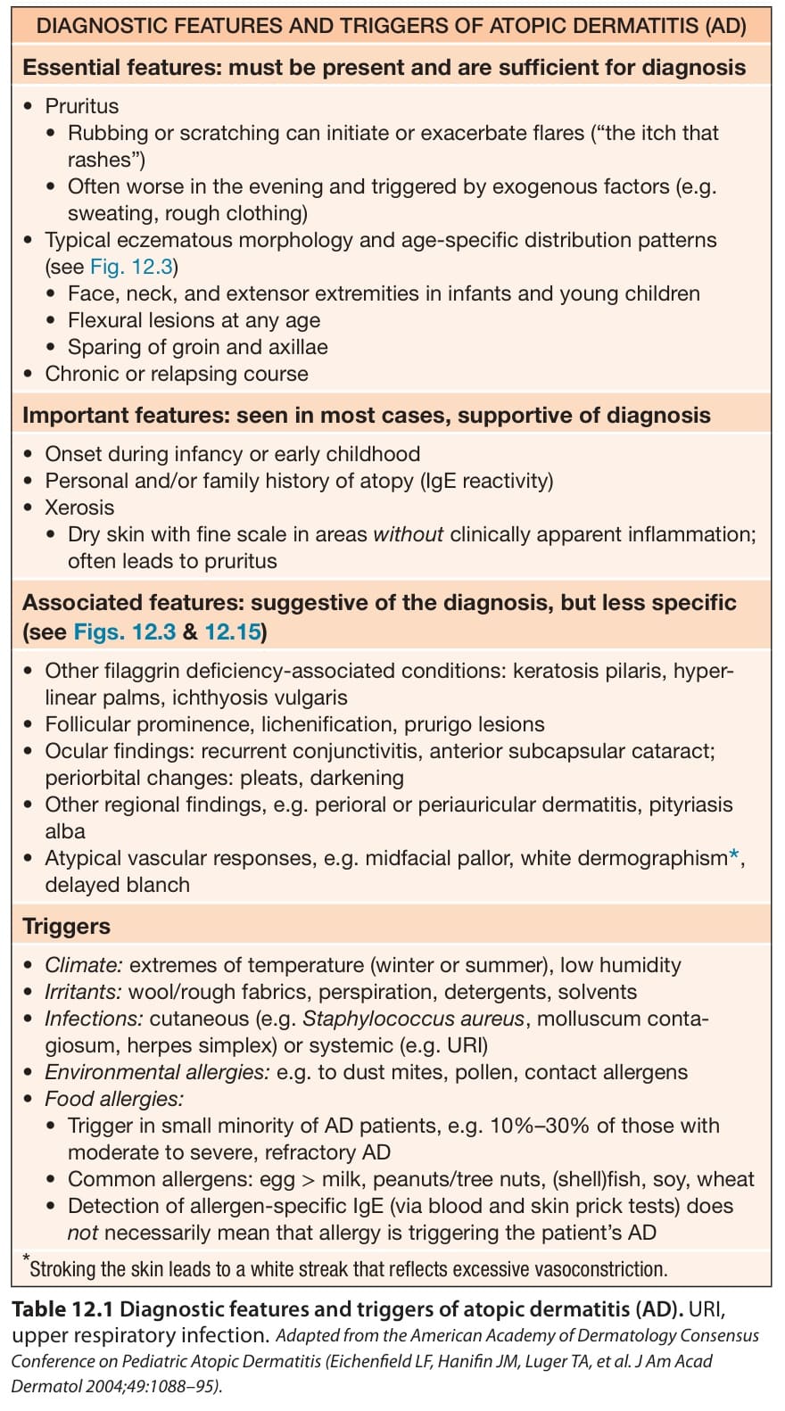
Table 12.1 Diagnostic features and triggers of atopic dermatitis (AD). URI, upper respiratory infection.Adapted from the American Academy of Dermatology Consensus Conference on Pediatric Atopic Dermatitis (Eichenfield LF, Hanifin JM, Luger TA, et al. J Am Acad Dermatol 2004;49:1088–95).
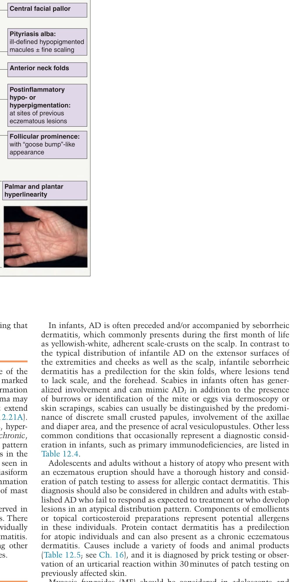
Table 12.4 Differential diagnosis of atopic dermatitis. A, adults; B, both; C, children/infants.
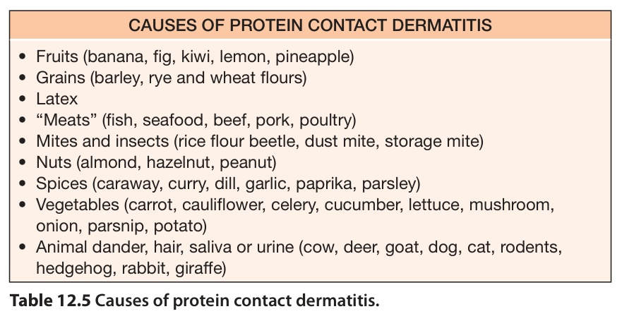
Table 12.5 Causes of protein contact dermatitis.
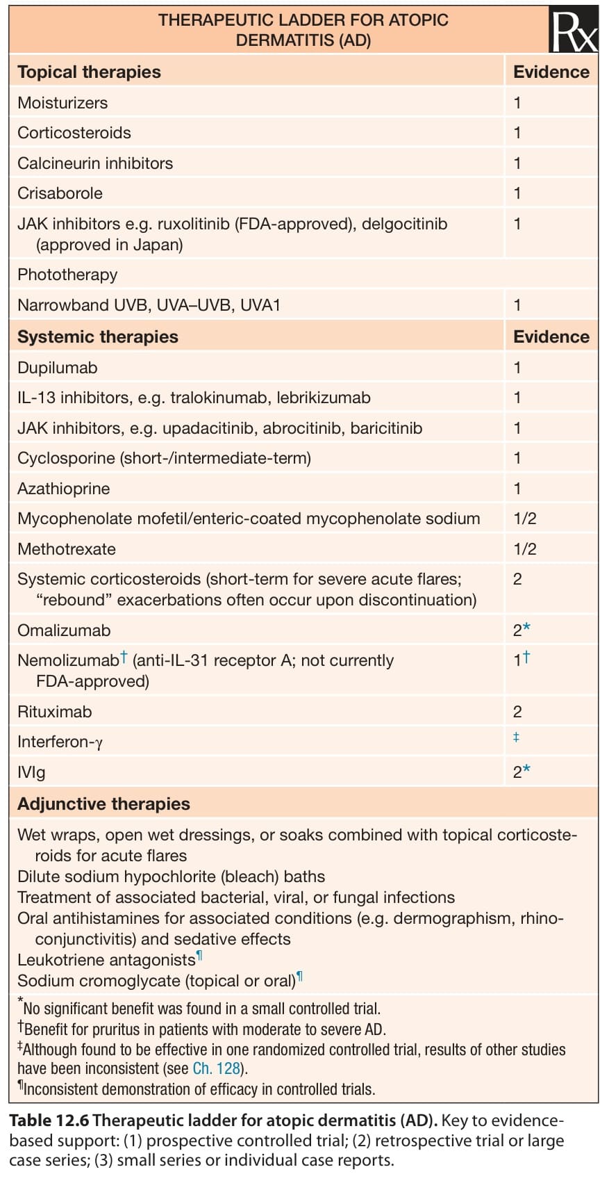
Table 12.6 Therapeutic ladder for atopic dermatitis (AD). Key to evidencebased support: (1) prospective controlled trial; (2) retrospective trial or large case series; (3) small series or individual case reports.
Association screening in the Epidermal Differentiation Complex (EDC) identifies an SPRR3 repeat number