PHOTOCHEMOTHERAPY WITH PSORALENS (PUVA)
Psoralen photochemotherapy (PUVA) combines the use of psoralens (P) and long-wave UV radiation (UVA). This combination results in a therapeutically beneficial phototoxic effect, which is not produced by either of the components alone. Psoralens can be administered orally or applied topically in the form of solutions, creams, or baths with subsequent UVA exposure.
Historical Aspects
Topical exposure to extracts, seeds, or parts of plants (e.g. Ammi majus, Psoralea corylifolia) that contain natural psoralens, followed by exposure to sunlight, was used as a remedy for vitiligo for thousands of years in ancient Egypt and India. In 1974, it was shown that the oral ingestion of 8-methoxypsoralen (8-MOP) and subsequent exposure to a new, high-intensity, artificial UVA radiation source was highly effective for the treatment of psoriasis. This was followed by recognition of its therapeutic effect for multiple skin disorders (Table 134.3). Over the past decade, the use of PUVA has decreased significantly, due in part to the increased risk of developing keratinocyte carcinomas and melanoma, especially with prolonged use in fair-skinned individuals, in addition to the effectiveness of NB-UVB and especially systemic
A Before treatment. B After treatment (15 exposures) with high-dose (130 J/cm, left side) versus medium-dose (65 J/cm, right side) UVA1 phototherapy. Note that there is no difference in the therapeutic result.
immunomodulators (“biologics”) in psoriasis. Even though PUVA often represents a second-line form of phototherapy, it is essential that dermatologists become familiar with treatment regimens, potential side effects, and indications, in particular since it is still the (see metaanalysis below) therapy of choice in early stage mycosis fungoides.
Psoralens
Psoralens are naturally occurring linear furocoumarins that are found in a large number of plants, and there are several synthetic psoralen compounds. For oral and topical (bath, cream) PUVA, mainly 8-MOP (methoxsalen) is used; it is of plant origin but also available as a synthetic drug. The synthetic compound, 4,5′,8-trimethylpsoralen (TMP, trioxsalen), is less phototoxic than 8-MOP after oral administration, but more phototoxic when delivered via bath-water. TMP had been used primarily in Scandinavia for bath PUVA. 5-Methoxypsoralen (5-MOP, bergapten) is also therapeutically effective when given orally; it is less erythemogenic and is not associated with gastrointestinal intolerance (Fig. 134.9). In some European countries, it was routinely used for PUVA, but it is
no longer commercially available. Oral preparations of 8-MOP contain crystals, micronized crystals, or solubilized psoralens in a gel matrix. The latter liquid preparation induces earlier, higher, and more reproducible peak plasma levels than the crystalline preparations.
The steps between the ingestion of a psoralen and its arrival in the skin include disintegration and dissolution of the drug, absorption, firstpass effect, blood transport, and tissue distribution. The absorption rate of a psoralen from the gut depends on the physicochemical properties of the molecule, the rate of dissolution, the galenic characteristics of the preparation, and the fat content of concomitantly ingested food. Peak serum levels are usually reliably and predictably achieved by liquid preparations, whereas wide time variability occurs with crystalline formulations. Before reaching the skin via the circulation, psoralens are metabolized during their passage through the liver. Plasma levels of 8-MOP administered orally at different doses show a strong non-linearity, indicating a saturable first-pass effect. Thus, small differences in the ingested doses and absorption rates of psoralens lead to large differences in plasma levels. As a practical consequence, small amounts of the drug are almost completely metabolized by the liver during the first pass and therefore may be therapeutically inactive.
8-MOP serum levels show a wide range of inter-individual differences. Even on different occasions in the same patient, serum levels may differ; however, the levels are usually sufficiently constant to provide for relatively reproducible therapeutic results. This unpredictable pharmacokinetic behavior is probably due to inter- and intra-individual variations in intestinal absorption, first-pass effect, blood distribution in the body, and metabolism and elimination of the drug.
For a given patient, serum levels of 8-MOP correspond fairly well with skin reactivity, with the peak of skin phototoxicity coinciding with peak serum levels. A correlation between 8-MOP serum levels and epidermal concentrations does exist. Whether there is a significant correlation between maximum psoralen blood concentrations and the minimal phototoxicity dose (MPD) remains equivocal.
The pharmacokinetics of topical 8-MOP depend on the mode of application. 8-MOP administered topically as a 0.15% emulsion or solution has been shown to result in plasma levels comparable to those obtained by oral delivery, when large areas of the body are treated. In contrast, due to the much lower concentrations that are typically used, plasma levels following bath-PUVA treatment of almost the entire body surface are very low. Bath-water-delivered psoralens are readily absorbed into the skin, but promptly eliminated without cutaneous accumulation.
Psoralen photochemistry
Psoralens react with DNA in three steps. First, in the absence of UV radiation, the psoralen intercalates into the DNA double strand. Via a photochemical reaction, absorption of photons in the UVA range results in the formation of a 3,4- or 4′,5′-cyclobutane monoadduct with pyrimidine bases of native DNA. The 4′,5′ monoadducts can absorb a second photon and this reaction leads to the formation of an inter-strand cross-link of the double helix with the 5,6 double bond of the pyrimidine base of the opposite strand (Fig. 134.10).
UVA-activated psoralens can also react with molecular oxygen. The reactive oxygen species formed by this reaction cause cell membrane damage by lipid peroxidation and may activate the cyclooxygenase and arachidonic acid metabolic pathways.
The cross-linking of psoralens with epidermal DNA inhibits DNA replication and causes cell cycle arrest. Although not proven, it is generally assumed that this effect may be the therapeutic mechanism in psoriasis. Psoralen photosensitization also causes an alteration in the expression of cytokines and cytokine receptors. However, DNA cross-linking does not appear to be a prerequisite for all the therapeutic effects of PUVA, and the successful treatment of other skin diseases is unlikely to be due directly to this molecular reaction. Psoralens also interact with RNA, proteins, and other cellular components and indirectly modify proteins and lipids via singlet oxygen-mediated reactions or by generating free radicals.
PUVA can reverse the pathologically altered patterns of keratinocyte differentiation markers and reduce the number of proliferating epidermal cells. Infiltrating lymphocytes are strongly suppressed by PUVA, with variable effects on different T cell subsets. PUVA is far more potent in inducing apoptosis in lymphocytes than in keratinocytes, which may
explain its efficacy in CTCL as well as in inflammatory skin diseases. Although much is known about pathways and mechanisms in psoralen photosensitization, the interactions and relative contributions to the clearing of a specific disease are not well understood.
Psoralens also stimulate melanogenesis. This involves the photoconjugation of psoralens to DNA in melanocytes, followed by mitosis and subsequent proliferation of melanocytes, increased formation and melanization of melanosomes, enhanced transfer of melanosomes to keratinocytes, and increased synthesis of tyrosinase via stimulation of cAMP activity (see Ch. 65).
Action Spectrum and Light Sources
The action spectrum for 8-MOP-induced delayed erythema has its maximum activity in the 330–335 nm range and the antipsoriatic activity of 8-MOP (plus UVA) appears to parallel this erythema action spectrum. Conventional therapeutic UVA fluorescent tubes and broad-spectrum metal halide lamps, which are filtered in the UVB and UVC range, effectively cover the psoralen action spectrum. The typical fluorescent UVA lamp used for PUVA therapy peaks at 352 nm and emits ~0.5% in the UVB range (Fig. 134.11). Major advantages of mercury halide units are the stability of output and their high irradiance, enabling shorter treatment times.
UVA doses are given in J/cm, instead of mJ/cm used for UVB, and are usually measured with a photometer with a maximum sensitivity at 350–360 nm. Within the phototherapy cabinet, the irradiance must be relatively uniform to ensure that the dose does not vary at different anatomic sites. UVB emission should be kept low enough to avoid erythemogenic UVB doses so that sufficient UVA is absorbed to activate the psoralen photosensitivity reaction.
Photobiologic Effects of PUVA
PUVA treatment produces an inflammatory response that manifests as delayed phototoxic erythema. The reaction depends on the dose of drug, the dose of UVA, and the individual’s sensitivity (skin type). More recently, it was shown that changes in 8-MOP dose had no detectable effect on the maximum slope of the PUVA erythema dose–response curve. Thus, 8-MOP dose changes within individuals, over a narrow but clinically relevant range, appear to significantly alter the threshold response to PUVA erythema but not the rate of increase in erythema with increasing UVA dose.
The time course for PUVA erythema differs markedly from sunburn or UVB erythema; the latter appears after 4–6 hours and peaks 12–24 hours after exposure to UVB. PUVA erythema does not occur before 24–36 hours and it peaks at 72–96 hours or even later after exposure. It has a shallower dose–response curve than UVB erythema (by a factor of ~2) and this difference is maintained even at the time of maximum erythema. PUVA erythema persists for longer periods of
time (lasting up to more than 1 week) and it consists of a deeper red, even a violaceous, hue. Severe reactions may lead to blistering and to superficial skin necrosis. Overdoses of PUVA are frequently followed by edema, intense pruritus, and sometimes by a peculiar stinging sensation in the affected skin area. At present, erythema is the only clinical parameter that allows for an assessment of the magnitude of the PUVA reaction and then determination of UVA dose adjustments.
Pigmentation, the second important effect of PUVA, may develop without clinically evident erythema, especially when oral TMP is used. In normal skin, PUVA pigmentation peaks about 7 days after exposure and may last from several weeks to months. As with sun-induced tanning, the individual’s ability to tan is genetically determined, but the dose–response curve is much steeper. A few PUVA exposures result in a much deeper tan than that produced by multiple exposures to solar radiation. When administering PUVA therapy for inflammatory and neoplastic disorders, an important strategy is to produce a clinical response prior to the induction of significant pigmentation.
Treatment Protocols
Oral PUVA
For oral PUVA, the general approach is to keep the dose of psoralen, as well as the interval between drug intake and UVA exposure, constant, and to vary the UVA dose according to the patient’s sensitivity. A dosage of 8-MOP of 0.6 to 0.8 mg/kg body weight is administered orally 1–3 hours before exposure, depending on the absorption characteristics of the particular drug formulation (see above).
The initial UVA doses are selected either by skin phototyping or by determining the minimal phototoxicity dose (MPD). In the US, skin reactivity to solar radiation is evaluated and patients are classified based on a skin phototype scheme according to their sunburn and tanning history. Therapeutic doses of UVA are then given according to an empirical scheme based on this classification (Table 134.4). In Europe, until recently, the most widely used approach consisted of MPD determination analogous to MED determination. The MPD is defined as the minimal dose of UVA (following psoralen ingestion) that produces a barely perceptible, but well-defined, erythema. The doses range from 0.5 to 5 J/cm if oral 8-MOP is used. Erythema readings are performed 72 to 96 hours after irradiation, when the phototoxicity reaction usually peaks. As with the MED, the MPD is determined on previously non-sun-exposed skin (lower back or buttocks). Since determining the MPD is rather time-consuming and can take up to a week to perform, it has been abandoned for practical reasons in most European phototherapy centers and replaced by phototyping. In several phototherapy guidelines, MPD testing is still recommended but not considered to be mandatory.
Oral PUVA should not be used in patients with hepatitis because a slower metabolism of psoralens may prolong photosensitivity. Impaired renal function may also reduce psoralen excretion.
Bath PUVA
Bath-water delivery of psoralens has become increasingly popular because it provides uniform drug distribution over the skin surface, very low psoralen plasma levels, and a rapid elimination of free psoralens from the skin, thereby reducing the period of photosensitivity. Due to the absence of systemic photosensitization, bath-water delivery of 8-MOP circumvents gastrointestinal side effects and potential ocular effects. Skin psoralen levels are highly reproducible and photosensitivity lasts no more than 2 hours. However, peak erythema is delayed as compared to the oral route; therefore erythema readings should be performed 96 to 120 hours after irradiation, which is the major reason why MPD determination has been replaced by phototyping in most phototherapy centers. The higher incidence of unwanted phototoxicity can be prevented by a lower starting dose and more cautious dosimetry during the initial treatment phase.
Originally, bath PUVA was performed with TMP, but 8-MOP is now being used as well. Bath PUVA consists of 15–20 minutes of whole body (or hand and foot) immersion in solutions of 0.5–5.0 mg of 8-MOP per liter of bath water. Irradiation has to be performed immediately thereafter, as photosensitivity decreases rather rapidly. However, one must take into account that, unlike with oral PUVA, the phototoxic threshold declines early on during treatment (this may be due to persistent psoralen adducts). Thus, at the onset, dose increments should be done more cautiously.
Topical PUVA
Application of 8-MOP (0.1%–0.01%) in creams, ointments, or lotions, followed by UVA irradiation, is effective but has several disadvantages. The non-uniform distribution on the skin surface may produce unpredictable phototoxic erythema reactions, and inadvertent application to surrounding uninvolved skin can lead to cosmetically unacceptable hyperpigmentation. Furthermore, if numerous lesions are present, the application is laborious and time-consuming, and the treatment does not prevent the development of new active lesions in previously unaffected, untreated areas. Finally, extensive application (“paint” PUVA) of a 0.15% 8-MOP emulsion was found to cause plasma levels comparable to those detected with oral ingestion. Therefore, topical PUVA utilizing psoralen creams, ointments, or lotions is now reserved primarily for dermatoses of the palms and soles.
Initial Treatment (Clearing) Phase
The initial treatment phase is defined as the treatment period until clearing of the disease is achieved; repeated exposures are required, with gradual dose increments as pigmentation develops. Doses that are too low frequently result in failure of treatment, except in those diseases in which induction of pigmentation is the desired objective. In most dermatoses amenable to PUVA, the frequency of treatments should be reduced after satisfactory control of the disease.
If MPD testing is performed, a safe initial therapeutic dose for oral PUVA is 50%–70% of the MPD. For bath PUVA, it is advisable to start at only 30% of the MPD, because photosensitivity is up to 10 times higher than with oral PUVA (Fig. 134.12). As noted previously, nowadays, most centers start with doses based on the patient’s skin phototype.
Irradiations are given two to four times weekly. Dose increments are performed no more often than twice a week (at least 72 hours apart) and never during the first week of treatment (if initial dose is based on MPD), in order to avoid an accumulation of delayed cutaneous phototoxicity. Although not necessary for therapeutic success, a minimally perceptible erythema is considered a clinical indicator of adequate dosimetry. There exists no rigid scheme for dose increments; the major parameter for dose adjustments should be the clinical response of the disease. It is essential to note that with bath PUVA, the MPD can even decrease during the first days after initiation of treatment by up to 50%, but then increase at later stages. This may be due to persistent psoralen adducts which are converted into cross-links upon subsequent exposures.
In the absence of erythema, the UVA dose can be increased safely by 30% in both oral PUVA and bath PUVA (see Fig. 134.12). However, because of erythema formation and/or an adequate treatment response, some patients will not require dose adjustments over prolonged periods of time.
Maintenance Phase
The purpose of maintenance therapy is to prolong the duration of clinical remission. In the original European regimen, maintenance therapy consisted of one month of twice-weekly treatments utilizing the final UVA dose administered during the clearing phase, followed by another month of once-weekly exposures. However, results of a more recent study indicated that short-term maintenance therapy was not effective in preventing early relapses of psoriasis. For MF, many institutions recommend some form of permanent maintenance therapy, but there is a lack of prospective studies to validate this recommendation. Perhaps a once-monthly treatment would be a feasible compromise.
Mild relapses during the maintenance phase are handled by temporarily increasing the frequency of treatments; in the case of a severe relapse, the original clearing phase schedule must be resumed until clearing is once again achieved. A disadvantage of maintenance therapy for patients in remission is the potential to overtreat the patient and to increase the potential for long-term toxicity that is related to the total cumulative dose of PUVA.
PUVA for Psoriasis
Basically, all types of psoriasis respond to PUVA (Fig. 134.13), although the management of erythrodermic or generalized pustular psoriasis is more difficult. Both the US and the European regimens have proven to be highly efficient and therefore have remained in use, although with the advent of systemic immunomodulators (“biologics”; see Ch. 128), phototherapy for all types of psoriasis has lessened.
Three studies have compared bath-water delivery of 8-MOP with oral administration. In two reports, initial doses were determined by skin phototyping and treatments were given two to three times weekly. Dosage increments were made with every treatment in the first study, whereas smaller increments were performed every third treatment in the second. Patients in the third study were treated according to the guidelines of the standard European regimen for oral PUVA; this group
Before treatment. B After 6 weeks of treatment.
had the lowest incidence of treatment failures and overdose episodes. Relative to the results obtained with oral PUVA, bath PUVA showed equal clearing rates with lower numbers of exposures. The greater therapeutic efficacy could be due to a higher penetration of psoralens through the abnormal stratum corneum overlying psoriatic plaques as compared with healthy perilesional skin where phototoxicity is monitored during the therapy. The incidence of erythema and pruritus was similar or lower with bath PUVA as compared to oral therapy. In all the investigations, episodes of systemic intolerance such as nausea and vomiting were recorded only with oral PUVA.
PUVA alone can produce a definite remission in many cases of psoriasis, but a considerable number of patients require combination therapy (see below).
Combination Treatments
Topical combination
As with UVB phototherapy, PUVA can be combined with other treatment modalities to improve efficacy and to reduce potential side effects. Topical adjuvant therapies include corticosteroids, anthralin, tar, and vitamin D analogues (see Chs. 125 & 129). However, some patients are reluctant to “retry” topical agents that previously proved unsuccessful when used alone.
Methotrexate, cyclosporine, systemic
The combination of PUVA and methotrexate (MTX) during the clearing phase reduces the duration of treatment, number of exposures, and cumulative UVA dose, and it is also effective in clearing patients unresponsive to PUVA or UVB alone. To date, no increased risk of keratinocyte carcinomas has been reported for this combination. In contrast, cyclosporine plus PUVA has been clearly shown to dramatically enhance cutaneous carcinogenesis. Thus, this latter combination should be categorically discouraged, including the use of cyclosporine subsequent to PUVA therapy. Presently, there are no studies evaluating the safety and efficacy of biologics plus PUVA.
Retinoids
The combination of PUVA with systemic retinoids (RePUVA) is one of the most potent therapeutic regimens for psoriasis. The therapeutic efficacy of PUVA therapy is significantly enhanced upon combination with oral retinoids (acitretin [0.5–0.75 mg/kg] or isotretinoin [0.75– 1 mg/kg]) that are administered daily for 5–10 days before PUVA is started, with the combination continued throughout the clearing phase. RePUVA reportedly can reduce the number of exposures by one-third and the total cumulative UVA dose by more than one-half. RePUVA can also clear “poor PUVA responders”.
The underlying mechanism of the synergistic action of retinoids plus PUVA may be a reduction in the inflammatory infiltrate and an accelerated desquamation of the psoriatic plaques, thereby optimizing the optical properties of the skin. In theory, retinoids may also reduce the long-term carcinogenic risk of PUVA by reducing the number of exposures and providing chemoprevention against skin cancers. Some evidence for this was presented in a study where patients with psoriasis treated with PUVA in combination with systemic retinoids showed a reduced risk of SCC but not a significantly altered incidence of BCC.
Short-term side effects of retinoids are completely reversible upon discontinuation, and long-term toxicity is not relevant because of the limited duration of their use (until clear). The potential teratogenicity of retinoids represents a serious concern. For women of childbearing age, the use of isotretinoin is an option, because contraception is only necessary for 1–2 months after discontinuation of therapy, as opposed to acitretin, which requires two subsequent years of contraception.
PUVA for Cutaneous T Cell Lymphoma, in Particular Mycosis Fungoides
Based on the empirical beneficial effect of natural sunlight on early stage MF, Gilchrest et al. first reported the successful use of PUVA for MF. Patients with unsatisfactory responses to other therapies and histologically confirmed plaque or tumor stage MF or the erythrodermic form of CTCL were subjected to PUVA. Complete clearing was observed, except in those areas that were shielded from UVA exposures, thus excluding the possibility of spontaneous remission. This also indicated that the therapeutic effect of PUVA was local rather than systemic.
Numerous studies followed which addressed the rates of initial response and average duration of remission in relation to the stage of MF, the efficacy of PUVA compared with other established treatment options, the efficacy of combination treatments, the mechanisms responsible for the therapeutic effects, and the risk of short- and long-term side effects.
Treatment schedules and dosimetry in the photochemotherapy of MF are essentially the same as for psoriasis, consisting of three phases – clearing, maintenance, and follow-up. Some authors suggest that remission should be confirmed by histologic examination of previously involved areas. In some institutions, maintenance therapy includes two exposures per week for one month and one exposure per week for another month. If still in remission, therapy is discontinued and the patient is monitored monthly and then bimonthly. If a relapse occurs, the patient is again treated with three or four PUVA exposures per week until complete clearing. However, other investigators advocate permanent maintenance treatment consisting of treatments once monthly or every other month, as it appears that PUVA treatment is not curative. Since the course of MF varies considerably from patient to patient, the final answer remains to be determined.
Clinical experience indicates that patients benefit most from individualized schedules. For example, a recent study described the administration of a low frequency regimen (2×/week) for early MF (stage IA–IIA). Oral PUVA was administered for up to 24 weeks in 27 patients; 19 complete and 8 partial remissions were observed and the average cumulative UVA dose was rather low (78.5 J/cm²). Eleven of the 19 patients with a complete remission underwent maintenance therapy (14 exposures over 9 months), with 7 of those still in remission after one year. In comparison, 7 of 8 patients who did not receive maintenance therapy experienced a relapse.
Several studies involving large patient cohorts have provided information on the initial response rates of PUVA treatment in relation to disease stage. Overall, the best initial complete response rates were obtained for stages IA (~90%), IB (~76%) and IIA (~78%), as compared to stages IIB and III (~60%). There was some heterogeneity in results and the use of different treatment protocols, psoralen preparations, and light sources may have contributed to this finding. Only a few patients with stage IV disease have been treated with PUVA monotherapy, because, in this stage, PUVA is generally considered to be only a palliative or adjunctive therapy. Relapses usually respond to PUVA in the same manner as the initial response. Clinical remissions appear to be directly related to phototoxic destruction of the malignant infiltrate. Thus, complete clearing may be induced when the cells are confined to the epidermis and the superficial dermis and do not exceed the depth of UVA penetration into the skin. Patients with tumor stage MF exhibit a high rate of early recurrences, which require permanent maintenance treatment; often only the combination of PUVA with local radiotherapy and/or systemic chemo-immunotherapy can result in complete tumor resolution.
In summary, the data thus far indicate that PUVA is an excellent treatment option for early stage (IA–IIA) CTCL (Fig. 134.14). High rates of complete clearing can be achieved and a substantial percentage of patients remain free of disease for many years. In a meta-analysis of seven studies that compared PUVA versus NB-UVB for the treatment of early MF (IA–IIA), 778 patients (527 PUVA, 251 NB-UVB) were analyzed. Complete remission was achieved in 74% of the patients in the PUVA group and 62% in the NB-UVB group; the remissionfree interval ranged from 10 to 45 months (mean 33 months) in the PUVA group compared to 5 to 24 months (mean 15 months) in the NB-UVB group. The significantly longer remissions rates may be one of the reasons why in some treatment guidelines PUVA is regarded as the therapy of choice in early stage MF.
In advanced stages (IIB–IVB) of MF, PUVA is not sufficient as monotherapy, but as an adjunctive treatment it can reduce the tumor burden in the skin and can improve the quality of life for patients. Presently, no therapeutic regimen is known to arrest eventual progression of advanced stages of MF to tumor formation, dissemination, and a fatal outcome. Prolonged remissions have been observed with combinations of PUVA plus systemic retinoids (e.g. bexarotene) or IFN-α-2a. Possible long-term side effects related to frequent
Before treatment. B After 12 weeks of treatment that was four times weekly.
PUVA treatments are probably less meaningful for patients with CTCL compared with patients who have benign disorders.
While PUVA therapy is very effective in inducing remission as long as the lymphoma is confined to the skin, the impact of this therapy on the natural course of CTCL and on patient survival is not yet determined.
PUVA for Vitiligo
As mentioned previously, vitiligo was the first disease treated with an ancient form of psoralen photochemotherapy in India and Egypt. PUVA in its modern form stimulates melanogenesis as well as melanocyte proliferation and migration, and it reconstitutes the normal skin color in more than 50% of vitiligo patients.
Treatment protocols and results
Nowadays, oral 8-MOP is the photosensitizer most frequently used, followed by exposure to sunlight or artificial UVA radiation. In the past, if sunlight were the radiation source, oral TMP was the preferred photosensitizer due to its weaker phototoxicity. To induce repigmentation, patients need constant long-term therapy. It is crucial that the patient understands that PUVA, like other vitiligo therapies, may require months or even years to achieve a satisfactory result. Overly optimistic prognoses regarding therapeutic efficacy and length of time to produce adequate repigmentation should be avoided. The patients must also be aware that the treatments stimulate the pigmentation of normal skin, which will intensify the contrast between normal and vitiliginous skin.
Treatments should be given at least twice a week, but not more than three times, with at least one day between treatments. If there is no response after 4–5 months or ~30–40 treatments, PUVA should be terminated. Responsiveness is defined as development of multiple perifollicular macules of repigmentation, or, in the case of small (<2 cm) lesions, contraction in size. If treatment is discontinued, reversal of acquired repigmentation may occur unless the lesion has completely repigmented. Completely repigmented areas can be stable for a decade or more without relapse.
Photochemotherapy with topical 8-MOP can be used in patients with small lesions (<5% total body surface area) or in children in whom one may hesitate to use systemic PUVA. The therapy must be done in an office setting, since home treatment involves a high risk of severe phototoxic side effects. Topical 8-MOP preparations (0.0005%–0.005% 8-MOP in a cream or ointment base) are applied evenly on the treatment area 30 minutes prior to irradiation. Exposure times (beginning at ≤0.25 J/cm) are increased weekly in increments of 0.25 J/cm unless marked erythema occurs. Once- to twice-weekly treatments are recommended. With topical PUVA, the vehicle plays a major role in delivery and resulting cutaneous psoralen levels. Solutions should be avoided as they can “run”, producing hyperpigmented streaks. Application of sunscreen to the surrounding uninvolved skin prior to application of the topical psoralen can reduce hyperpigmented rims.
Patient selection appears to be critically important. For oral PUVA, the patients should be ages 10–12 years or older, and for all forms, they must be available for 12–24 months of continuous therapy. Lips, distal dorsal hands, fingers and toes, palms, soles, and nipples are very refractory to treatment, as are large areas with only white hairs. Patients need to be made aware of this fact.
Stable disease, i.e. nonprogressive for at least 6 months to a year, is usually easier to treat. On average, a complete treatment regimen consists of at least 150 exposures. However, because of the different response rates in different body areas, total repigmentation is only rarely achieved, and ~30% of patients do not respond at all, despite months of therapy. As mentioned above, in a trial involving patients with non-segmental vitiligo, NB-UVB therapy resulted in a better color match when compared to oral PUVA. As a result, the use of NB-UVB has increased while PUVA therapy for vitiligo has waned in most phototherapy centers.
PUVA for Atopic Dermatitis (See Ch. 12)
Many patients with moderate, severe, and even erythrodermic forms of atopic eczema can benefit from PUVA therapy. The treatment guidelines are essentially the same as for psoriasis, but patients may develop a temporary flare of their dermatitis during the initial treatment phase. Also, atopic dermatitis is more difficult to treat than psoriasis and quite often a higher number of treatments is required. Although atopic dermatitis may be cleared by PUVA, recurrence rates are high and rapid, requiring frequent maintenance exposures. However, in young patients, long-term maintenance therapy should be avoided. A combination of PUVA with topical corticosteroids appears to be superior to PUVA alone in maintaining remissions, and in a more recent study involving patients with severe atopic dermatitis, PUVA provided better short- and long-term responses than did medium-dose UVA1. These latter results require further confirmation. Due to the introduction of systemic immunomodulators such as dupilumab to treat moderate-to-severe atopic dermatitis, the importance of PUVA as a therapeutic approach will likely decrease in the future.
PUVA for Lichen Planus (See Ch. 11)
In generalized lichen planus, PUVA can provide an effective alternative to systemic corticosteroids. Studies have pointed to an overall response rate between 50% and 90%, suggesting that lichen planus is less responsive than psoriasis to PUVA. In addition, more treatment sessions and higher cumulative UVA doses were generally necessary for complete remission and not all patients responded satisfactorily. Marked postinflammatory hyperpigmentation may impair the final cosmetic result. An exacerbation during PUVA treatment has been reported in a few patients. Bath PUVA also works in lichen planus, and a combined RePUVA regimen (as is used for psoriasis) may accelerate clearing in disseminated and particularly keratotic forms of lichen planus. Compared to NB-UVB, oral PUVA led to a better initial clinical response rate, but both were effective treatments and produced similar long-term outcomes.
PUVA for Graft-Versus-Host Disease (see Ch. 52)
Due to clinical and histologic similarities between idiopathic lichen planus and lichenoid GVHD, PUVA treatment was evaluated for the latter. Beneficial effects were observed in patients who had not responded to conventional immunosuppressive therapy alone. PUVA can also improve acute GVHD. Results of PUVA therapy for scleroderma-like variants of cutaneous GVHD are controversial. In our experience, the more localized and circumscribed morpheaform lesions appear to respond to PUVA fairly rapidly, with softening of the associated sclerosis. However, the more advanced and widespread sclerodermoid form hardly responds to PUVA.
In contrast to other conditions, PUVA may exert not only local but also systemic effects, since improvement of mucosal erosions was observed during treatment of chronic lichenoid GVHD with PUVA, but this could have also been coincidental. Apparently, there is no improvement of GVHD of other organs, e.g. liver and gut. Of note, deterioration of liver GVHD has not been reported thus far. This is important, since psoralens are metabolized by the liver.
The therapeutic regimen used for the treatment of chronic GVHD is basically the same as for psoriasis. UVA doses should not be increased too aggressively, in order to avoid erythema and possible (re)activation of GVHD. The UVA dosage is increased by no more than 0.5 J/cm every second to fourth exposure and patients are treated three to four times weekly. After clearing of skin lesions, exposures are reduced according to the maintenance schedule outlined for psoriasis.
There may be a slight overall increased risk of cutaneous malignancies in hematopoietic stem cell recipients, but much less than in solid organ transplant patients who receive long-term immunosuppression. If patients are receiving long-term prophylactic treatment with voriconazole, switching to posaconazole should be considered in order to reduce possible enhancement of SCC formation.
PUVA for Urticaria Pigmentosa (See Ch. 118)
PUVA causes a temporary involution of cutaneous mastocytosis. The treatment results in loss of Darier’s sign, relief of itching, and flattening and sometimes disappearance of cutaneous macules and papules. Surprisingly, even systemic symptoms such as histamineinduced migraines and flushing fade gradually as treatment is continued. In most patients, the clinical manifestations recur several months after discontinuation of PUVA. The recurrences respond as well as the original lesions, and clearing of signs and symptoms can again be achieved. In children, PUVA is also often helpful, and appears to be justified in widespread, disabling mastocytosis. To avoid sudden mediator release from mast cells, a PUVA regimen of gradually increasing doses, especially during the initial phase, seems advisable, although the therapy should otherwise be undertaken according to the usual guidelines.
PUVA for Miscellaneous Dermatoses
Both acute and chronic pityriasis lichenoides respond to photochemotherapy and favorable results have been reported for lymphomatoid papulosis. Nevertheless, the experience with these conditions is limited to anecdotal cases. In pityriasis rubra pilaris, the results are quite inconsistent. Some patients seem to respond well, while others may flare and some require combination treatment with retinoids or methotrexate. Generalized granuloma annulare has been reported to clear completely, but long-term maintenance treatment was required to maintain remissions. There were initial reports of both topical and systemic PUVA leading to regrowth of hair in patients with alopecia areata in whom exposures were localized to the alopecia areas. The number of treatments required for regrowth varied widely, and circumscribed lesions responded better than did total alopecia. However, follow-up studies of larger patient groups concluded that PUVA is generally not effective in alopecia areata, which is in accordance with our own experience. Localized scleroderma and pansclerotic morphea have been successfully treated with bath PUVA and oral PUVA.
PUVA for Photodermatoses
Tolerance to sunlight can be induced in several photodermatoses with NB-UVB (see above) or PUVA. Although NB-UVB is tried first due to safety concerns, PUVA represents a very effective preventive treatment for polymorphous light eruption (PMLE; via hardening). In ~70% of patients with this condition, a 3- to 4-week course of PUVA suffices to suppress the disease upon subsequent exposure to sunlight. PUVA induces pigmentation rapidly and intensively at relatively low (suberythemogenic) UVA doses that usually remain well below the threshold doses for eliciting the PMLE. About 10% of the patients develop typical lesions during the initial phase of PUVA, and as with UVB phototherapy, interventions include interruption of treatment, reduction in the UVA dose, and application of topical corticosteroids.
The therapeutic value of preventive photochemotherapy for PMLE has to be balanced against its potential long-term side effects. Given the fact that very few treatments and low cumulative UVA doses (ranging from 15 to 40 J/cm per treatment course, depending on skin type and photosensitizer) are required for photoprotection and that PMLE can severely restrict outdoor activities, PUVA treatment of PMLE is justified when other preventive measures (e.g. sunscreens, NB-UVB) fail.
There is also some experience with PUVA prophylaxis of other photodermatoses. In solar urticaria, PUVA therapy is an effective preventive treatment and is certainly better than antihistamines. Sensitivity to sunlight can be decreased by 10-fold or more after a single treatment course. The suppressive effect may last throughout the summer, depending on regularly repeated sunlight exposures. Problems may occur during the first several PUVA exposures, since in some patients the urticaria threshold dose appears to be lower than their MPD. In these cases, stepwise UVA irradiation of single quadrants of the body a few hours before each PUVA treatment has proven useful. PUVA is then administered during the refractory period. Of note, UVA rush hardening appears to be equally effective as PUVA, with the advantage of inducing the hardening effect and providing protection within a few days.
Successful photochemotherapy has also been reported in occasional patients with chronic actinic dermatitis and hydroa vacciniforme.
Side Effects and Long-Term Hazards of PUVA
As discussed above, oral 8-MOP has a high incidence of nausea (30% of patients) and vomiting (10% of patients), and this may occasionally require discontinuation of the medication. These side effects are more common with liquid than with crystalline preparations, probably because of higher psoralen serum levels.
The short-term side effects of the combined action of psoralens plus UVA radiation consist of redness, swelling, and occasionally blister formation as can be seen with excessive sunburns. Generalized pruritus or tingling sensations may herald phototoxic side effects. When large areas of skin are affected, systemic symptoms of excess phototoxicity such as fever and general malaise may occur. Nonsteroidal anti-inflammatory drugs and topical and systemic corticosteroids may be required to alleviate these symptoms, but they have to be given early. Because of high epidermal psoralen concentrations, such overdosage phenomena are more common after topical psoralen application. Accumulation of phototoxic effects after several consecutive UVA exposures is also more common with topical PUVA.
Some patients experience persistent pruritus during PUVA treatment, particularly after slight UVA overdosage, and in rare cases, a stinging pain may develop in circumscribed areas. PUVA-induced skin pain unrelated to actual phototoxic burns occurs rarely, but it may necessitate discontinuation of treatment. The mechanism is unknown and the symptoms are unresponsive to antihistamines. Usually, these complaints subside slowly upon continuation of treatment.
Ophthalmologic effects
Eye protection is mandatory during UVA exposure, and UVA-opaque glasses are required for ambient exposure until the evening of the treatment day. UVA penetrates into the ocular lens and potentially could induce cataracts by forming psoralen-protein photoproducts. The higher permeability of the ocular lens at a younger age led to the relative contraindication of oral PUVA in children younger than 12 years of age. Despite the experimental data indicating a risk of premature cataract formation, clinical evaluation has shown no increase in lens opacities even in patients who neglected to practice careful eye protection. Obviously, there is no risk with topical or bath PUVA.
Laboratory data
Because psoralens can cause liver damage in laboratory animals when given in excessive doses, concern was expressed in the past regarding possible hepatotoxic effects in humans. Several large-scale studies demonstrated no significant abnormal laboratory findings in patients receiving PUVA over prolonged periods of time. In particular, serial laboratory examinations performed over a period of several years have not revealed any substantial evidence for an impairment of hepatic function. Liver biopsies after 1 year of therapy did not show signs of hepatotoxicity. Anecdotal case reports of hepatitis during PUVA treatment were most likely unrelated to therapy. Several large-scale studies have negated a possible relationship between PUVA therapy and the occurrence of anti-nuclear antibodies.
Potential long-term risks of PUVA
Repeated phototoxic injury to the skin can be expected to result in cumulative actinic damage regardless of whether it is induced by sunlight, artificial UV radiation, or PUVA. Although the precise action spectrum of actinic damage has not been determined, epidermal changes are attributed to UVB and dermal changes more to UVA, because the latter penetrates more deeply into the skin. Thus, chronic exposure to PUVA may produce changes in the skin that generally resemble those known as dermatoheliosis and may add to the injury induced by sunlight. High cumulative doses of whole body UVB or PUVA result in pigmentary changes, xerosis, loss of elasticity, wrinkle formation, and actinic keratoses. Additionally, PUVA may induce profuse formation of dark stellate lentigines (Fig. 134.15), termed PUVA lentiginosis. These lentigines result from repeated and prolonged treatment and are commonly associated with high cumulative doses of UVA and a high number of treatments. So far, no increased risk of cutaneous melanoma
associated with these lentigines has been recorded, but the cosmetic effect may be quite disturbing.
The major concern with prolonged and repeated phototherapeutic regimens is photocarcinogenesis. Therefore, from the onset, PUVA-treated patients were carefully monitored for the development of precursors and malignant skin tumors. Almost all data were obtained from psoriatics, since they represent the largest group of patients receiving PUVA.
The risk is certainly related to DNA damage, but PUVA-induced immunosuppression may play a role as well. In PUVA patients, the risk of SCC, but not of BCC, is significantly increased in comparison with matched controls, and the magnitude of the increase appears to be dosedependent. However, there is uncertainty regarding PUVA being the sole factor; many of the affected patients had previous exposure to excessive sunlight and to treatments with carcinogenic potential, including arsenic, UVB, and antimetabolite therapy. Specifically, high levels of UVB exposure appear to increase the risk of keratinocyte carcinomas in PUVA-treated patients. Of note, in non-White individuals who received long-term oral PUVA therapy, an increased risk of skin cancers was not observed. In a more recent update from the original PUVA study, exposure to <150 PUVA treatments had, at most, modest effects on SCC risk and even high-dose exposure to PUVA did not greatly increase BCC risk.
According to a single study, the genitalia of male patients previously treated with tar and UVB appeared to be particularly susceptible to the carcinogenic effects of PUVA, but the risk seemed not to be increased if there had only been exposure to PUVA. In a retrospective study from France comprised of 5400 patients treated between 1978 and 1998, no case of genital skin cancer was found, despite the fact that the genital area had not been protected during UVA exposure, raising the question as to whether genital shielding is absolutely necessary. No increased risk of cutaneous carcinoma has so far been reported for patients treated with PUVA for vitiligo.
Only a few anecdotal cases of cutaneous melanoma have been described in long-term PUVA-treated psoriatics, and, to date, with one exception, no increased risk of melanoma has been observed in any reported large-scale study. However, Stern et al. reported on the cohort of 1380 patients enrolled in the PUVA Follow-up Study (16-center study), and since 1975, 23 patients had developed 26 invasive or in situ cutaneous melanomas. Beginning 15 years after the initial exposure to PUVA, an increased risk of melanoma was observed in their cohort of PUVA-treated patients. However, the lead author concluded that perhaps PUVA has been judged unfairly. Its long-term risks have been subject to much greater scrutiny than the risks of other therapies utilized for severe psoriasis, such as methotrexate and particularly immunosuppressive therapies such as cyclosporine (CSA). Notably, several other US studies, as well as European studies, have not observed an increased risk of melanoma in patients treated with PUVA.
An increased risk of cutaneous SCC has been observed in solid organ transplant recipients who have received long-term CSA and in patients who have received CSA subsequent to PUVA therapy. UVA-sparing, aggressive regimens without prolonged maintenance therapy may be safer than continuous non-aggressive regimens (see Table 134.4).
In a study of 944 Swedish and Finnish patients, bath PUVA with TMP appeared to have no relevant risk of carcinogenesis. In addition, no association between cutaneous carcinoma and 8-MOP bath PUVA was found in 158 Finnish psoriatic patients. Perhaps related to lower cumulative UVA doses, these data on the long-term safety of bath PUVA are encouraging, but no premature conclusions should be drawn.
UVB and PUVA in HIV-Infected Patients
Treatment of psoriasis with oral PUVA has not induced progression of HIV disease nor was there an increase in side effects. The conclusion is that PUVA may be safe for HIV-infected patients with psoriasis. Theoretical modeling of UVB- and PUVA-induced HIV promoter activation in human skin indicated that UVB was more likely than PUVA to activate viral transcription in vivo. Although further studies are needed to unequivocally establish the safety of both UVB and PUVA phototherapy in HIV-associated psoriasis, the benefits are considerable, particularly in those with severe psoriasis.
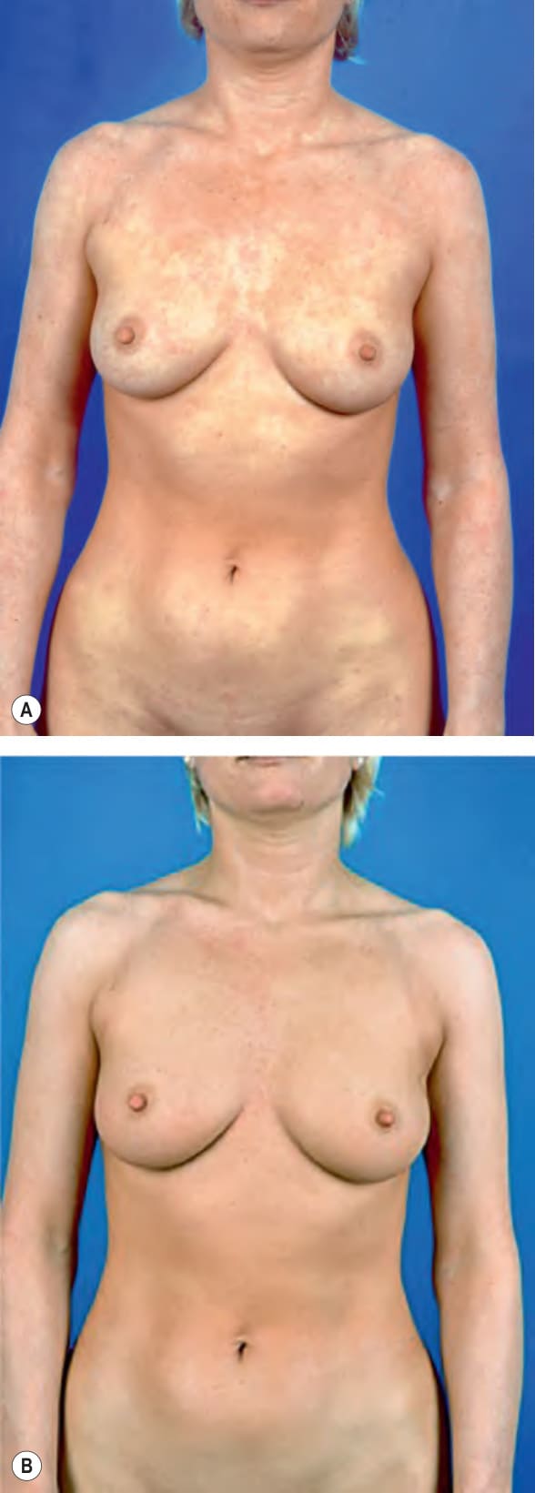
Fig. 134.8 UVA1 phototherapy of atopic dermatitis (splitbody comparison).
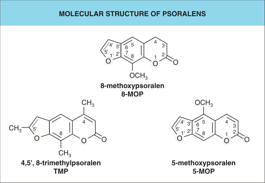
Fig. 134.9 Molecular structure of psoralens.
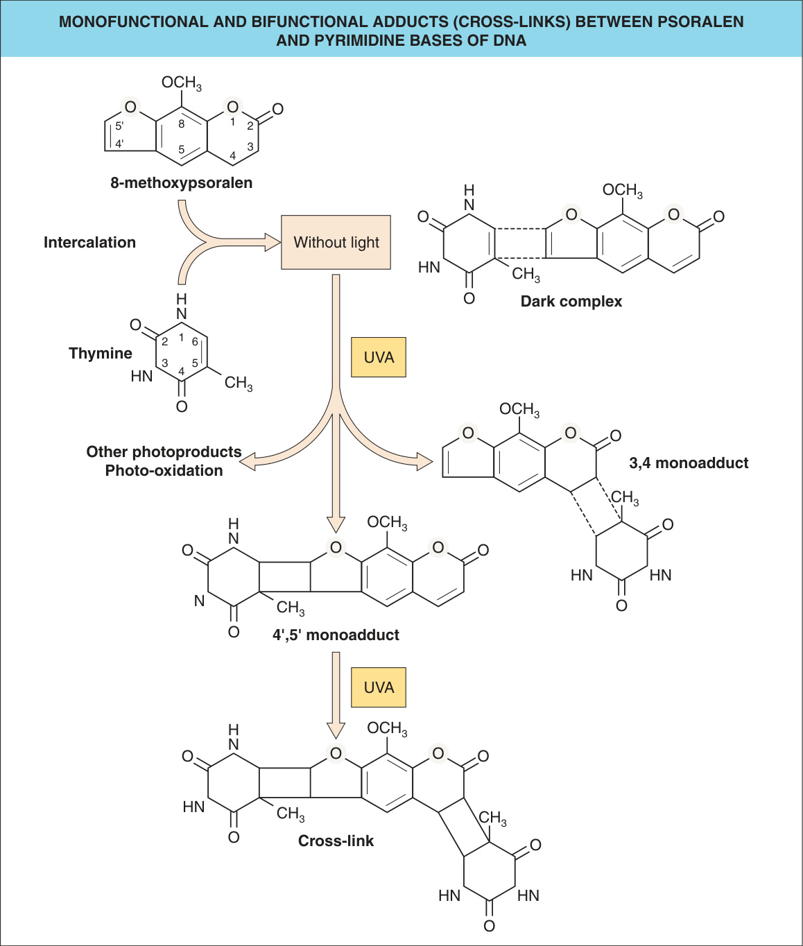
Fig. 134.10 Monofunctional and bifunctional adducts (cross-links) between psoralen and pyrimidine bases of DNA.
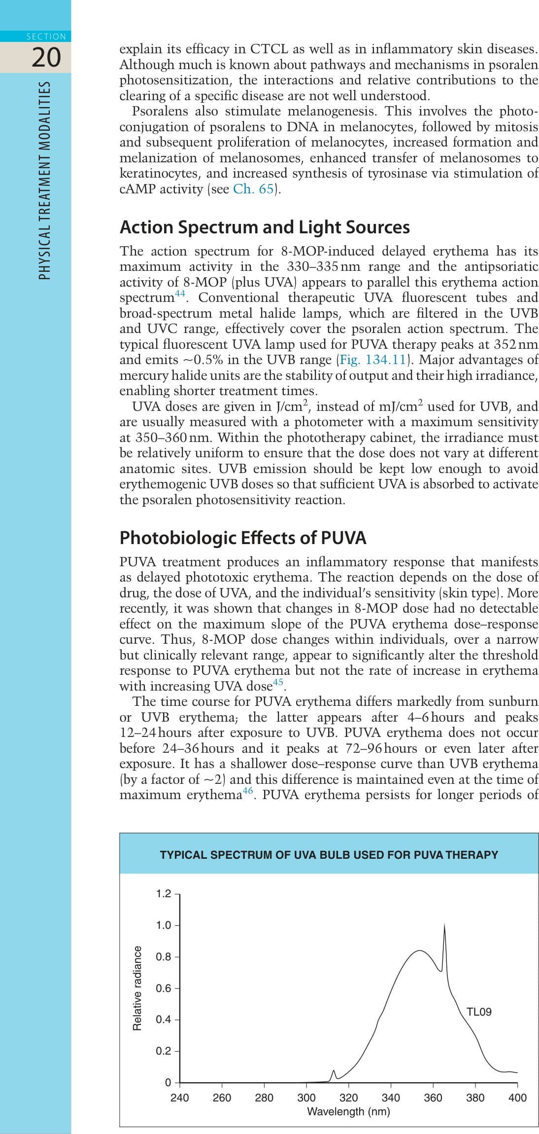
Fig. 134.11 Typical spectrum of a UVA bulb (Philips TL09) used for PUVA therapy.
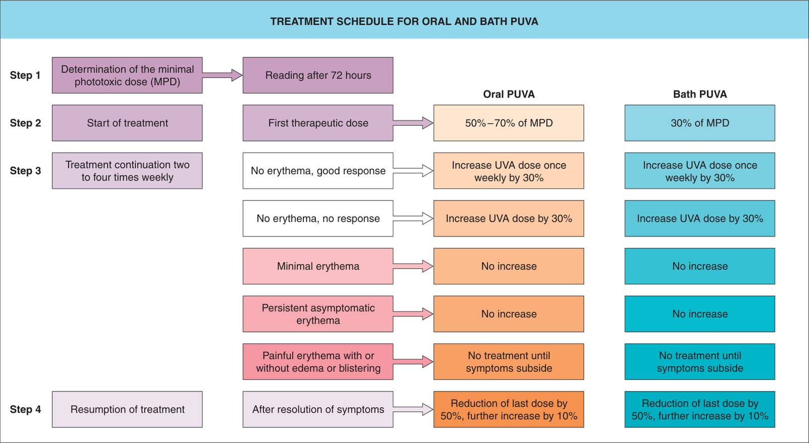
Fig. 134.12 Treatment schedule for oral and bath PUVA.
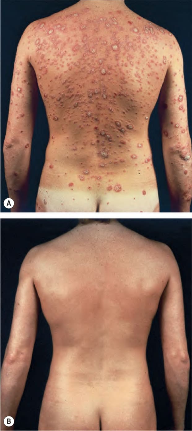
Fig. 134.13 PUVA treatment for psoriasis (four times weekly).A
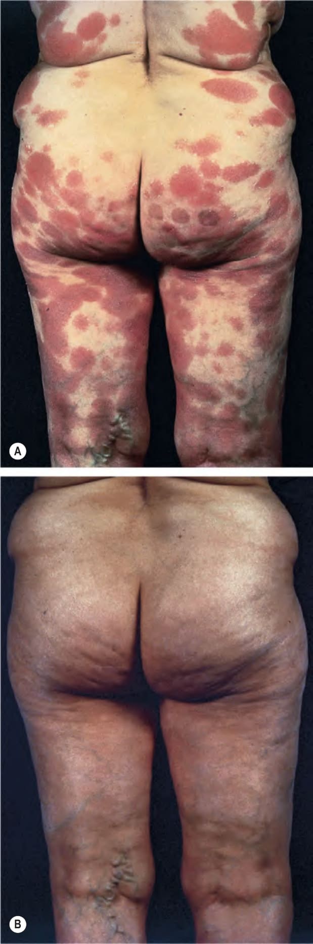
Fig. 134.14 PUVA treatment for cutaneous T cell lymphoma (mycosis fungoides).A
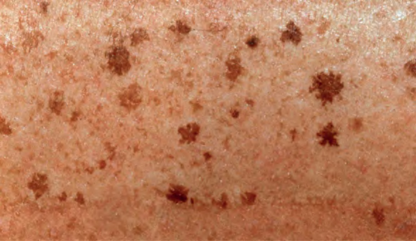
Fig. 134.15 PUVA lentigines. These develop after repeated PUVA treatments over several years and usually within a background of photodamaged skin.
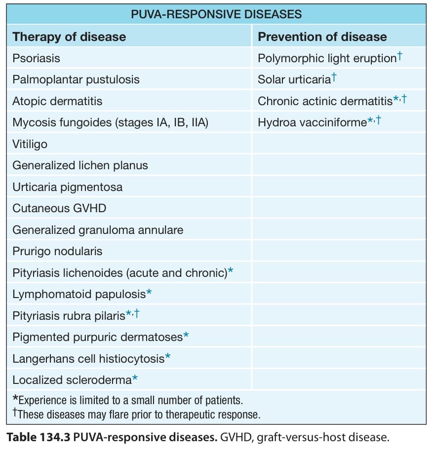
Table 134.3 PUVA-responsive diseases. GVHD, graft-versus-host disease.
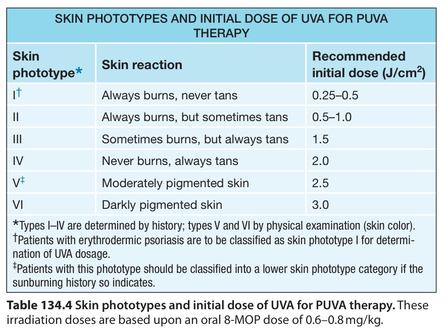
Table 134.4 Skin phototypes and initial dose of UVA for PUVA therapy. These irradiation doses are based upon an oral 8-MOP dose of 0.6–0.8 mg/kg.