DERMATOLOGIC APPLICATIONS
PDT agents can photosensitize neoplastic, inflamed, and vascular tissues, modulate immunologic processes, localize to the pilosebaceous unit, and induce lethal damage to microbes. To date, good-quality clinical evidence outside of cutaneous oncology is limited; for the most part, non-oncologic uses of PDT are off-label or investigational. Dermatologic applications for PDT are summarized in Table 135.4.
Oncologic
PDT is an accepted and approved treatment for AKs. There are also multiple controlled trials on the use of topical PDT for superficial carcinomas such as superficial BCC and Bowen disease (squamous cell carcinoma in situ) with licensed approvals varying according to country. PDT is not indicated for cutaneous melanoma.
Based on biophysical considerations alone, AKs can be treated with either blue or red light, whereas red light should be used for super-ficial keratinocyte carcinomas because of its deeper tissue penetration. Multiple studies have documented that cosmesis following PDT can be superior to that following conventional therapies. PDT may be particularly practical and well suited for patients with multiple synchronous AKs or larger, superficial keratinocyte carcinomas, as well as those with evidence of field cancerization. A single PDT session may be sufficient for AKs, with retreatment if required, whereas at least two separate PDT sessions are usually recommended for the treatment of superficial BCC and Bowen disease.
Actinic keratoses
Clinical trials have demonstrated that up to 90% of face and scalp AKs can be cleared with topical PDT (Fig. 135.5), with a one-year sustained clearance of ~80%. ALA and mALA appear to have similar clearance rates. The PDT clearance rate is superior when compared to cryotherapy, 50% trichloroacetic acid, topical diclofenac, and CO laser, but less effective than topical 5% 5-fluorouracil; it is comparable to topical imiquimod for AKs on the head. PDT combined with imiquimod is more effective than either treatment alone, and fractionation of light exposures is associated with improved outcomes.
On acral sites, cryotherapy may perform better than PDT (88% vs 78% clearance in one study), which in turn may clear more lesions than imiquimod (58% vs 37%). Patients also report higher satisfaction with PDT than with imiquimod. Resistance to PDT may be explained by the hyperkeratotic nature of AKs in acral sites, thus making them more difficult to eradicate due to drug and light penetration issues. Consequently, prior to PDT, gentle curettage debridement of scale is often recommended. Several other methods for lesion preparation to enhance PDT efficacy have been described (see Table 135.3). For hypertrophic AKs, multiple treatment sessions may be required to achieve a satisfactory overall result. PDT can also be used in combination or sequentially with other topical treatments for AKs.
In solid organ transplant recipients, the response rate to PDT appears to be lower than in immunocompetent patients (48% vs 72%). This supports a role for immune activation as part of the mechanism of action of PDT. That said, possibly due to its major effect being direct cytotoxicity, PDT has been shown to be superior to topical 5-fluorouracil or imiquimod for clearance of AKs in transplant recipients.
Daylight treatment of AKs has been studied extensively. In randomized controlled trials, mALA D-PDT for AKs was as effective, less painful, and scored higher for patient satisfaction compared to red LED-activated mALA PDT. In general, two hours of daylight exposure are sufficient to deliver an effective light dose throughout the year at latitudes <45° (see above regarding higher latitudes). As expected, mALA D-PDT clears thin AKs more readily than hypertrophic AKs. D-PDT with either mALA or ALA nanoemulsion gel have been used successfully to treat AKs.
Actinic cheilitis represents a useful potential PDT application since alternative treatment approaches such as surgical vermilionectomy or laser vaporization are more complicated and have associated morbidities.
Basal cell carcinomas (BCCs)
Superficial BCCs respond well to PDT, with an expected primary clearance rate of 92%–97% at 12 weeks and 9% recurrence rate at 12 months (Fig. 135.6). Although less effective than surgery, PDT has the advantage of improved cosmetic outcome over both surgery and cryotherapy. Longer term follow-up with a two-session protocol found that the 5-year response rate for imiquimod (80%) was superior to either 5-fluorouracil (70%) or PDT (63%) for clearance of superficial BCCs. More than one session of PDT is recommended to treat a given BCC and two separate illuminations per treatment session (fractionation) increases the long-term clearance rate.
Nodular BCCs do not appear to be as well suited to PDT in that lower and variable response rates of 35%–90% have been observed. This type of BCC is best treated surgically when possible. Similarly, difficultto-treat BCCs (e.g. high risk areas of the face, recurrent, sclerosing type) are probably best treated by Mohs micrographic surgery because of the importance of achieving deep margin control. Multiple superficial BCCs arising in the context of cancer-prone genodermatoses such as basal cell nevus syndrome are a good indication for PDT since multiple lesions can be treated during one session and multiple surgeries thereby minimized.
D-PDT has also been studied for the treatment of superficial and nodular BCCs, with ~75% of lesions treated with two sessions having a complete response at 12 months. Because daylight activation of photosensitizers may be due predominantly to the superficially penetrating blue wavelengths within natural outdoor light, it is not yet known if daylight is a sufficiently effective light source for treating BCCs.
Squamous cell carcinomas (SCCs)
Bowen disease is confined, by definition, to atypical keratinocytes that span the entire epidermis, and is thus eminently within the therapeutic range of PDT. Many PDT experts consider Bowen disease to be an ideal application since PDT can be used to treat multiple lesions as well as large, extensive plaques. A PDT clearance rate for Bowen disease of ~90% can be achieved with a 2-year sustained clearance rate of ~70%. For clearance of Bowen disease, PDT outperforms topical 5-fluorouracil, imiquimod, and cryotherapy. Bowen disease of the anogenital region and lower extremity can also be an appropriate target for PDT. In the case of lower extremity Bowen disease, a single session of ablative fractional laser-assisted PDT demonstrated superior long-term efficacy compared to two standard mALA sessions, with a 5-year clearance rate of 85%. In this setting, the fractional laser is not used for photosensitizer activation, but rather for treatment site preparation prior to topical drug application (see Ch. 136). PDT is not recommended for recurrent Bowen disease or microinvasive or invasive SCC.
PDT photochemoprevention of AKs and keratinocyte
PDT is an effective intervention for managing field cancerization beyond just clearing visible AKs. There is increasing evidence that skin sites treated with topical PDT may develop new lesions at a slower rate, raising the possibility of a photodynamic prevention effect. Photochemoprevention with PDT may prove especially beneficial in reducing the incidence of AKs and SCCs in solid organ transplant recipients, particularly when periodic “maintenance” PDT is performed.
Extramammary Paget disease
Treatment of in situ extramammary Paget disease is complicated given the difficult anatomic locations of these lesions, the high recurrence rate due to unpredictable subclinical and adnexal extension, and significant morbidity. Although PDT is a potential treatment that would allow anatomic preservation, variable success has been reported; the absence of conclusive evidence precludes its recommendation as first-line therapy.
Other neoplasms
Because activated and malignant T cells can be selectively sensitized by exogenous ALA, PDT has been used to treat the mycosis fungoides form of cutaneous T cell lymphoma. Patch- or plaque-stage lesions may be more responsive than tumor-stage mycosis fungoides. While PDT’s historical roots in dermatology lie in its initial use during the 1970s to treat intracutaneous metastases, it is now widely accepted that PDT is far more appropriate for treating superficial carcinomas rather than advanced disease.
Non-Oncologic PDT Applications
Acneiform disorders
Not only did early studies demonstrate that exogenous ALA could induce PpIX formation within the skin, they also noted the preferential accumulation of active photosensitizer and selective phototoxicity within the pilosebaceous unit. In addition, singlet oxygen exerts antibacterial and anti-inflammatory effects. These effects, in addition to antibiotic resistance and the challenges surrounding the use of oral isotretinoin, have provided an impetus to developing PDT regimens for acne. A reduction in inflammatory acne lesions of 60%–70% was reported with both ALA and mALA PDT, with the two photosensitizers producing similar effects. Use of higher red light fluences and longer incubation times are associated with longer remission, but also with more pain and local reactions (Fig. 135.7). Currently, there is no consensus as to the optimum PDT parameters for treating acne.
It should be noted that Cutibacterium (formerly Propionibacterium) acnes produces cutaneous porphyrins, which can be readily demonstrated with Wood’s lamp examination of any sebaceous-dense area, including normal skin. Exposure to high-intensity blue light alone,
in the absence of any exogenous PDT prodrug, can lead to clinical improvement of inflammatory acne lesions.
PDT has also been used to treat other acneiform disorders. It was superior to topical 1% clindamycin for the treatment of perioral dermatitis in a small split-face trial. Rosacea and hidradenitis suppurativa represent other diseases for which PDT was reported as being useful. In patients with rosacea, ALA-PDT reduced papulopustular lesions as well as flushing, pruritus, burning, and prickling sensations.
Photoaging
When used to treat AKs and keratinocyte carcinomas, PDT is thought to produce cosmetically favorable results, including improvement of sun-damaged skin. Well-designed trials directly comparing PDT to non-PDT treatments in which photoaging is the primary focus are sparse relative to the extent that this treatment approach is promoted and used in clinical practice (see Fig. 135.5). A randomized trial of repetitive D-PDT to prevent AKs found a significant reduction in fine lines, pigmentation, skin roughness, and erythema. Optimal PDT parameters for treatment of photoaging have not yet been determined. Histologically, PDT appears to increase collagen deposition and decrease elastosis.
Cutaneous infections
There are several mechanisms by which PDT can treat infectious diseases. Singlet oxygen may directly destroy microbes or it may activate host immunologic reactions against the microorganisms. In addition, PDT can induce necrosis of infected tissue, for example, in the treatment of verrucae or condylomata. Conditions that have been shown, in controlled trials, to improve with PDT include verrucae vulgares, condylomata acuminata, and leishmaniasis. The emergence of drug-resistant bacteria plus the need for better antibiotic stewardship have also motivated investigations into employing PDT for bacterial skin infections and colonized chronic leg ulcers. In addition, PDT has been used on superficial fungal infections such as tinea pedis and onychomycosis.
Vascular anomalies
Systemic photosensitizers are biodistributed via the peripheral circulation and it stands to reason that intravascular drug activation with light would be useful in inducing the involution of abnormal vascular structures. Vascular malformations such as port-wine birthmarks have been shown to respond to systemic PDT with results that appear to be at least equivalent to those achieved with conventional pulsed dye laser therapy. Experience with this technique is largely from China, where hematoporphyrin-type preparations are used systemically. Topical ALA/pulsed dye laser-activated PDT has also been described, taking advantage of combined photodynamic and photothermal effects.
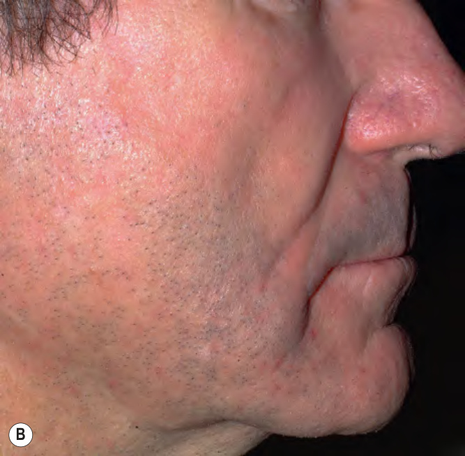
Fig. 135.5 Photodynamic therapy (PDT) of facial actinic keratoses and photoaging.A Pretreatment. B Following 3 monthly PDT sessions utilizing topical methyl aminolevulinate (mALA), clearing of actinic keratoses and a smoother, more even skin surface texture and color. Courtesy Luis Torezan, MD.
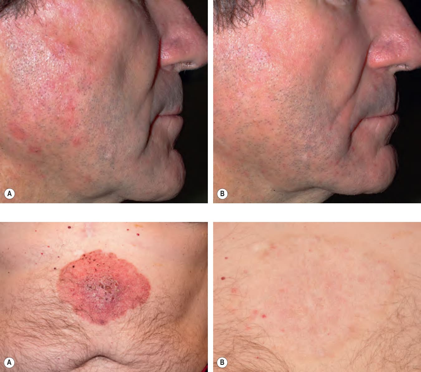
Fig. 135.6 Photodynamic therapy (PDT) of a large superficial basal cell carcinoma (BCC).A Superficial BCC measuring 11.5 by 9 cm on the upper abdomen. B Six months following 6 sessions of PDT using topical methyl aminolevulinate (mALA) followed by irradiation with red light from an LED (light-emitting diode), there is complete clinical clearing (closer view).
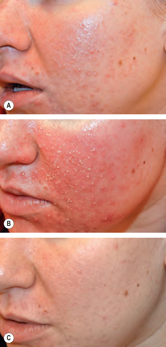
Fig. 135.7 Use of photodynamic therapy (PDT) for moderate but recalcitrant acne vulgaris. This patient declined the use of oral isotretinoin and had failed therapy with topical agents and oral antibiotics. A Baseline. B Three days after PDT utilizing topical 20% 5-aminolevulinic acid (ALA; 3 hours incubation) followed by irradiation with red light from an LED (light-emitting diode; 630 nm; 37 J/cm; 70 mW/cm); intense inflammation and pustulation is limited to treated sites. C Two months after single treatment session.
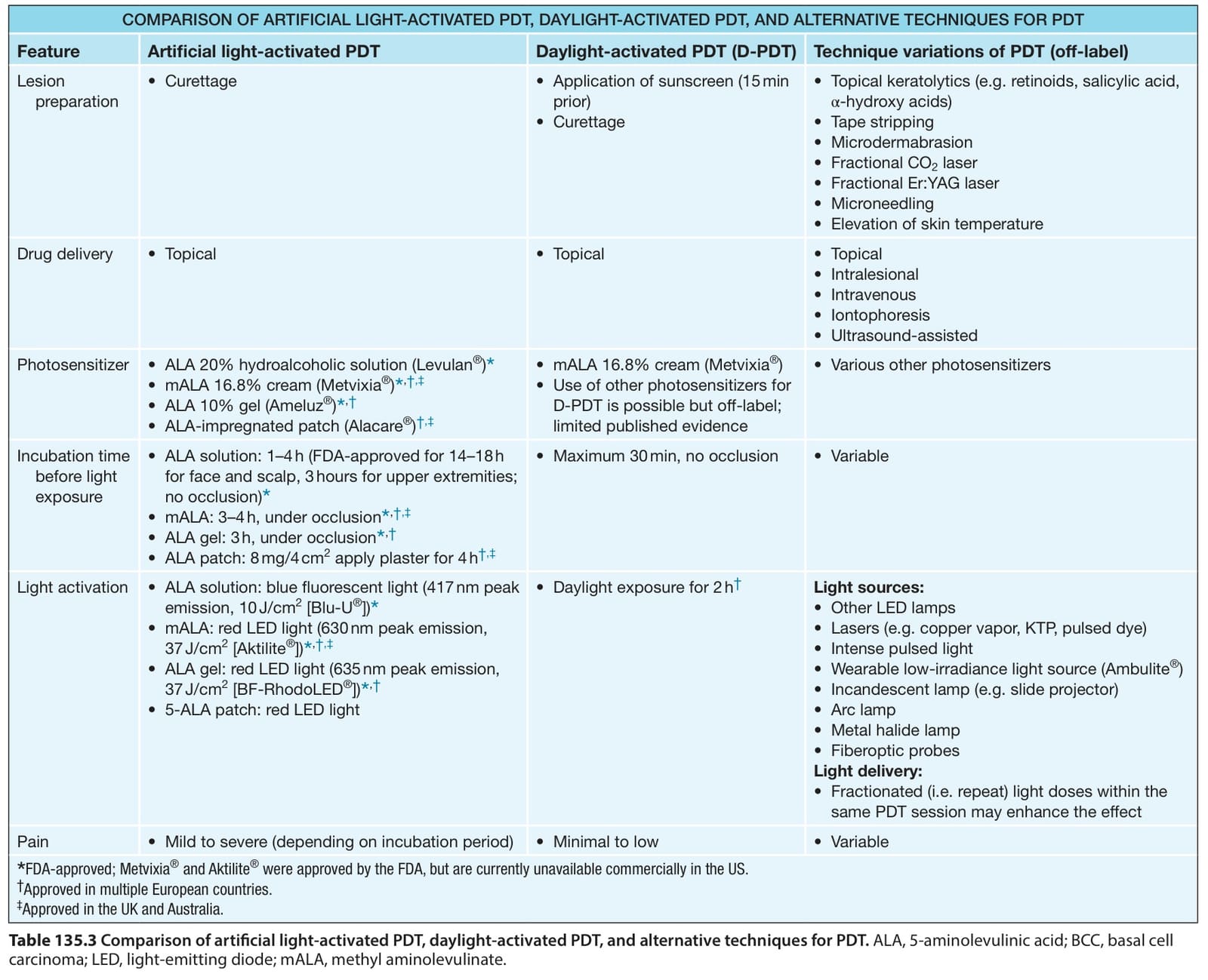
Table 135.3 Comparison of artificial light-activated PDT, daylight-activated PDT, and alternative techniques for PDT. ALA, 5-aminolevulinic acid; BCC, basal cell carcinoma; LED, light-emitting diode; mALA, methyl aminolevulinate.
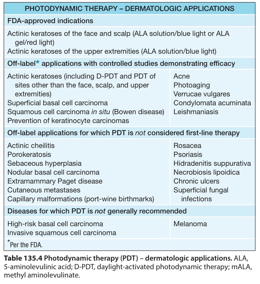
Table 135.4 Photodynamic therapy (PDT) – dermatologic applications. ALA, 5-aminolevulinic acid; D-PDT, daylight-activated photodynamic therapy; mALA, methyl aminolevulinate.