INSTRUMENTS AND TECHNIQUES
Cryosurgical techniques can be subdivided into several types (Table 138.2):
●Open (spray, open-spray; Fig. 138.1A). This is the most frequently used technique. The instrument consists of a metal container with spraying tips or openings through which the cryogen is released. This technique is suitable for lesions that are flat or elevated as well as benign or malignant; the key factor is achieving the correct freezing temperature. Larger openings deliver more LN and freeze tissue faster, so they are ideal for larger, bulky lesions. Smaller openings freeze tissue slower and allow for a more precise freezing. Therefore, they are ideal for small benign lesions, such as lentigines or seborrheic keratoses.
●Semi-open (confined spray, cone, confined; Fig. 138.1B). This refers to the use of a polycarbonate plate with holes, neoprene cones, or otoscope specula to restrict sprayed LN to a limited area. All of these items are composed of non-conducting materials. Freezing is faster than with the open technique as the LN is confined to the lesion. As splattering of LN is avoided and normal surrounding tissue is spared, it tends to be less painful.
●Semi-closed (chamber; Fig. 138.1C). One end of a metal cone is attached to the cryogen unit while the rubber-protected distal end is held firmly against the skin. This system generates potent and deep freezing and should be reserved for skin cancers and cutaneous metastases.
●Closed (probe, contact, cryoprobe; Fig. 138.1D,E). The cryogen is delivered via a closed system, i.e. from the unit through metal probes in contact with the skin; the remaining cryogen exits by way of a rubber releasing hose. Probes come in different shapes and diameters. To ensure homogeneous freezing, they need to be applied to flat tumor surfaces. In addition, when treating hemangiomas, the probe should be applied with pressure in order to “press out” the blood and lower the final temperature. For delicate procedures, such as freezing cysts near the eye, ultrasound gel or water can be placed on the skin under the probe.
●Tweezers (Fig. 138.1F). A previously frozen Teflon® forceps is used to grasp pedunculated lesions. The freezing front is then allowed to advance just to the skin surface. This is an ideal technique for filiform lesions, with sparing of normal surrounding skin as well as minimization of post-treatment hypo- or hyperpigmentation.
●Intralesional (Fig. 138.1G). While originally developed for keloids, this technique consists of passing a thick needle through the tumor. One end of the needle is attached to the cryogen unit (screwed on or via a Luer lock), while the other end that has exited the skin allows for LN release. It can be used for large nodular tumors where freezing originating from the center of the mass provides an advantage. It is ideal for preserving surrounding tissue.
●Dipstick (Fig. 138.1H). A LN-saturated, cotton-tipped applicator is placed directly onto the lesion. This technique is still used by some physicians for verrucae and solar lentigines.
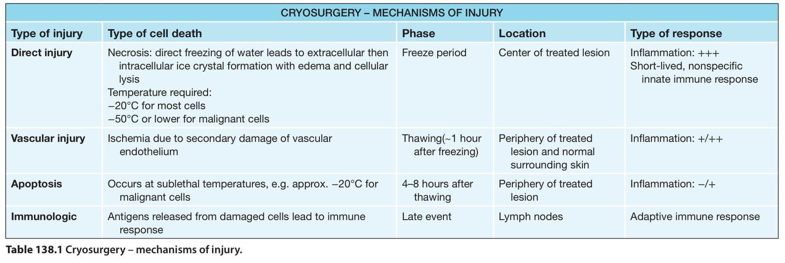
Table 138.1 Cryosurgery – mechanisms of injury.
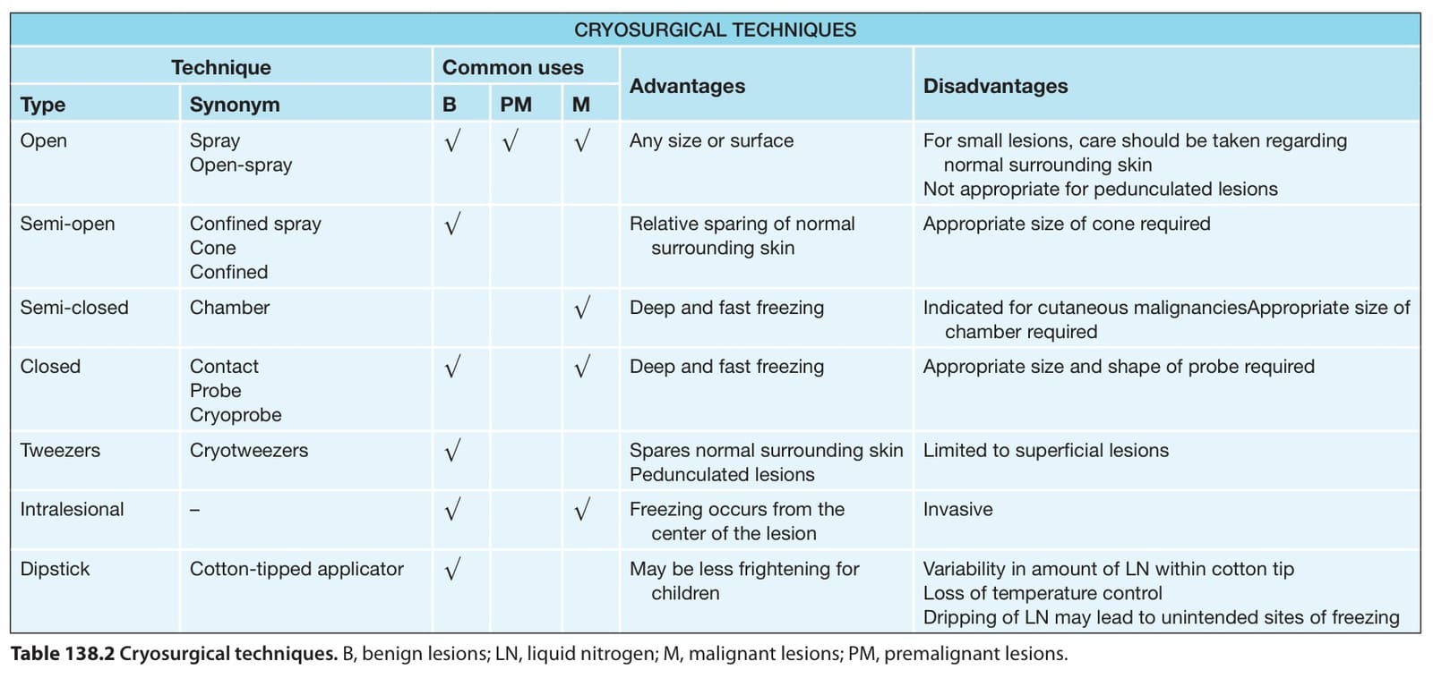
Table 138.2 Cryosurgical techniques. B, benign lesions; LN, liquid nitrogen; M, malignant lesions; PM, premalignant lesions.