TREATMENT
Cryosurgery should not be performed without first establishing the correct diagnosis via clinical examination, dermoscopy, and/or histologic examination. The appropriate technique is then chosen (see Table 138.2).
Benign Lesions
Table 138.4 outlines benign conditions that are amenable to cryosurgical management. For some, such as verrucae, it is a first-line option. The open (spray) technique is used for most benign lesions, including verrucae and acne cysts, while the semi-open technique or cryotweezers may be preferred for filiform warts and acrochordons (Video 138.1). The closed (contact, probe) technique can be employed for small adnexal cysts, pseudocysts (e.g. mucoceles), and sebaceous hyperplasia as well as for vascular lesions (e.g. hemangiomas, spider telangiectasias, pyogenic granulomas) (Fig. 138.3). A single freeze–thaw cycle is usually sufficient for benign lesions and cryogens other than LN can be used (see above). Whenever using the spray technique, the use of small diameter apertures and intermittent spraying leads to a more delicate spray and less damage to normal surrounding skin.
For verrucae, removal of excess keratin and moistening of the lesion can improve the effectiveness of cryosurgery (see above). When removing verrucae and seborrheic keratoses, the freezing front should extend a few millimeters beyond the clinical margin to avoid a doughnut wart or residual lesion. If the closed or contact technique is used to treat hemangiomas and other vascular lesions, it is best to prefreeze the probe to prevent it from sticking to the skin surface and to press firmly
(see Fig. 138.3). These recommendations are reflected in the PPPP rule: probe • pressure • prefreezing • patience. If the probe does become frozen to the skin lesion, simply allow for thawing before removing the probe. This approach is also recommended for mucosal lesions. Mucoceles may be drained prior to treatment with the closed technique.
Lentigines are lightened with a brief 3- to 4-second freeze and either the open spray or the dipstick technique can be employed. For keloids, the open spray technique – either alone or immediately prior to corticosteroid injection – or the intralesional technique is preferred. In darker skin phototypes, the anticipated cryosurgical technique may initially be performed in a test site that is in a cosmetically insensitive (“hidden”) location. The site is then examined for hypo- or hyperpigmentation prior to cryosurgery in other locations.
Premalignant Lesions, Including Actinic Keratoses
Cryosurgery is a very effective therapeutic option for AKs and other premalignant conditions (Fig. 138.4). One 20- to 40-second freeze– thaw cycle is usually sufficient for a 1 cm lesion. The surface temperature reaches −15° to −20°C and, once frozen, the lesion can be grasped between two fingers (Fig. 138.5). Initial freezing should be in the center of the AK, with the freezing front then spreading laterally ~2–3 mm
beyond the clinical margin. Some clinicians treat each lesion with two 15-sec freeze–thaw cycles. Recurrences should be assessed to determine if a squamous cell carcinoma needs to be excluded.
Within areas of significant cutaneous photodamage, there are rarely single AKs but rather multiple AKs as well as subclinical epidermal dysplasia. This is often referred to as “field cancerization” and is best treated by a combination of cryosurgery plus a topical agent (e.g. 5-fluorouracil, imiquimod) or photodynamic therapy. The latter are used to treat subclinical disease and thin lesions while cryosurgery is used to treat thicker keratotic AKs. It is more logical to first treat the field and then use cryosurgery for residual lesions.
Cryopeeling refers to the treatment of large areas of photodamaged skin, i.e. field treatment via cryosurgery. The areas of involvement are superficially frozen for 5–10 seconds, and given the associated pain, it is best to apply a topical anesthetic before and immediately after the procedure (see Ch. 143). Individual thicker lesions can be treated at the same time with a longer freeze time (see above). The freeze time for actinic cheilitis ranges from 10 to 20 seconds.
Cutaneous Malignancies
Cryosurgery is a reasonable alternative for the treatment of skin cancer (Table 138.5), but the lesions and techniques need to be carefully selected. Because a double freeze–thaw cycle is more destructive, it is the recommended cryosurgical procedure for skin cancer. Except for very small lesions, local anesthesia is administered. In addition, since keratin is a poor conductor of cold, removal of excess keratin via curettage or paring will assist in reaching the correct freezing temperature at the base of the tumor, thus improving effectiveness.
The most commonly used techniques are open (spray) and closed (contact, probe); the closed technique is preferred as it leads to deeper and faster freezing (see Table 138.2). However, it is important to use a probe that corresponds to the size and shape of the tumor to assure complete apposition. The presence of air pockets (“interphase”) between the probe and the tumor surface can prevent achievement of the desired final temperature. If the open (spray) technique is chosen, the freezing
should begin in the center of the tumor followed by lateral spread of the freeze front to 5 mm beyond the clinical margin of the tumor. A similar margin should be obtained with the closed technique.
Because cold advances as isotherms (see Fig. 138.1D,E), temperatures should be measured at the external margin. The temperature can be monitored by measuring (via an infrared thermometer) the surface temperature at the external margin and thereby inferring the temperature at the base of the tumor. More recently, high frequency ultrasound (HFUS; ~22 MHz) has been employed to determine the width, depth, and shape of tumors, thereby providing very useful information for selecting the optimal cryosurgical technique. For example, when basal cell carcinomas (BCCs) are thin, they can be safely treated with either the spray or the probe technique whereas for deeper nodular BCCs, the probe technique is preferred (Fig. 138.6; Video 138.2).
HFUS can also be used to monitor the freezing procedure. An initial black area is seen when the tumor is completely frozen. Re-appearance of the tumor is indicative of thawing, which first occurs at its surface and is last seen at the tumor base. By combining this information with measurement of the temperature at the outer edge of the frozen tumor, the temperature at the base of the tumor can be implied.
For patients with multiple BCCs, dermoscopic diagnosis and HFUS of each lesion can be followed by a double freeze–thaw cycle, all in one treatment session. BCCs in several sites, including the nose, ears, and eyelids, have been treated successfully with cryosurgery. Immunocryosurgery, consisting of a 5-week course of topical imiquimod with a double freeze–thaw cycle at week two, has also been utilized for skin cancer (Fig. 138.7). Lastly, melanocytes are known to be very sensitive to cold and cryosurgery is an option for selected cases of lentigo maligna (Fig. 138.8). However, it is important to ensure that melanocytes within the follicular epithelium are destroyed. Of note, cryosurgery has also been used, in combination with imiquimod, as treatment for locoregional cutaneous metastases of melanoma.
Palliative Treatment
Cryosurgery has long been used for palliative (compassionate) treatment of skin cancer (Table 138.6). Detailed discussion with the patient and/ or family/caretakers should be undertaken before instituting palliative treatment. The goal is to reduce pain, bleeding, infection, tissue destruction, foul smell, and/or tumoral masses that cause personal and social rejection. Palliative cryosurgery can improve quality of life in individuals with inoperable tumors or in those who reject conventional therapies or are poor surgical candidates due to underlying medical conditions.
Large tumoral masses can be treated by either: (1) the segmental technique; or (2) the fractional technique. In the segmental technique, the tumor is divided by imaginary lines into portions. Each segment is treated independently, allowing the freezing fronts to overlap. The aim is to avoid excessive freezing in the center of the tumor prior to the freeze front reaching the periphery. In the fractional technique, the center of the tumor is treated and then allowed to heal. As the central
A Prior to treatment. B Eight months later, following a 5-week course of topical imiquimod with a double freeze–thaw cycle at week two.
scar contracts, it reduces the tumor diameter and residual disease can then be treated. In some patients, repeated treatment of the central tumor is performed to further reduce tumor burden.
The open (spray) technique is most commonly employed, especially in tumors with an irregular surface or in patients with a bleeding diathesis in whom curettage should be avoided. The closed (probe) technique, although ideal for skin cancer treatment, requires probes whose size appropriately matches that of the tumor, and since these tumors are usually very large, proper probes can be difficult to obtain. The semi-closed (chamber) technique, which is very destructive, is ideal for cutaneous metastases as long as the tumor fits inside the chamber. For very large tumors, the intralesional technique is an option.
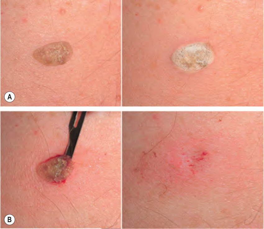
Fig. 138.2 Shave removal of a seborrheic keratosis following the use of liquid nitrogen as an anesthetic.A A seborrheic keratosis (SK) is frozen with a 2–3 mm margin. B Once the tissue starts thawing, a swift stroke with a curette or scalpel will make the SK “pop out”. A hemostatic solution (aluminum chloride) is applied before complete thawing.
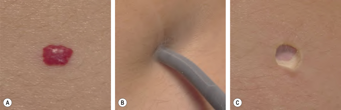
Fig. 138.3 Removal of a cherry angioma via the closed (probe, contact, cryoprobe) technique.A Prior to treatment. B A previously frozen probe is pressed into the lesion. C The frozen angioma and a transient depression can be seen.
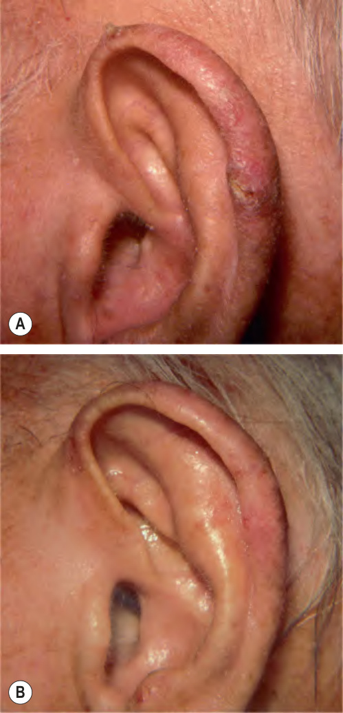
Fig. 138.4 Actinic keratosis treated with the open- spray technique.A Prior to treatment. B Three weeks later. Courtesy Emanuel G. Kuflik, MD.
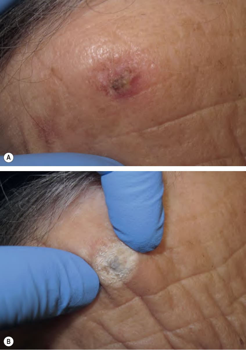
Fig. 138.5 Cryosurgery of a hypertrophic actinic keratosis.A Prior to treatment. B The open-spray technique should produce a block of frozen tissue that can be grabbed with both fingers; the surface temperature is approximately −15°C.
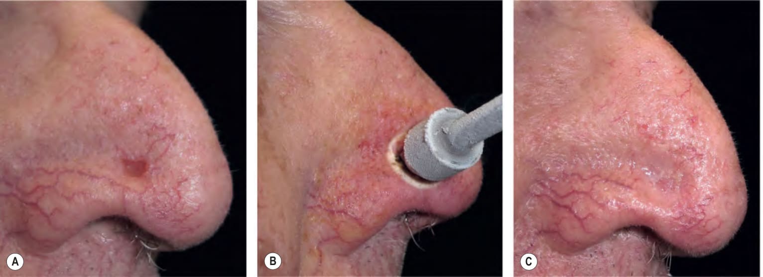
Fig. 138.6 Treatment of a basal cell carcinoma on the nose with the closed (probe, contact, cryoprobe) technique.A Prior to treatment. B An appropriately sized frozen cryoprobe is pressed against the tumor; a double freeze–thaw cycle is performed. C Healing is nearly complete a few weeks later.
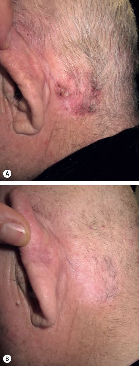
Fig. 138.7 Treatment of a large, ill-defined, postauricular basal cell carcinoma with immunocryosurgery.
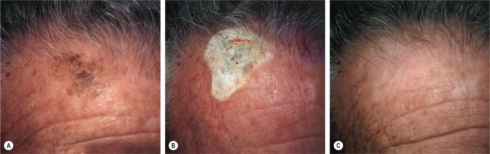
Fig. 138.8 A lentigo maligna on the forehead of a 72-year-old man.A The lesion measures 3.1 × 2.5 cm. B Cryosurgery to the entire lesion. Note the wide lateral spread of freeze. C The healed cryosurgical site 5 months after treatment. Courtesy Emanuel G. Kuflik, MD.
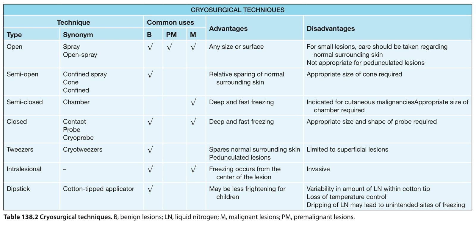
Table 138.2 Cryosurgical techniques. B, benign lesions; LN, liquid nitrogen; M, malignant lesions; PM, premalignant lesions.
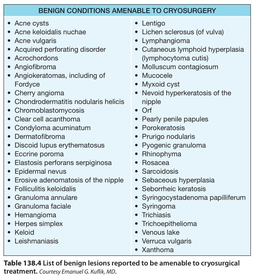
Table 138.4 List of benign lesions reported to be amenable to cryosurgical treatment.Courtesy Emanuel G. Kuflik, MD.
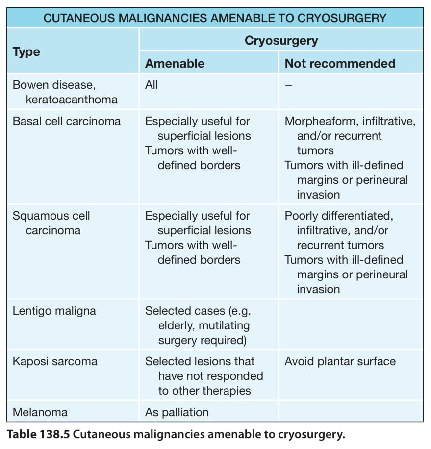
Table 138.5 Cutaneous malignancies amenable to cryosurgery.
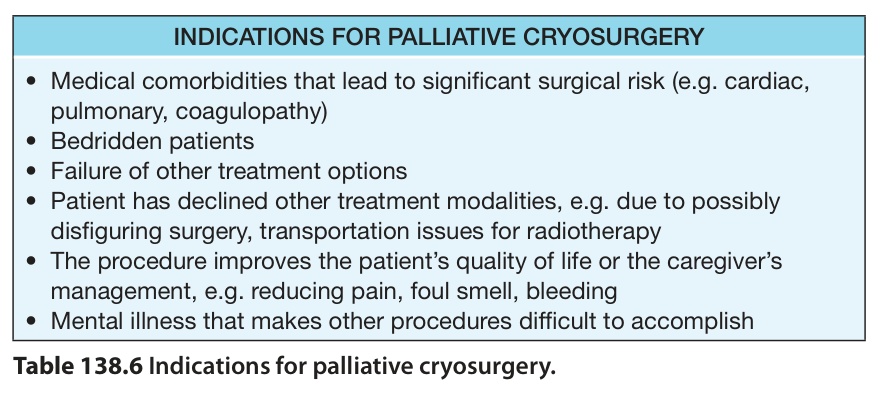
Table 138.6 Indications for palliative cryosurgery.