REACTIONS AND COMPLICATIONS
Acute Reactions (Acute Radiodermatitis)
Acute morbidity from skin-directed radiotherapy is usually minimal (in-field cutaneous erythema and desquamation) and limited to the irradiated area (Table 139.4). Systemic side effects such as fatigue, nausea, and vomiting do not occur.
Daily washing with water (± mild soap or soap-free cleansers) is recommended while receiving radiotherapy, including non-skindirected. Use of prophylactic topical corticosteroids (e.g. mometasone) may reduce the severity of acute radiodermatitis and improve comfort and pruritus. Given the limited time period of application, cutaneous atrophy is usually not an issue. No particular dressing has proven to be superior.
Most patients can expect to have local discomfort and moist desquamation by the conclusion of radiation therapy (Fig. 139.10), which then lasts for a few weeks. Hemorrhagic crusting can also develop and re-epithelialization usually occurs by week 3 or 4 following completion of radiotherapy. Delayed healing (beyond 6–8 weeks) is uncommon, as is associated soft tissue infection.
In-field radiation recall can occur following administration of antineoplastic drugs, including chemotherapeutic agents (e.g. anthracyclines, taxanes, antimetabolites; see Table 21.15), immunotherapies, and targeted therapies (e.g. inhibitors of EGFR, mTOR, and BRAF). Rarely, it develops following other medications or vaccines (e.g. SARS-CoV-2). Radiation enhancement characterized by higher grade, in-field skin
toxicity and mucositis can also occur, including with the concurrent use of epidermal growth factor receptor inhibitors (EGFRi). This may be complicated by secondary staphylococcal infections.
Late Reactions
The late effects of radiotherapy (e.g. telangiectasia, epidermal atrophy, dyspigmentation, fibrosis; see Table 139.4 & Fig. 139.10) often appear months to years later. They are more likely to appear with continued unprotected sun exposure, when a large dose per fraction (>3–4 Gy) was delivered, if the total dose was >55 Gy, and when large fields were irradiated. Chronic non-healing, radiation-induced ulcers represent poorly vascularized tissue that requires the importation of well-vascularized tissue to enable healing. The latter usually consists of local skin flaps or sometimes staged skin–muscle or axial pedicle flaps of non-irradiated tissue; grafts do not take well on irradiated tissue due to the poor vascular bed.
In a review of over 400 patients treated with radiotherapy, 92% were considered to have a good or excellent cosmetic result. However, 36% of those receiving >60 Gy in ≤2 Gy fractions had only a fair (16%) or poor (20%) cosmetic result. Smaller treatment fields (2–3 cm) tolerate hypofractionation better than do larger areas, but, even so, if cosmesis is an important consideration, larger fractions should be avoided. Patients should also be warned of the small risk (<5%) of late soft tissue and cartilage necrosis, which are related to larger tumor size and larger doses per fraction.
Younger Patients
When irradiating younger patients, better long-term cosmetic results are more likely if a low dose per fraction (2 Gy/fraction) is administered. A typical dose for a small BCC would be 50–54 Gy delivered in
25–27 daily fractions. However, even with this dose fractionation schedule, patients can still expect some degree of in-field hypopigmentation and telangiectasia; the latter can be addressed via pulsed dye laser treatment and camouflaging cosmetics (see Ch. 153). Other options, therefore, should be considered prior to recommending radiotherapy to younger patients. The risk of a radiotherapy-induced in-field malignancy arising decades after high-dose, therapeutic, skindirected radiotherapy is rare and poorly documented in the literature (see Table 139.4).
Cutaneous Diseases Induced by Radiation Therapy
Table 139.5 outlines the cutaneous disorders that are induced by radiotherapy, with some occurring outside the field of radiation as well as others limited to the site of irradiation.
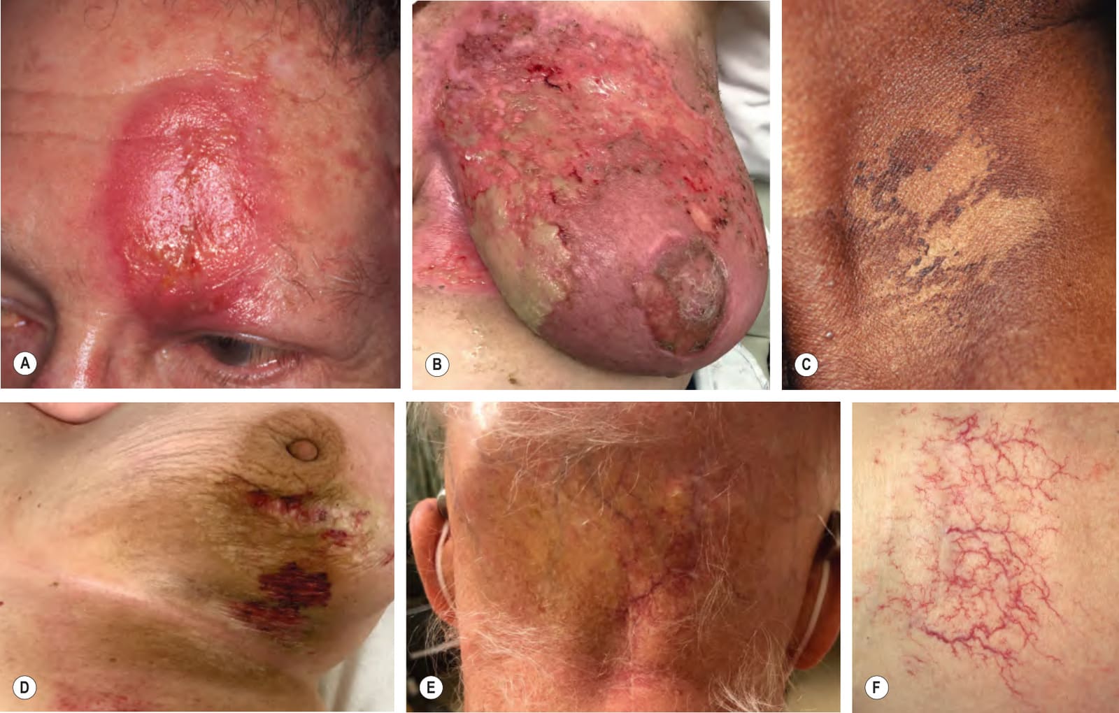
Fig. 139.10 Acute and chronic cutaneous side effects of radiation therapy.A Confluent moist desquamation and marked edema in a patient near completion of radiotherapy for a squamous cell carcinoma with neurotropism (grade 2 acute radiodermatitis). B Confluent moist desquamation and superficial ulcerations on the breast after three weeks of radiotherapy for breast cancer (grade 3). C Dry desquamation of the neck several weeks following radiation therapy for a pharyngeal squamous cell carcinoma (grade 1). D Development of morphea and lichen sclerosus within irradiated skin; note the purpura that accompanies lichen sclerosus. E Permanent scarring alopecia and telangiectasias of the occipital scalp after radiotherapy for anaplastic cerebellar astrocytoma. F In-field telangiectasias; note the square distribution pattern. A, F, Courtesy Jean L. Bolognia, MD; B, D, E, Courtesy Jonathan Leventhal, MD.
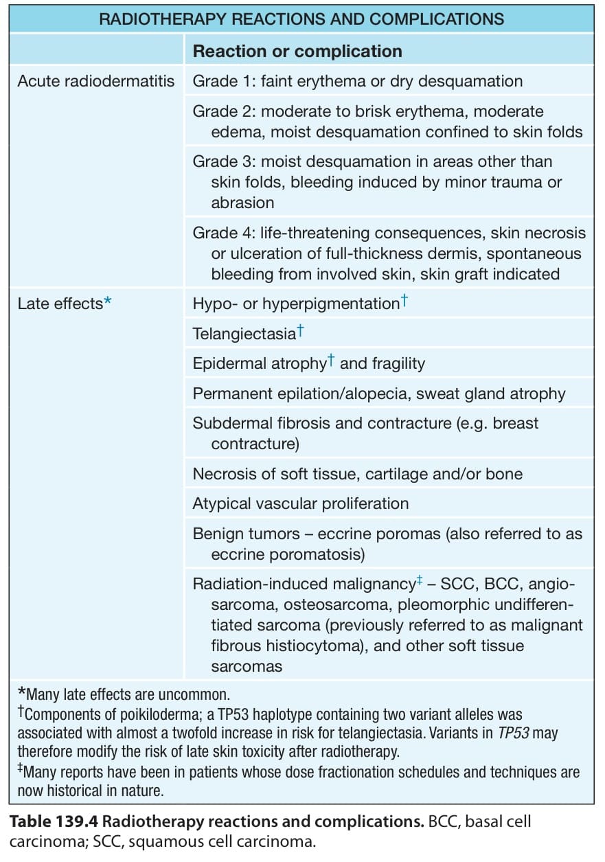
Table 139.4 Radiotherapy reactions and complications. BCC, basal cell carcinoma; SCC, squamous cell carcinoma.
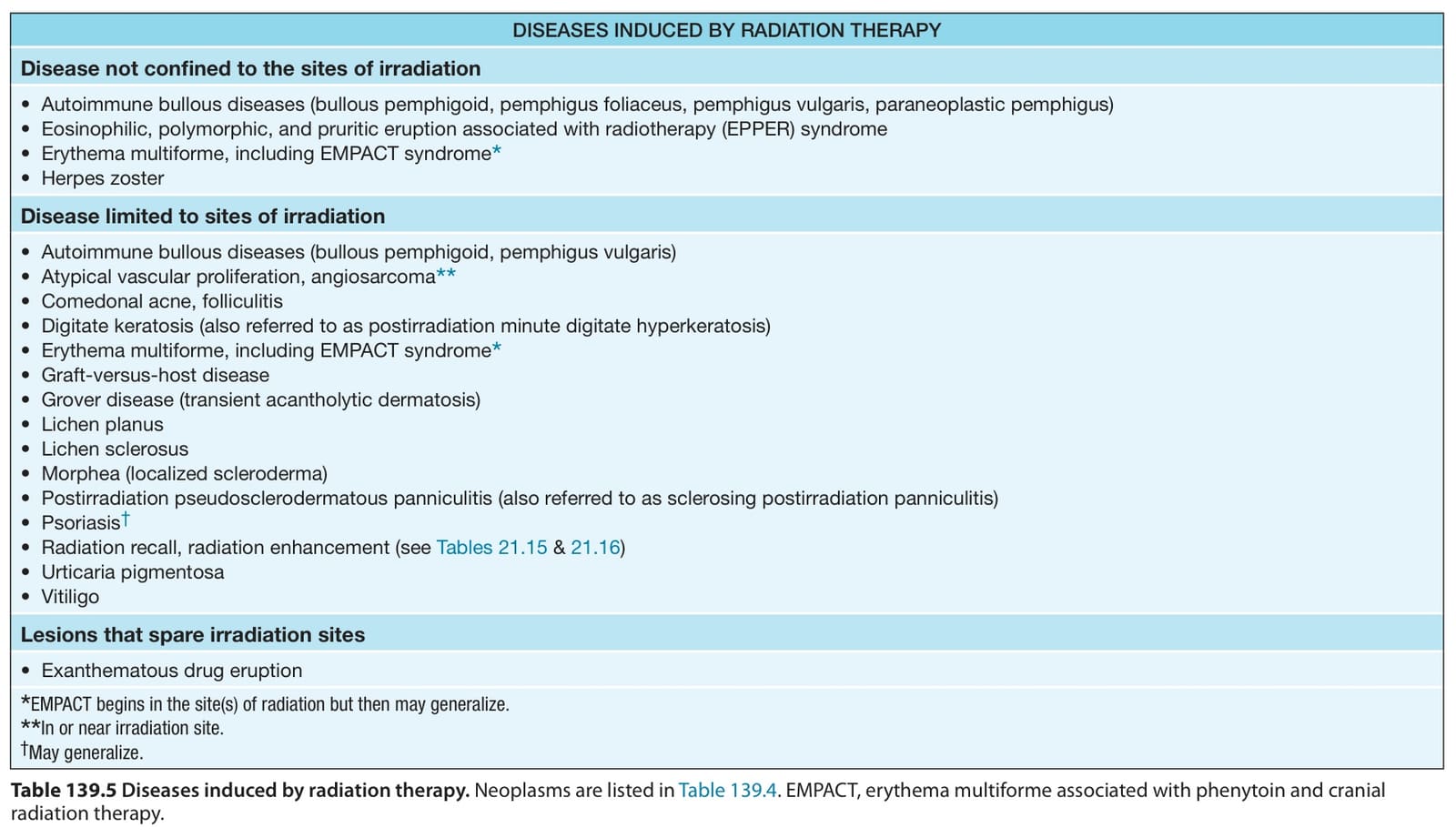
Table 139.5 Diseases induced by radiation therapy. Neoplasms are listed in Table 139.4. EMPACT, erythema multiforme associated with phenytoin and cranial radiation therapy.