SEBORRHEIC DERMATITIS
Key features
Infantile and adult forms
Lesions favor the scalp, ears, face, central chest, and intertriginous areas
Etiologic links with active sebaceous glands, abnormal sebum composition, and Malassezia (Pityrosporum) spp.
Can be a cutaneous sign of HIV infection
Introduction
Seborrheic dermatitis is a common, mild, chronic eczema typically confined to skin regions with high sebum production and the large body folds. Although its pathogenesis is not fully elucidated, there is a link to sebum overproduction (seborrhea), the commensal yeast Malassezia, and individual susceptibility.
History
Seborrheic dermatitis was first described by Unna, who also suspected Malassezia furfur (Pityrosporum ovale) as a causative factor. The nosologic position of seborrheic dermatitis was widely discussed for decades, the focus resting on dysfunction of the sebaceous glands and the high amounts of M. furfur present in scales of seborrheic dermatitis. In 1984 it was shown that seborrheic dermatitis could be suppressed by systemic ketoconazole. This finding was corroborated by later studies, and it became clear that seborrheic dermatitis was strongly linked to these yeasts. Today, it is accepted that several species of Malassezia, including M. furfur, play a direct role in seborrheic dermatitis (see Table 77.3).
Epidemiology
There are infantile and adult forms, with the former being self-limited and most prevalent the first 3–4 months of life, while the latter is chronic with a peak in the fourth to sixth decades. The prevalence of seborrheic dermatitis is estimated to be 5%, but its lifetime incidence is likely much higher. Men are afflicted more often than women. Extensive and therapy-resistant seborrheic dermatitis is an important cutaneous sign of HIV infection. It is more commonly observed in patients with neurologic diseases (e.g. Parkinson disease, cerebrovascular accidents) or who are immunosuppressed. Additional contributing factors include nutritional deficiencies, impaired epidermal barrier, and occasionally medications (e.g. neuroleptics, atypical antipsychotics). Seborrheic dermatitis-like eruptions have also been reported with dupilumab and antineoplastic agents (e.g. tyrosine kinase inhibitors).
Pathogenesis Malassezia
The genus Malassezia consists of lipophilic yeasts that are part of the normal resident skin flora. Studies on the predominance of certain Malassezia spp. have revealed conflicting results. M. furfur may behave as an opportunistic pathogen (e.g. fungemia in neonates receiving intravenous lipid emulsions) or cause or aggravate a spectrum of skin diseases: pityriasis versicolor, Malassezia (Pityrosporum) folliculitis, seborrheic dermatitis, and possibly atopic dermatitis.
M. furfur and related species can be regularly isolated from lesions of seborrheic dermatitis, including infantile seborrheic dermatitis (whereas healthy prepubertal children are not colonized to a significant degree). This coincides with the presence of enlarged sebaceous glands during the neonatal period. There is no simple quantitative relationship between yeast number and severity of seborrheic dermatitis, and unaffected skin may carry a load of organisms similar to seborrheic dermatitis lesions. Even in the scalp, where M. furfur predominates amongst the resident flora, only twice as many yeasts may be recovered from areas of seborrheic dermatitis as from normal controls. Amongst patients with AIDS, harboring of Malassezia spp. is equivalent, irrespective of the presence or absence of seborrheic dermatitis clinically; however, there is a difference in the amount of yeasts.
Active sebaceous glands and seborrheic dermatitis
Seborrheic dermatitis occurs predominantly in areas of the skin with active sebaceous glands and is often associated with sebum overproduction, which in turn can facilitate the growth of Malassezia. In infants, sebum is produced for a few weeks after birth, and the adult form of seborrheic dermatitis does not develop before puberty. This argues for a role for sebaceous gland activation by androgens. However, patients with seborrheic dermatitis may have normal sebum production and those with excessive sebum production are often free of seborrheic dermatitis. Thus, the amount of sebum produced alone does not appear to be the decisive risk factor.
It has been proposed that the composition of the skin surface lipids is the relevant factor. In patients with seborrheic dermatitis, triglycerides and cholesterol are elevated but squalene and free fatty acids are significantly decreased. Free fatty acids (which have a known antimicrobial effect) are formed from triglycerides by bacterial lipases produced by the lipolytic Cutibacterium (formerly Propionibacterium) acnes. A major constituent of the resident microbial skin flora, C. acnes has been found to be greatly reduced in seborrheic dermatitis. Seborrheic dermatitis may thus be linked to an imbalance of the microbial flora.
An etiologic role for immune mechanisms against Malassezia – particularly in view of the prominence of seborrheic dermatitis in HIV-infected individuals – has been suspected but never convincingly proven. Intrinsic host factors, such as changes in sebum production and a defective epidermal barrier, could favor overgrowth of Malassezia spp. which then incites a host inflammatory response. However, additional studies on cellular immunity have yielded contradictory results. The inflammation seen in seborrheic dermatitis might be irritant, caused by toxic metabolites, lipase, and reactive oxygen species.
Clinical Features
Seborrheic dermatitis is defined by clinical parameters, including:
●sharply demarcated patches or thin plaques that vary from pink– yellow to dull red to red–brown with bran-like to flaky “greasy” scales; vesiculation and crusting may occur but are rare and mostly due to irritation
●a predilection for areas rich in sebaceous glands – scalp, face, ears, presternal region – and, less often, the intertriginous areas
●a mild course with little or moderate discomfort. Seborrheic dermatitis is most often limited in extent, but generalized and even erythrodermic forms can occur, albeit rarely.
Infantile seborrheic dermatitis
This form usually begins about one week after birth and may persist for several months. Initially, mild greasy scales adherent to the vertex and anterior fontanelle regions arise which may later extend over the entire scalp. Inflammation and oozing may finally result in a coherent scaly and crusty mass covering most of the scalp (“cradle cap”; Fig. 13.1A). Lesions of the axillae, inguinal creases, neck, and retroauricular folds are often acutely inflamed, oozing, sharply demarcated, and surrounded by satellite lesions (Fig. 13.1B). Superinfection with Candida spp. or occasionally bacteria (e.g. group A Streptococcus) can occur. A disseminated
A Involvement of the scalp with thick adherent yellow scale overlying mild inflammation is often referred to as “cradle cap”. B Glistening red plaques of the neck, axillary and inguinal folds as well as the penis and umbilicus. Note disseminated lesions on the trunk and extremities. A, Courtesy Antonio Torello, MD; B, Courtesy Robert Hartman, MD.
eruption of scaly papules with a psoriasiform appearance (“psoriasiform id reaction”) may develop on the trunk, proximal extremities, and face in association with exuberant or superinfected seborrheic dermatitis, especially of the diaper area.
Adult seborrheic dermatitis
In adults, seborrheic dermatitis is generally found on the scalp and, usually of milder intensity, on the face; less often, lesions occur on the central upper chest and the intertriginous areas. Erythrodermic seborrheic dermatitis has been described as a rarity.
Pityriasis simplex capillitii (dandruff) is defined as a diffuse, slight to moderate, fine white or greasy scaling of the scalp and terminal hairbearing areas of the face (beard area), but without significant erythema or irritation. Scales accumulate visibly on dark clothing. This common condition may be considered the mildest form of seborrheic dermatitis of the scalp.
In seborrheic dermatitis of the scalp, there is inflammation and pruritus in addition to dandruff. The vertex and parietal regions are predominantly affected, but in a more diffuse pattern than the discrete plaques of psoriasis. Towards the forehead, the erythema and scaling are usually sharply demarcated from uninvolved skin, with the border either at the hairline or slightly transgressing beyond it. Pruritus is usually moderate but may be intense, particularly in patients with male pattern alopecia; folliculitis, furuncles, and meibomitis are not uncommon complications, elicited by scratching and rubbing.
Seborrheic dermatitis of the facial skin is often strikingly symmetric, affecting the forehead, medial portions of the eyebrows, upper eyelids, nasolabial folds and lateral aspects of the nose, retroauricular areas, and occasionally the occiput and neck (Fig. 13.2). Lesions are yellow-ish-red, with a typical bran-like scale. Non-purulent otitis externa is often observed. If present, lesions of the trunk are preferentially found in the presternal and intertriginous areas (Fig. 13.3); those on the central chest can have a petaloid appearance. Seborrheic dermatitis, like inverse psoriasis, is a cause of intertrigo (Fig. 13.4).
In patients with seborrheic dermatitis, the skin is sensitive to irritation, and exposure to sun or heat, febrile illnesses, and overly aggressive topical therapy may precipitate flares and dissemination. Rebound flares can also follow tapers of systemic corticosteroids. Seborrheic dermatitis is more common during the winter in patients with skin phototypes I–II, most likely a reflection of increased xerosis and decreased epidermal barrier function.
Irritated seborrheic dermatitis lesions can become bright red and erosive. Malassezia (Pityrosporum) folliculitis is another complication characterized by pruritic erythematous follicular papules, sometimes pustules, typically in sites rich in sebaceous glands.
Adult seborrheic dermatitis has a chronic relapsing course. Patients feel well and systemic signs are absent. Extensive and severe seborrheic dermatitis, however, should raise the suspicion of underlying HIV infection. Among patients with seborrheic dermatitis tested for HIV infection, 2% were found to be positive, frequently in a late stage of their disease. In patients with Parkinson disease, seborrheic dermatitis is a common finding, along with seborrhea. Its severity, however, is not correlated with that of the Parkinson disease. The facial immobility of patients with Parkinson disease might result in a greater accumulation of sebum on the skin, resulting in a permissive effect on the growth of Malassezia. Seborrheic dermatitis may also be more common in patients with other causes of immobility such as cerebrovascular accidents.
Pathology
Acute seborrheic dermatitis displays spongiosis with a superficial perivascular and perifollicular lymphocytic infiltrate composed mainly of lymphocytes. Older lesions show irregular acanthosis and focal parakeratosis. The latter may appear similar to psoriasis, but exocytosis of neutrophils, Munro microabscesses, and confluent parakeratotic horny layers are absent.
Differential Diagnosis
Infantile seborrheic dermatitis is distinguished from atopic dermatitis by its earlier onset, different distribution pattern, and, most importantly, by the absence of pruritus, irritability, and sleeplessness. In contrast to
atopic dermatitis, infants with seborrheic dermatitis generally feed well and are content. Irritant diaper dermatitis is confined to the diaper area and tends to spare the skin folds (see below). Candidiasis of the diaper area can result from colonization with fecal yeast and some infants have
seborrheic dermatitis with a superimposed candidal infection. The differential diagnosis also includes streptococcal intertrigo (see Fig. 13.12). Infantile psoriasis may be difficult to distinguish from psoriasiform seborrheic dermatitis. Although psoriasiform diaper dermatitis can represent the initial manifestation of psoriasis, many affected infants do not subsequently develop psoriasis elsewhere.
Rare conditions to be considered are Langerhans cell histiocytosis, “nutritional dermatitis” (e.g. acrodermatitis enteropathica; see Fig. 51.13), and Leiner disease. The last-mentioned is a questionable entity, once thought to be the maximal variant of infantile seborrheic dermatitis, but nowadays viewed as erythroderma in the setting of underlying immunosuppression (see Chs. 10 & 60).
When scalp scaling is present in prepubertal, especially Black, children, the possibility of tinea capitis due to Trichophyton tonsurans should be considered. In pityriasis amiantacea, thick asbestos-like scales adhere to tufts of scalp hairs; up to a third of the affected children and adolescents eventually develop psoriasis.
A number of entities are included in the differential diagnosis of adult seborrheic dermatitis. Distinction of seborrheic dermatitis of the scalp from psoriasis can be difficult, and there may be an overlap in some patients (“sebopsoriasis”). However, the plaques of psoriasis tend to be thicker, with silvery white scale, more discrete, less pruritic, and unassociated with seborrhea. In addition, features of psoriasis may be found elsewhere. Dry scaling of the scalp, along with dry brittle hair (as opposed to greasy hair), is a symptom of xerotic skin (e.g. in atopic dermatitis), frequently mistaken for (and mistreated as) seborrheic dermatitis. Mild erythema and scale of the posterior scalp, often with demonstrable hair loss, can be seen in dermatomyositis.
Seborrheic dermatitis of the face may closely resemble both early rosacea and the butterfly lesions of systemic lupus erythematosus. Lupus erythematosus rarely affects the nasolabial folds and often has a clearly demonstrable photodistribution. Notably, seborrheic dermatitis and rosacea frequently coexist. The facial dermatitis seen in patients receiving dupilumab may resemble seborrheic dermatitis. The differential diagnosis of seborrheic dermatitis of the trunk includes pityriasis rosea (but in this latter entity the lesions are ellipsoid in shape, have collarette-like scaling, and there is no predilection for the central chest) as well as superficial eruptive psoriasis and subacute cutaneous lupus erythematosus.
Seborrheic dermatitis of the intertriginous areas must be distinguished from inverse psoriasis, erythrasma, intertriginous dermatitis, candidiasis, and, rarely, Langerhans cell histiocytosis (see Fig. 13.4).
Treatment
Infantile seborrheic dermatitis
Infantile seborrheic dermatitis usually responds satisfactorily to bathing and application of emollients. Ketoconazole cream (2%) is indicated in more extensive or persistent cases. Short courses of low-potency topical corticosteroids may be used initially to suppress inflammation. Mild shampoos are recommended for the removal of scalp scales and crusts. Avoidance of irritation (e.g. the use of strong keratolytic shampoos or mechanical measures to remove the scales from the scalp) is important.
Adult seborrheic dermatitis
The mainstay of therapy is the use of topical azoles (e.g. ketoconazole), either as shampoos (scalp) or as creams (body). The high response rate (75%–90%) of this treatment has been documented in double-blind trials. Ciclopirox olamine has antifungal and anti-inflammatory activities and has also been shown to be effective as a shampoo or cream in double-blind, randomized trials. Seborrheic dermatitis tends to relapse
if a maintenance regimen is not instituted. As M. furfur has a slow proliferation rate, an interval of two to several weeks will pass until relapses appear. The intervals of topical therapy should follow this rhythm.
Additional measures, particularly in the initial stages of treatment, include emollients and low-potency topical corticosteroids; the latter were found to be equally efficacious as topical azoles in a Cochrane analysis. Second-line treatment options include zinc pyrithione, selenium sulfide, and tar shampoos as well as topical calcineurin inhibitors. Systemic antimycotics (e.g. weekly fluconazole) should only be used in cases of severe findings or frequent recurrences.
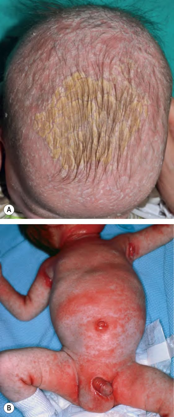
Fig. 13.1 Infantile seborrheic dermatitis.
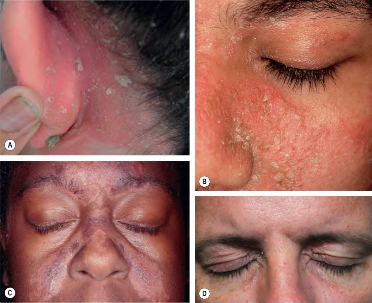
Fig. 13.2 Adult seborrheic dermatitis of the face, ear and scalp.A Rather sharply demarcated pink plaque with flaky white and greasy scale. Note the fissure in the retroauricular sulcus. B Sharply demarcated pink–orange thin plaques with yellow, greasy scale, especially in the nasolabial fold. When this degree of severity is seen, the possibility of underlying HIV infection needs to be considered. C Symmetric red–brown to violet plaques of the central forehead, nasal bridge, and medial cheeks with an associated hypopigmented peripheral and figurate rim. D Involvement of the upper eyelids, medial canthal regions, and infraorbital creases; seborrheic dermatitis confined to the major crease in the outer canthal region can also be seen. C, Courtesy Jeffrey P. Callen, MD; D, Courtesy Kalman Watsky, MD.
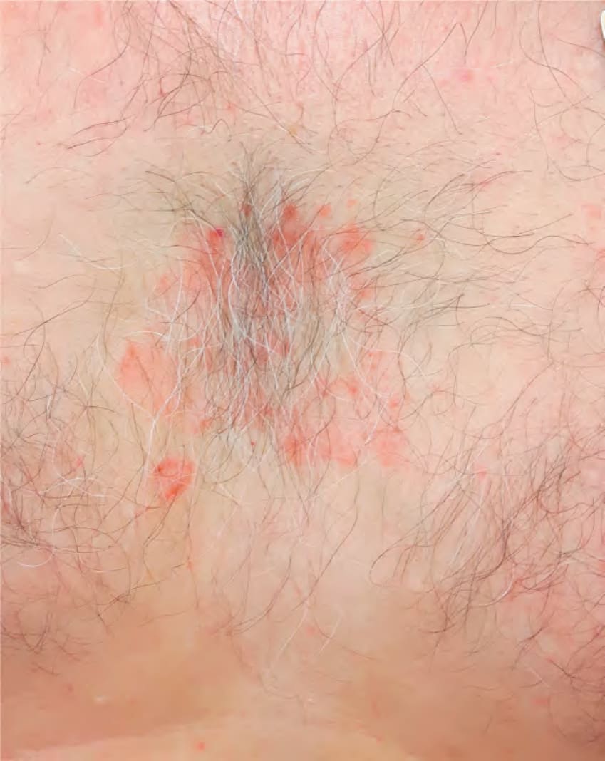
Fig. 13.3 Seborrheic dermatitis of the presternal region in an adult. It sometimes resembles the petals of a flower.
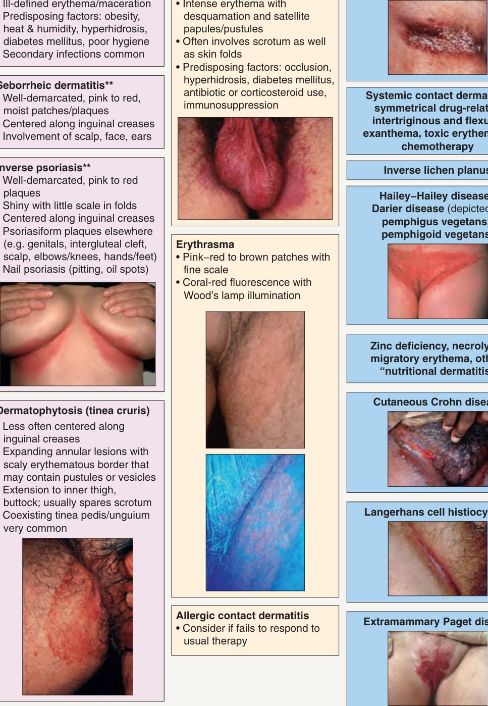
**Fig. 13.4 Differential diagnosis of intertriginous dermatoses in adults. Individual patients often have multiple disorders superimposed upon one another. Bullous impetigo and streptococcal intertrigo are considerably more common in children than adults. *Also referred to more nonspecifically as intertriginous dermatitis or intertrigo. The term “sebopsoriasis” may be used when features of both seborrheic dermatitis and psoriasis are present. Insets: Courtesy Luis Requena, MD; Eugene Mirrer, MD; Louis A. Fragola, Jr, MD; David Mehregan, MD; Julie V. Schaffer, MD.
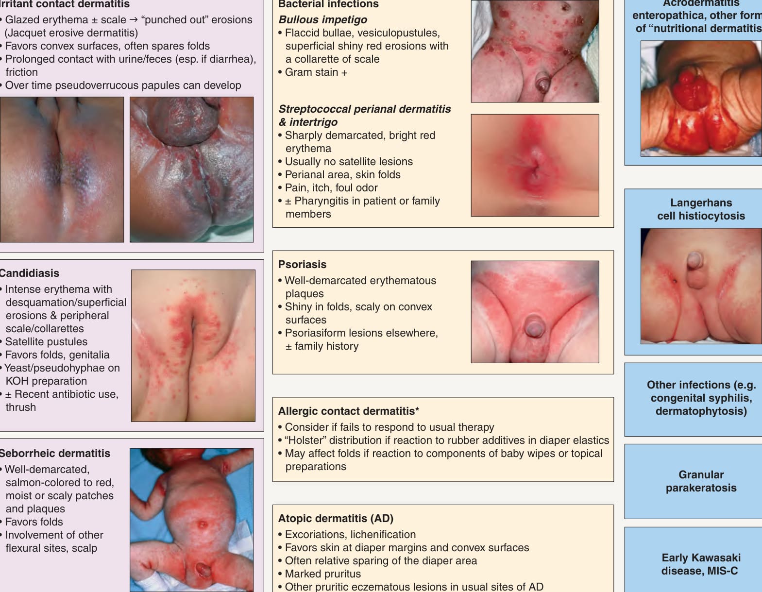
*Fig. 13.12 Differential diagnosis of diaper dermatitis. While the most common etiologies are irritant contact dermatitis, cutaneous candidiasis and seborrheic dermatitis, patients often have a combination with one disorder superimposed on another. The spectrum of “erosive papulonodular dermatosis” due to chronic irritation in the diaper area includes granuloma gluteale infantum, pseudoverrucous papules, and Jacquet erosive dermatitis. Discrete papules or nodules are seen in scabies, whereas congenital syphilis often presents with erosions and even ulcerations, MIS-C, SARS-CoV-2-related multisystem inflammatory syndrome in children. Potential allergens include sorbitan sesquioleate (an emulsifier in diaper balms), fragrances, disperse dyes, rubber additives (e.g. mercaptobenzothiazole), and preservatives in baby wipes (e.g. iodopropynyl butylcarbamate). Insets: Courtesy Robert Hartman MD; Julie V. Schaffer, MD.