ASTEATOTIC ECZEMA
Synonyms: Eczema craquelé Winter eczema Winter itch Desiccation dermatitis
Key features
Dry, rough, scaly, and inflamed skin with superficial cracking that resembles a “dried riverbed”
Sites of predilection are the shins, lower flanks, and posterior axillary line
Associated with aging, xerosis, low relative humidity, and frequent bathing
Introduction
Dry skin (xerosis, asteatosis, exsiccosis) may result from both exogenous and endogenous causes: a dry climate or low indoor humidity; excessive exposure to water, soaps, and surfactants; marasmus and malnutrition; chronic kidney diseases and hemodialysis; hypothyroidism; HIV infection; and heritable conditions such as ichthyosis vulgaris and atopic dermatitis. Asteatotic eczema also arises in patients with reduced sweat gland function (e.g. chronic graft-versus-host disease) or in sites of edema due to hypoalbuminemia from hepatic cirrhosis or nephrotic syndrome. That said, the most common cause of xerosis is aging. Rarely, but especially when widespread and refractory to therapy, asteatotic eczema may be related to an underlying neoplasm, including lymphoma.
History and Epidemiology
Asteatosis as the cause of “nummular eczema” was first mentioned by Gross in the late 1940s. Dry skin probably occurs in everyone over the age of 60 years, but its severity is strongly linked to the exogenous and endogenous factors mentioned above.
Pathogenesis
Xerosis of aging skin is not caused by deficient sebum production, but by a complex dysfunction of the stratum corneum (see Ch. 124). There is a decrease of intercellular lipids with a deficiency of all key stratum corneum lipids and an altered ratio of esterified fatty acids to ceramide 1; this, plus a persistence of corneodesmosomes and premature expression of involucrin and formation of the cornified envelope, results in corneocyte retention and marked impairment of barrier recovery. The water-binding capacity of the stratum corneum is reduced owing to decreased synthesis of “natural moisturizing factor” (NMF) as well as decreased intercellular lipids and glycerol content. Consequently, the stratum corneum desiccates, loses its pliability, and forms small cracks, which render the skin surface dull, rough, and scaly.
Mild xerosis is asymptomatic, but if more pronounced, the skin conveys unpleasant sensations such as pruritus and stinging. Inflammation is enhanced by the release of proinflammatory cytokines secondary to barrier perturbation, mechanical factors (scratching, rubbing), and the application of irritating or sensitizing substances in topical preparations and skin care products.
Occasionally, eczema craquelé can appear in the setting of acute edema, e.g. from congestive heart failure or the re-feeding of patients with anorexia nervosa. One theory is that this is related to the rate of distention of the skin.
Clinical Features
Xerosis first arises on the shins. Later it may spread to the proximal extremities and trunk, but spares the face and neck as well as the palms and soles. It develops insidiously over many years, whereas asteatotic eczema often has a more subacute to acute onset.
Xerotic skin is dry and dull, with fine bran-like scales that may be released as powdery clouds when patients take off their stockings. If more advanced, the skin exhibits a criss-cross pattern of superficial cracks and fissures of the horny layer (“crazy-paving”, eczema craquelé, “dried riverbed”) and appears pink to light red in color (Fig. 13.5). The skin becomes rough, and it may develop an appearance similar to ichthyosis vulgaris (“pseudo-ichthyosis”). In more advanced stages of asteatotic eczema, there is a dull erythema as well as oozing, crusting, and abundant excoriations; disseminated nummular lesions are frequently seen. Vesiculation and lichenification are not regular features except when irritant or allergic contact dermatitis is superimposed. Hemorrhage into the fissures is occasionally observed.
Pathology
Histologically, xerotic skin appears rather normal except for a compact and slightly irregular stratum corneum. Asteatotic eczema in addition exhibits mild focal spongiosis, focal parakeratosis, and a sparse inflammatory infiltrate in the superficial dermis.
Differential Diagnosis
Conditions that need to be distinguished from asteatotic eczema include stasis dermatitis, adult atopic dermatitis (which may overlap with asteatotic eczema), allergic contact dermatitis, nummular dermatitis, and scabies.
Treatment
Asteatotic eczema usually clears within a few days of the application of topical corticosteroid ointment. Proper attention must be given to the care of xerosis in order to avoid relapses: regular use of petrolatum, water-in-oil emollients, urea-, ceramide- or lactic acid-containing preparations, use of bath oils, and the elimination of factors that aggravate dry skin (see above). Topical calcineurin inhibitors have also been used. Coexisting stasis dermatitis should be treated as well (see below).
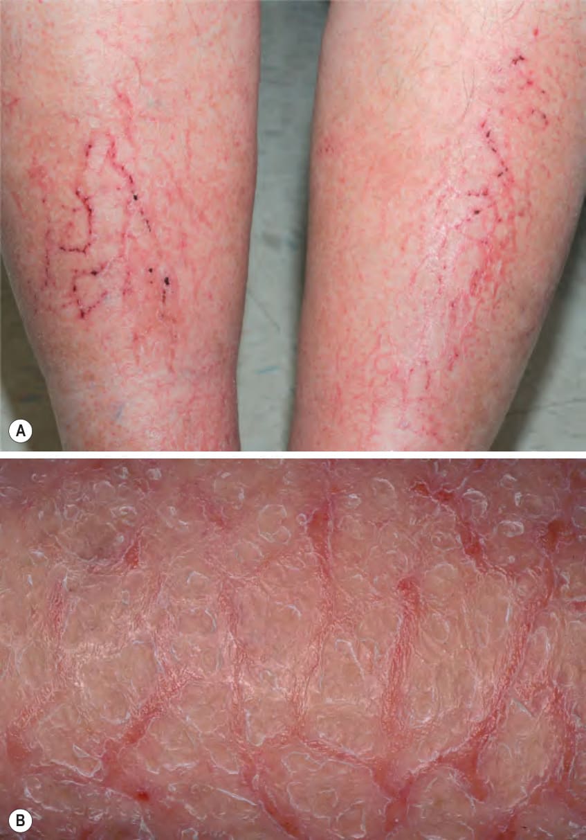
Fig. 13.5 Asteatotic eczema (eczema craquelé).A On the shins, a criss-cross pattern of superficial cracks, fissures, and hemorrhagic crusts said to resemble a dried riverbed. B Xerosis with adherent white scales (pseudo-ichthyosis) and inflammation as well as the criss-cross pattern of superficial cracks and fissures. A, Courtesy Kalman Watsky, MD; B, Courtesy Louis A. Fragola, Jr, MD.