DISSEMINATED ECZEMA (AUTOSENSITIZATION)
Synonyms: Autosensitization dermatitis Autoeczematization Generalized eczema Id reaction
Key features
Secondary lesions of eczema distant from the primary site of exposure or involvement
Symmetric distribution pattern
Most often associated with allergic contact dermatitis and stasis dermatitis
Introduction
Dermatitis caused by exogenous agents initially arises at the site of contact. Not infrequently, additional patches of eczema develop at distant sites. This phenomenon, termed secondary dissemination, has puzzled dermatologists for decades. It is most often observed in allergic contact dermatitis, particularly if associated with stasis dermatitis (see Fig. 13.8), but may occur with uncomplicated stasis dermatitis, other forms of eczema, and, occasionally, severe tinea pedis.
Disseminated eczema appears later than the primary lesions by a few days to weeks, tends to follow a strikingly symmetric distribution pattern, and shows a predilection for analogous anatomic sites (e.g. extensor aspects of the lower and upper extremities, palms, and soles). It may even arise in the absence of and without a preceding “primary” eczema, e.g. in nummular dermatitis (see below). Disseminated eczema must be distinguished from atopic dermatitis, which arises a priori in a disseminated fashion.
History and Pathogenesis
The phenomenon of secondary dissemination of eczemas was first described by Whitfield, but its pathogenesis is still not fully elucidated. The orderly and symmetric distribution pattern may reflect systemic (hematogenous) dissemination and argues against simple spread of contact irritants or allergens on the body surface. It is unclear, however, as to exactly what is disseminated via the bloodstream. It could be allergens; for example, the ingestion of allergens such as nickel has been shown to elicit disseminated eczema in sensitized individuals.
Hematogenous dissemination of microbial products leading to a variety of (non-infectious) manifestations distant from the site of infection, such as “tuberculids” and “bacterids”, was an accepted pathogenic model in the first half of the twentieth century and was extrapolated to the phenomenon of disseminated eczema. Dyshidrotic eczema of the soles, for example, was interpreted as an “id” reaction associated with tinea pedis, and nummular eczema as an “id” reaction caused by “focal” infections of the tonsils. Because disseminated eczema could hardly be attributed solely to infections, attention shifted to an “autosensitization” to epidermal antigens mediated by cytotoxic autoantibodies. This hypothesis, however, has never been verified.
Instead, it became clear both from animal experiments and from routine patch testing (“excited skin syndrome”, “angry back”) that inflammatory processes of the skin, both allergic and irritant or caused by infections, lower the irritancy threshold of distant skin and thus facilitate the development of an eczematous reaction. Obviously, circulating activated memory T cells may play an additional role in disseminated eczema associated with allergic contact dermatitis, including that seen with rebound poison ivy dermatitis following a short, rapid taper of systemic corticosteroids. It remains to be determined which factors regulate the symmetric distribution of disseminated eczema.
Epidemiology
An estimated two-thirds or more of patients with contact dermatitis associated with stasis dermatitis develop episodes of disseminated eczema. The incidence is much lower in the other types of eczema or tinea pedis.
Clinical Features
Disseminated eczema associated with allergic contact dermatitis is characterized by moderately to poorly demarcated patches of eczema, most often on the extremities (Fig. 13.6A). Lesions are also found on the face, and less so on the trunk (Fig. 13.6B). The areas of involvement
A Multiple flat-topped papules, several of which have been excoriated, in addition to patches of eczema. B Square-shaped area of ACD due to nickel in a buckle with an associated id reaction. The latter consists of edematous crusted papules that are separate from the area of direct nickel contact. Courtesy Julie V. Schaffer, MD.
vary greatly in size and number and may consist of discrete papules which are often excoriated. Disseminated eczema in patients with seborrheic and asteatotic eczema differs slightly in predilection sites and morphology (see above).
Pathology
In biopsy specimens of disseminated eczema, the histologic findings are those of an acute or subacute eczematous dermatitis (see section “Stasis dermatitis”).
Differential Diagnosis
Conditions to be distinguished are those eczemas that can arise in a widespread or disseminated fashion: atopic dermatitis, airborne contact dermatitis, contact dermatitis caused by constituents of textiles, photoallergic dermatitis, and eczematous drug eruptions (e.g. calcium channel blockers). Other conditions to be considered are mycosis fungoides and Sézary syndrome.
Treatment
Topical corticosteroids and systemic antihistamines are the mainstay of therapy. Short courses of systemic corticosteroids may be required, but identification and aggressive topical treatment of the inciting dermatosis is necessary to help prevent recurrences or a rebound flare.
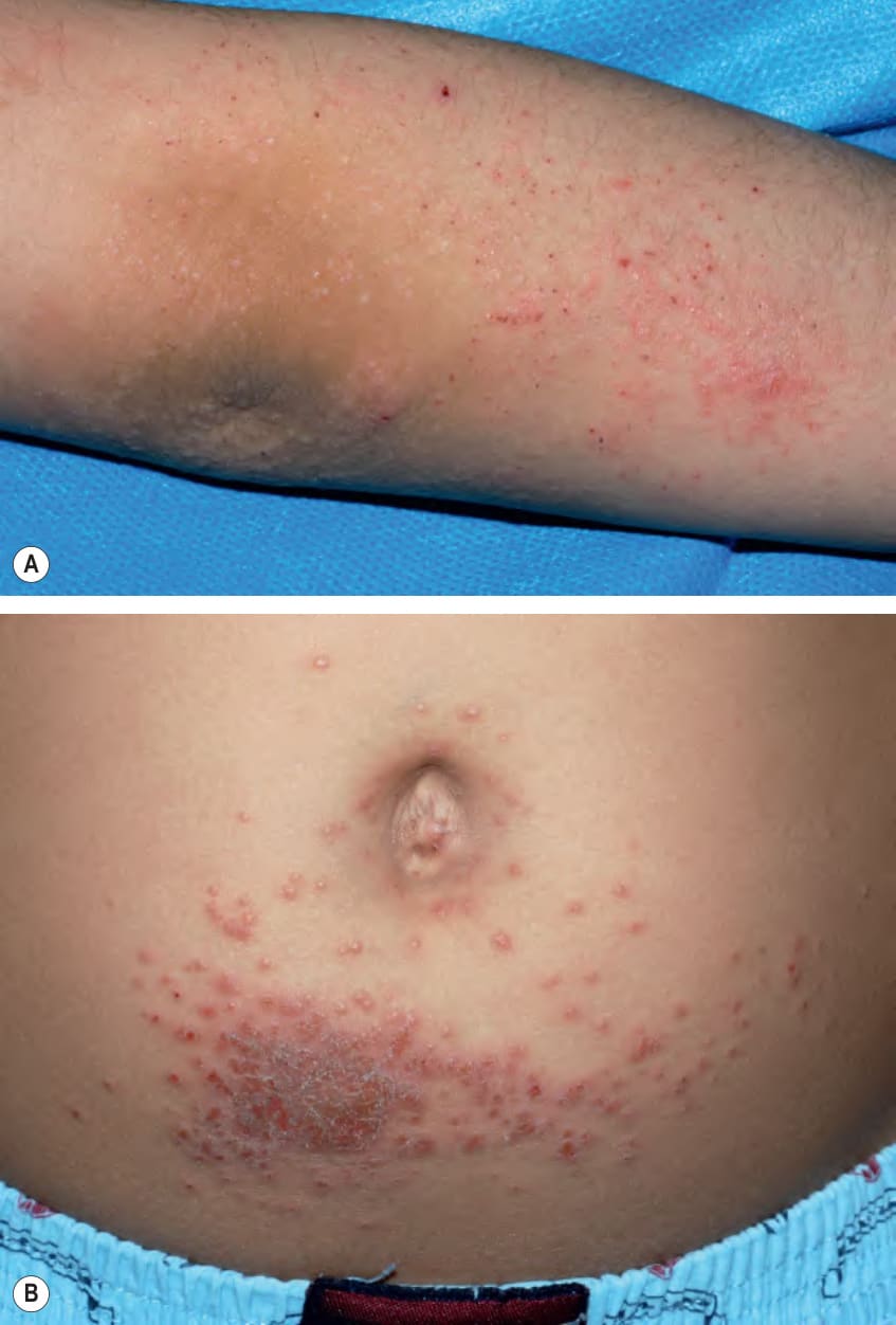
Fig. 13.6 Id reactions in children due to allergic contact dermatitis (ACD) to nickel.
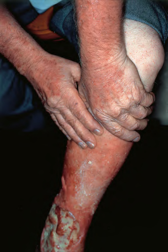
Fig. 13.8 Autosensitization dermatitis in a patient with venous ulceration.