NUMMULAR DERMATITIS
Synonyms: Nummular eczema Discoid eczema Microbial eczema
Key features
Coin-shaped, disseminated, eczematous lesions
Usually very pruritic
Chronic course
Introduction
Nummular dermatitis is an uncommon, disseminated eczema characterized by its coin-shaped lesions. Because such lesions can occur as a feature of atopic dermatitis, asteatotic eczema, and stasis dermatitis, the nosologic position of nummular dermatitis as an independent clinical entity has been questioned.
Epidemiology
Nummular lesions of eczema are not uncommon. However, in the literature there are widely discrepant data on the prevalence of nummular dermatitis, ranging from 0.1% to 9.1%. Some of this variability may reflect the degree to which a distinction is made between nummular dermatitis and coin-shaped lesions observed in patients with other forms of eczema. Men are affected slightly more often and at a later age than women (>50 vs <30 years, respectively).
Pathogenesis
Nummular lesions of dermatitis are not infrequently associated with contact sensitization as well as with xerosis and venous hypertension. In a study of ~30 000 consecutive patients with different types of eczema who underwent patch testing, 3.5% were diagnosed as having nummular dermatitis; 32.5% of these individuals had at least one positive patch test reaction, most often to nickel sulfate, potassium dichromate, and cobalt chloride. Clinical and laboratory signs of atopy are absent in “true” nummular dermatitis.
Clinical Features
Nummular dermatitis is defined as an eruption of round (discoid) eczematous patches almost exclusively of the extremities, often the lower legs in men and the forearms and dorsal aspects of the hands in women (Fig. 13.7A). The lesions are well demarcated and measure 1–3 cm, only occasionally being larger (Fig. 13.7B,C). They may be acutely inflamed with vesicles and weeping, but are often lichenified and hyperkeratotic. Pruritus may be intense and excoriations are often prominent. Nummular dermatitis usually takes a very chronic course. A particularly therapy-resistant variety has been termed “oid-oid” disease, also referred to as exudative discoid and lichenoid chronic dermatitis or Sulzberger–Garbe syndrome.
Pathology
Histologically, the changes of subacute to chronic eczematous dermatitis are seen (Fig. 13.7D; see section “Stasis dermatitis”).
Differential Diagnosis
Nummular dermatitis must be distinguished from nummular lesions of atopic dermatitis and dissemination secondary to contact dermatitis or stasis dermatitis. Other conditions to be considered are psoriasis, inflammatory tinea corporis, mycosis fungoides, and Bowen disease.
Treatment
Options include medium- to high-potency topical corticosteroid ointments, topical tacrolimus or pimecrolimus, and emollients. Tar preparations have also been used successfully. However, a number of patients
Multiple coin-shaped lesions of acute and subacute dermatitis on the leg that are fairly well demarcated. There is often marked pruritus. B, C Coin-shaped eczematous plaques with scale-crust in two patients with different skin phototypes. Patients may report oozing of lesions. D Irregular acanthosis of the epidermis is accompanied by marked spongiosis as well as parakeratotic scale and crust. A lymphohistiocytic infiltrate is present in the dermis with a few lymphocytes within the epidermis. Edema is present in the papillary dermis. B, C, Courtesy Kalman Watsky, MD; D, Courtesy Lorenzo Cerroni, MD.
will require phototherapy to clear the lesions. Methotrexate has led to long-term improvement in children, and dupilumab is a therapeutic option for recalcitrant disease.
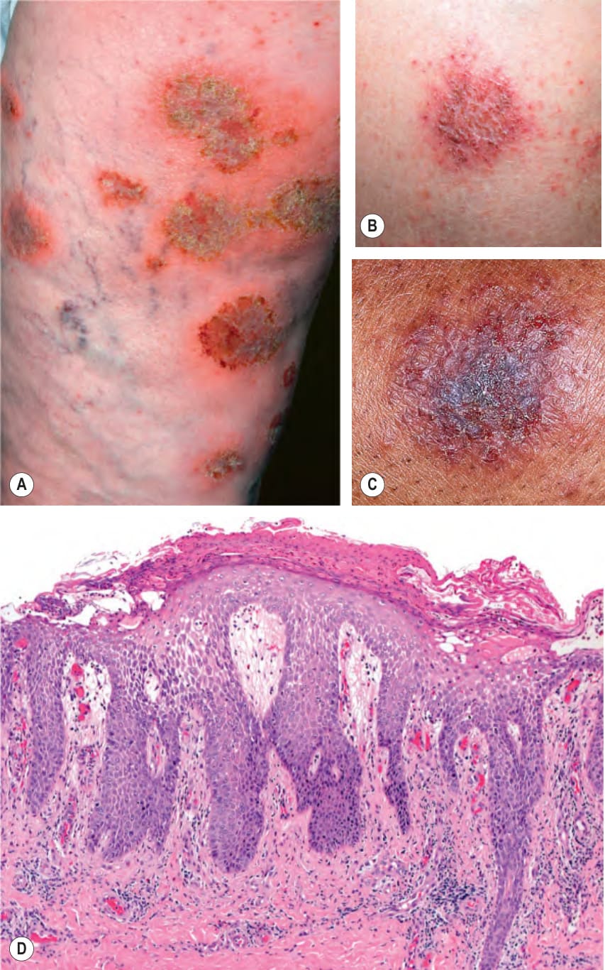
Fig. 13.7 Nummular dermatitis – clinical and histopathologic features.A