HTLV-ASSOCIATED INFECTIVE DERMATITIS
Key features
Rare dermatologic disorder of childhood and adolescence
Association with human T-cell lymphotropic virus type I (HTLV-1)
Eczema of the scalp, axillae, and groin; watery discharge of the nose
Prompt response to antibiotics
Introduction and Pathogenesis
Human T cell lymphotropic virus type I (HTLV-1), also referred to as human T cell leukemia virus type I and adult T cell lymphoma virus type 1, was first detected in 1980, and it can infect T cells, B cells, and monocytes. HTLV-1 is transmitted primarily via breastfeeding, but also by sexual intercourse, blood transfusion, and needle sharing in intravenous drug users. It has been associated with adult T cell leukemia/lymphoma (ATLL), myelopathy/tropical spastic paraparesis, and infective dermatitis. Presumably, HTLV-1-induced dysregulation of the immune system leads to immunosuppression and subsequent infections with Staphylococcus aureus or β-hemolytic streptococci. There is also a potential role for genetic factors, low socioeconomic status, and malnutrition.
The activity of HTLV-1 in infected subjects appears to be low – more than 90% of carriers remain asymptomatic and the majority of disease symptoms (other than infective dermatitis) emerge during adulthood. HTLV-1 induces spontaneous T cell proliferation independent of exogenous stimuli, followed by interleukin (IL)-2 receptor expression (CD25), increased IL-2 secretion, and induction of interferon-γ, IL-5, and IL-10. In symptomatic patients, levels of tumor necrosis factor and IL-6 are elevated. In addition to bacterial infections, affected individuals are more susceptible to parasitoses such as scabies and strongyloidiasis.
History and Epidemiology
Major endemic areas of HTLV-1 infection include sub-Saharan Africa, northern South America, the Caribbean basin, southern Japan, and Iran, affecting 10–20 million people worldwide. Infective dermatitis was first described in 1966 by Sweet, and a year later Walshe suggested an underlying immune defect was present because relapses of the dermatitis frequently occurred, even after a prompt initial response to antibiotic treatment. In 1990, the association with HTLV-1 was first reported. The probability of developing infective dermatitis was estimated to be 2% amongst HTLV-1-infected children, with cutaneous manifestations typically appearing only after protective maternal antibodies had disappeared.
Clinical Features
Infective dermatitis presents as an exudative and crusty eczematous dermatosis, affecting primarily the head and neck region, including the scalp, ears, eyelid margins, and paranasal skin, as well as the axillae and groin. A generalized fine papular rash is occasionally observed and there is usually secondary infection with Staphylococcus aureus or β-hemolytic streptococci. Complications include parasitic infestations, corneal opacities, and progression to more severe HTLV-1-associated disorders (e.g. ATLL). Infective dermatitis rarely persists into adulthood. However, in a series of adult patients in Bahia with HTLV-1- associated ATLL, 44% had a history of infective dermatitis. Diagnosis is established by major and minor criteria (Table 13.1)
Differential Diagnosis
The major entity in the differential diagnosis is atopic dermatitis. Apart from serologic evidence of HTLV-1 infection, lesions of infective dermatitis are more exuberant and obviously infected, but pruritus is less intense. Crusting of the nasal vestibule and blepharoconjunctivitis are more prominent than in atopic dermatitis. Furthermore, it is not associated with atopy. Other disorders to consider include seborrheic dermatitis and impetigo.
Treatment
Antibiotics (e.g. sulfamethoxazole–trimethoprim, cephalexin, erythromycin) result in improvement of the skin lesions but do not prevent relapses. Topical treatments include antibiotics (including intranasal), corticosteroids, and bleach baths.
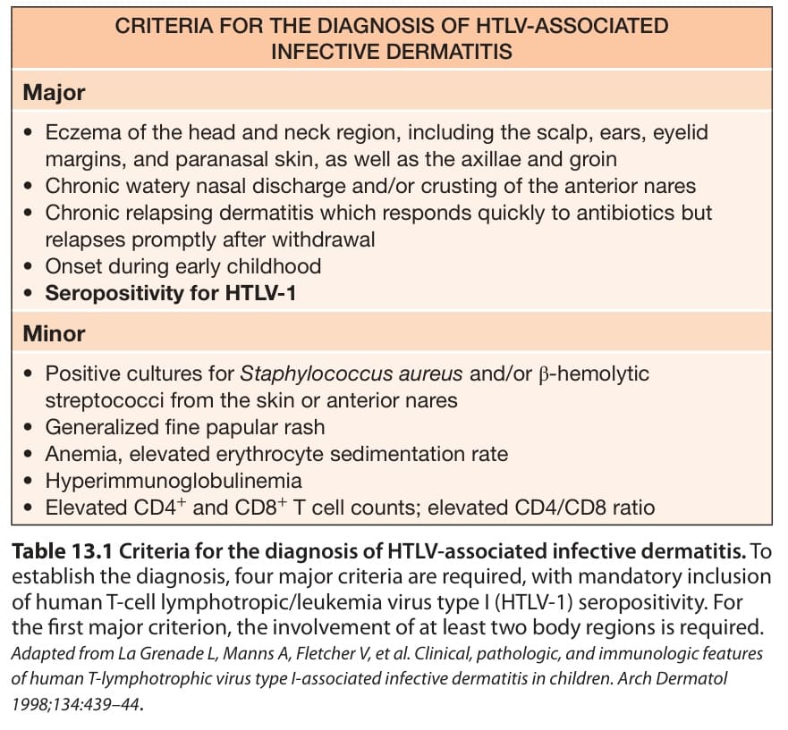
Table 13.1 Criteria for the diagnosis of HTLV-associated infective dermatitis. To establish the diagnosis, four major criteria are required, with mandatory inclusion of human T-cell lymphotropic/leukemia virus type I (HTLV-1) seropositivity. For the first major criterion, the involvement of at least two body regions is required. Adapted from La Grenade L, Manns A, Fletcher V, et al. Clinical, pathologic, and immunologic features of human T-lymphotrophic virus type I-associated infective dermatitis in children. Arch Dermatol 1998;134:439–44.