REGIONAL ECZEMATOUS DISORDERS
Stasis Dermatitis
Synonyms: Gravitational dermatitis Varicose eczema Congestion eczema
Key features
Often associated with other signs of venous hypertension
Can be complicated by allergic contact dermatitis
One of the most common causes of disseminated eczema
(autosensitization dermatitis)
Introduction
Stasis dermatitis is a common component of the clinical spectrum of chronic venous insufficiency of the lower extremities (see Ch. 105). It may arise as an early sign of chronic venous insufficiency, but can persist or recur throughout all stages and is often most prominent when ulcers are present. There is little doubt that chronic venous hypertension per se is the initial trigger for stasis dermatitis. Over time, additional etiological factors may act in concert, most importantly contact sensitization to ingredients of topical therapies. Stasis dermatitis is one of the most common causes of secondary dissemination of dermatitis, and is therefore a complex and multifactorial condition.
History
The term “stasis dermatitis” was introduced by Pillsbury, and the term “gravitation dermatitis” by Belisario.
Epidemiology
The prevalence of chronic venous insufficiency is variable among different ethnic groups and societies. In Central Europe, ~15% of the adult population has symptoms of chronic venous insufficiency and about 1% suffers from venous ulcers. Prevalence rates clearly rise with age.
Pathogenesis
Venous hypertension of the lower limbs is linked to the upright position and is caused by multiple factors, most importantly valvular incompetence of the deep leg veins (see Fig. 105.2). With venous hypertension, there is a slowing of blood flow within the microvasculature, distension of capillaries, and damage to the capillary permeability barrier, allowing the passage of fluid and plasma proteins into the tissue (edema) and extravasation of erythrocytes (stasis “purpura” and hemosiderin deposition). These processes lead to microangiopathy, with serious consequences (Table 13.2).
Both microangiopathy and chronic inflammation are likely to be responsible for stasis dermatitis. Stasis dermatitis typically occurs in the same (i.e. the medial supramalleolar) regions where microangiopathy is most intense, and the patches of dermatitis arise preferentially
over dilated varicose veins. Also, dermal inflammation is known to induce epidermal dysfunction, including barrier impairment.
Sometimes, patients report intense pruritus that even develops before the eczema. Pruritus may be caused by repeated congestion and decongestion, as well as by the release of inflammatory mediators within the dermis. Scratching or rubbing worsens and perpetuates the dermatitis. In addition, patients often apply topical agents to combat the pruritus and xerosis, increasing the risk of contact dermatitis (see Table 13.2).
Clinical features
The first sign of chronic venous insufficiency is usually a cushionlike pitting edema of the medial aspects of the shin and calf around and proximal to the ankle, corresponding to the location of the major communicating veins (Table 13.3). Edema is more pronounced in the
Note the involvement of the extensor surfaces of the upper extremities in this patient with an allergic contact dermatitis to neomycin, as well as stasis dermatitis and venous ulceration. Courtesy Jean L. Bolognia, MD.
evening and resolves overnight. Bouts of stasis purpura lead to areas of hemosiderin deposition. At this stage, stasis dermatitis is mild or absent; the skin may be dry and pruritic. Later, the edema extends to the distal third of the calf and subfascial edema arises, often accompanied by inflammation which may mimic cellulitis (acute lipodermatosclerosis; “pseudoerysipelas”). Over a period of years, the skin, subcutaneous adipose tissue, and deep fascia become progressively indurated and mutually adherent (chronic lipodermatosclerosis). A firm circular cuff is formed which appears to strangle the distal calf, creating an inverted wine bottle appearance. The skin may show intense hemosiderin pigmentation and changes of atrophie blanche. In this setting, venous ulcers develop spontaneously or are triggered by scratching or other trauma. They arise most often in the supramalleolar region, but may later expand (see Ch. 105).
Often stasis dermatitis appears when lipodermatosclerosis develops (see Fig. 105.5). Erythema and scaling are most pronounced around the inner malleoli, but may extend to involve the entire distal lower extremity. Stasis dermatitis is markedly pruritic, as evidenced by multiple excoriations which lead to oozing and crusting. Episodes of vesiculation occur infrequently, always raising the suspicion of superimposed contact sensitization. Chronic lesions of stasis dermatitis invariably exhibit considerable lichenification. Once ulcers form, stasis dermatitis frequently becomes highly irritated, oozing, and erosive (see Fig. 105.4).
Contact sensitization often leads to secondary dissemination. Patches of eczema arise in a strikingly symmetric distribution pattern, particularly on the anterior aspect of the contralateral leg, the anterior thighs, and the extensor surface of the upper extremities (Fig. 13.8); lesions may generalize to involve the trunk and face.
Pathology
The histologic features of eczema, including stasis dermatitis, vary according to the stage of the lesion. In acute eczematous eruptions, intraepithelial edema (spongiosis) predominates, with focal fluid accumulation as micro- or macrovesicles, accompanied by lymphocytes in a perivascular array in the superficial dermis and exocytosis of lymphocytes into the epidermis. There is usually focal parakeratosis. In the subacute phase, spongiosis is still evident, but may be more subtle and intraepidermal vesicles are less prominent. The epidermis thickens variably and parakeratosis is present focally (see Fig. 13.7D). Lymphocytes persist in the dermis and epidermis. In both acute and subacute stages, edema may be present in the papillary dermis.
In the chronic phase, thickening of the epidermis (acanthosis) is more pronounced and it occurs in a regular, psoriasiform pattern or, more often, has a more irregular profile. Inflammation and spongiosis are mild and may be absent. Changes in the granular layer are variable, from thickening, simulating lichen simplex chronicus, to thinning, where the pattern more closely approaches psoriasis; the latter is seen more often in nummular eczema. Ascribing a specific cause of the dermatitis based upon this constellation of histologic findings is impossible.
In stasis dermatitis, biopsy specimens display the histologic features just outlined, as well as signs of venous hypertension: an increased number of capillaries which are dilated and surrounded by cuffs of fibrin, hemosiderin deposits, and hyperplastic (and at times thrombotic) venules (Fig. 13.9). The increase in vessel number can be marked so as to mimic a vascular proliferation. In later stages, there is fibrosis of the dermal connective tissue and sclerosis of the adipose tissue (see Ch. 100).
Differential diagnosis
Stasis dermatitis is a straightforward diagnosis if the cutaneous signs of venous hypertension are present (see Table 13.2). Difficulties may arise in differentiating the individual etiologic components, such as asteatotic eczema and irritant or allergic contact dermatitis. Patch testing is required to exclude the latter. Plaques of stasis dermatitis may be mistaken for nummular eczema, psoriasis, or even mycosis fungoides.
Treatment
The major aim of therapy is the management of venous hypertension – a complex goal which is reviewed in Chapter 105. Basic measures include the regular use of adequate compression bandages or stockings to improve venous return, lifestyle changes, and exercise of the calf muscles. If indicated, surgical strategies are undertaken (see Ch. 155).
However, these surgical approaches do not replace the need for ongoing compression therapy. Topical treatment is the same as for other types of eczemas: judicious use of topical corticosteroids and emollients.
Dyshidrotic Eczema
Synonyms: Pompholyx (larger vesicles and bullae) Acute and recurrent vesicular hand dermatitis
Key features
Firm, pruritic vesicles of the palms, soles, and lateral and medial aspects of the fingers and toes
Association with atopic dermatitis and contact dermatitis (allergic and irritant)
No disturbance of sweat gland function
Introduction
Dyshidrotic eczema is a not-infrequent, chronic, relapsing palmoplantar eczematous dermatosis characterized by firm, pruritic vesicles and bullae. While the vesicles are due to spongiosis within the epidermis, their intact nature is explained by the thick tear-proof horny layer in these sites. Dyshidrotic eczema is not an independent disease entity because it is often a manifestation of other types of eczema, especially atopic dermatitis and irritant or allergic contact dermatitis.
Pathogenesis
Although the formation of vesicles is not linked to sweat gland dysfunction or trapping of sweat within the epidermis (as the term may suggest), hyperhidrosis can be an aggravating factor in some patients. Notably, treatment of hyperhidrosis with onabotulinumtoxin A may ameliorate dyshidrotic eczema.
Dyshidrotic eczema is frequently an expression of atopic dermatitis, particularly as a late-stage manifestation. In these patients, its intensity is usually low to moderate with a protracted and relapsing course, and it is accompanied by only a few other signs of atopic dermatitis. Less often, dyshidrotic eczema is a manifestation of acute or subacute allergic contact dermatitis. Flares can also result from exposure to known irritants (see Ch. 15).
Occasionally, administration of IVIg is followed by acute episodes of dyshidrotic eczema. The role of ingestants, in particular nickel and cobalt, has been debated, but improvement with a low-nickel diet (in nickel-sensitive patients with a positive oral provocation test) has been observed. Lastly, flares can also follow periods of emotional stress as well as exposure to hot climates and rarely sunlight.
Clinical features
Dyshidrotic eczema is characterized by symmetric, firm, deep-seated vesicles of the palms, the lateral and medial aspects of the fingers, and less often the soles and toes (Fig. 13.10A). The size of the vesicles may vary from pinhead-sized to several centimeters (“pompholyx”). When smaller lesions are clustered, they have been likened to tapioca pudding (Fig. 13.10B). While the markedly pruritic vesicles initially contain clear fluid, they have a tendency for purulent superinfection. Dyshidrotic eczema resolves via desquamation of characteristically thick scales.
Dyshidrosis lamellosa sicca (also referred to as keratolysis exfoliativa) is a minimal variant which displays no blisters but only small annular collarettes of white scale (see Fig. 39.13).
Pathology
Histologically, a spongiotic dermatitis is seen with the formation of micro- and macrovesicles within the epidermis (Fig. 13.10C). There is no association with sweat glands.
Differential diagnosis
Inflammatory tinea manuum and pedis, scabies, and palmoplantar pustular psoriasis need to be considered as well as dyshidrosiform pemphigoid, dyshidrotic cutaneous T cell lymphoma, erythema
Firm vesicles are seen along the side of the thumb and thenar eminence. Some of them are deep-seated. B Clusters of tiny vesicles and annular collarettes of scale where the vesicles have ruptured superimposed on plaques of dermatitis. C Histologically, spongiosis within the epidermis is accompanied by macrovesicles; the thick stratum corneum points to an acral location. A, Courtesy Louis A. Fragola, Jr, MD; B, C, Courtesy Lorenzo Cerroni, MD.
multiforme, fixed drug eruption, and, in children, infantile acropustulosis. Distinguishing between an id reaction involving the palms or soles and dyshidrotic eczema can sometimes be difficult and some clinicians view them as overlapping entities.
Treatment
Topical emollients and corticosteroids are the mainstay of treatment. Other topical treatments such as calcineurin inhibitors, retinoids, and PUVA (bath > systemic) may also be helpful. An underlying allergic or irritant contact dermatitis needs to be considered and addressed. In chronic relapsing or recalcitrant cases, systemic therapy (e.g. retinoids, oral corticosteroids, or other immunosuppressants such as cyclospor ine) may be considered.
Infectious Eczematous Dermatitis
This entity is regarded as an eczematous reaction to a localized infection with purulent drainage, most often due to Staphylococcus or Streptococcus spp. It is thought to result from a sensitization to bacterial antigens. Clinically, pustules and vesicles spread from an infectious nidus. More advanced stages are characterized by crusting, scaling, and oozing. In children, the nares, ears, and face are the most common sites of involvement, whereas in adults, the distal lower extremities are most often affected. Milder forms can be treated with open wet dressings, topical antibiotics, topical corticosteroids, and emollients whereas more severe disease usually requires the addition of oral antibiotics and sometimes oral corticosteroids.
Infectious eczematous dermatitis should be distinguished from eczema due to autosensitization, in which lesions develop at a distance from the primary site. Lastly, this disease should not be confused with HTLV-associated infective dermatitis (see above).
Juvenile Plantar Dermatosis
Synonyms: Atopic winter feet Forefoot dermatitis Dermatitis plantaris sicca Moon-boot foot syndrome Sweaty sock dermatitis
Key features
Dry, scaly, glazed and fissured plantar surface of the forefoot
Prepubertal children are affected
Association with atopy and exogenous factors
Introduction
The feet are a fairly common site for various eczematous dermatoses (Table 13.4). Juvenile plantar dermatosis is a characteristic condition that occurs primarily (but not exclusively) in children with an atopic diathesis.
History
Juvenile plantar dermatosis was first reported by Mackie and Husain.
Epidemiology
Juvenile plantar dermatosis occurs in prepubertal children, from the age of 3 years and upwards, when shoes are worn for longer periods of time. It is only occasionally seen in adults. There is seasonal variation, with worsening during the winter, and boys are affected slightly more often than girls.
Pathogenesis
An atopic disposition is clearly a risk factor, but exogenous factors play an equally important role, including plastic- and rubber-constructed sports shoes that youngsters often wear all day long. The humid environment leads to hydration of the horny layer, making it much less resistant to wear and tear. When the stratum corneum is rubbed off by friction, this leads to a characteristic glazed and thinned appearance of the skin. As involved areas become xerotic, cracks are formed. The inherently dry skin of atopic individuals may be a predisposing factor. Adults have a much thicker horny layer of their plantar skin and are thus less at risk of developing the condition.
Clinical features
The balls of the feet and the toe pads exhibit strikingly symmetrical changes: fairly well demarcated shiny, reddish, tender, dry lesions with
some scaling (Fig. 13.11), and often accompanied by painful cracks and fissures. The dorsa of the feet, the interdigital spaces, and the instep are typically spared. Less often, similar lesions are found on the hands.
Pathology
Histologically, the features of a chronic eczematous dermatitis are seen (see section “Stasis dermatitis”).
Differential diagnosis
Juvenile plantar dermatosis needs to be distinguished from allergic contact dermatitis to chemicals contained in leather (e.g. chromates, dyes) or rubber. However, the latter is uncommon in children and, if present, often involves the dorsa of the feet as well. Tinea pedis is equally uncommon in children, and the interdigital spaces are usually predominantly affected. Patch testing and KOH preparations may help in establishing the diagnosis.
Treatment
Juvenile plantar dermatosis is a chronic but self-limiting condition. Patients should be advised to avoid wearing impermeable socks and shoes, and the application of emollients, keratolytics, and/or paraffintype ointments is beneficial. When shoes are removed, socks, if damp, should also be removed and replaced by dry socks.
Diaper Dermatitis
Key features
Frequently, an irritant dermatitis of the diaper area due to occlusion and prolonged exposure to urine and feces
Secondary infection with Candida albicans often occurs
May be associated with seborrheic dermatitis and psoriasis
Epidemiology
Diaper dermatitis develops in at least 50% of infants, and it is responsible for a considerable percentage of dermatologic consultations in infants and toddlers. Seborrheic dermatitis is a predisposing factor.
Pathogenesis
Diaper dermatitis is the cumulative result of several factors, in particular dampness and exposure to urine and feces. In the past, ammonia derived from the urea in urine was held as primarily responsible for diaper dermatitis. More recently, the blame has been placed on the (alkaline) pH of the urine and the role of fecal bacteria. Enzymes produced by fecal bacteria, as well as residual pancreatic proteases and lipase in the stool, act as irritants and these enzymes are also activated in the
alkaline milieu. In addition, ureases are produced by fecal bacteria, resulting in a further increase of the urinary pH. This explains why diaper dermatitis is more likely to appear in cow milk-fed than in breastfed infants: cow milk formulas are colonized by a greater number of urease-producing bacteria.
Prolonged use of diapers, dampness, and the factors detailed above lead to the breakdown of the horny layer barrier function. An alkaline pH also facilitates the development of secondary C. albicans infection.
As diaper dermatitis tends to be most prominent on the inner parts of the thighs, genitalia, and buttocks, friction between the skin and the diaper material likely acts as a physical factor that leads to further irritation. In addition, chemical constituents of the diaper and/or topical preparations and baby wipes may lead to contact sensitization.
Clinical features
Diaper dermatitis is strictly confined to the diaper area, presenting with mild to pronounced erythema, erosions, and scaling. In the common form due to irritant contact dermatitis, genitocrural folds are typically spared. Depending on whether there is a secondary infection or an underlying dermatosis (e.g. seborrheic dermatitis, psoriasis), the clinical picture can vary (Fig. 13.12).
Differential diagnosis
The differential diagnosis is outlined in Fig. 13.12.
Treatment
In the acute phase, mild corticosteroid preparations are helpful. Topical imidazole creams are added for secondary infection with Candida spp. The major goal of long-term management is avoidance of the causative factors. Frequent changing of highly absorbent disposable diapers is associated with a lower incidence and severity of diaper dermatitis, and it leads to a more physiologic pH. Emollients containing white paraffin (Vaseline®) or soft zinc pastes provide both protective and soothing effects.
Additional figures available in our eBook (see inside front cover for access code).
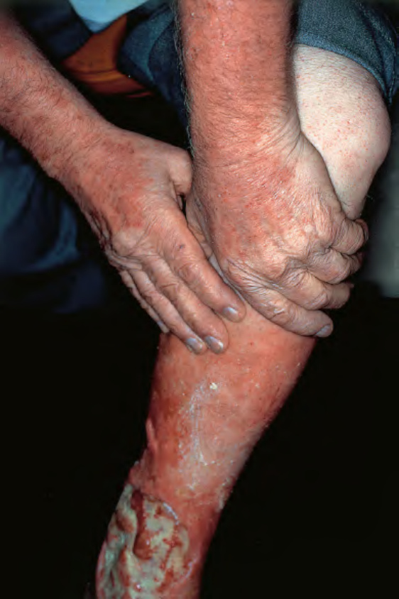
Fig. 13.8 Autosensitization dermatitis in a patient with venous ulceration.
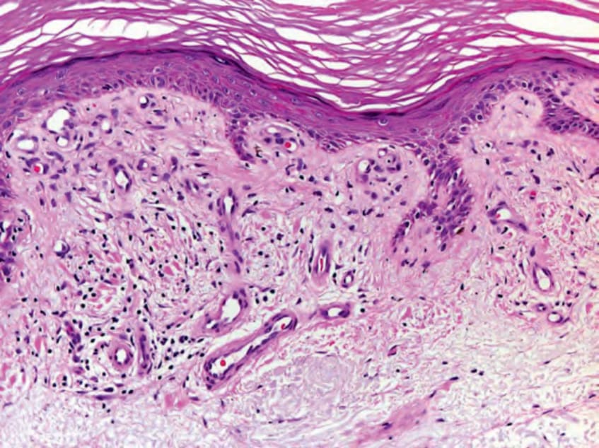
Fig. 13.9 Stasis dermatitis – histologic features. Orthohyperkeratosis and minimal spongiosis in the epidermis. There is an increased number of blood vessels surrounded by a fibrin cuff in a slightly fibrotic papillary dermis. Note also extravasation of erythrocytes and a mild lymphocytic infiltrate. Courtesy Lorenzo Cerroni, MD.
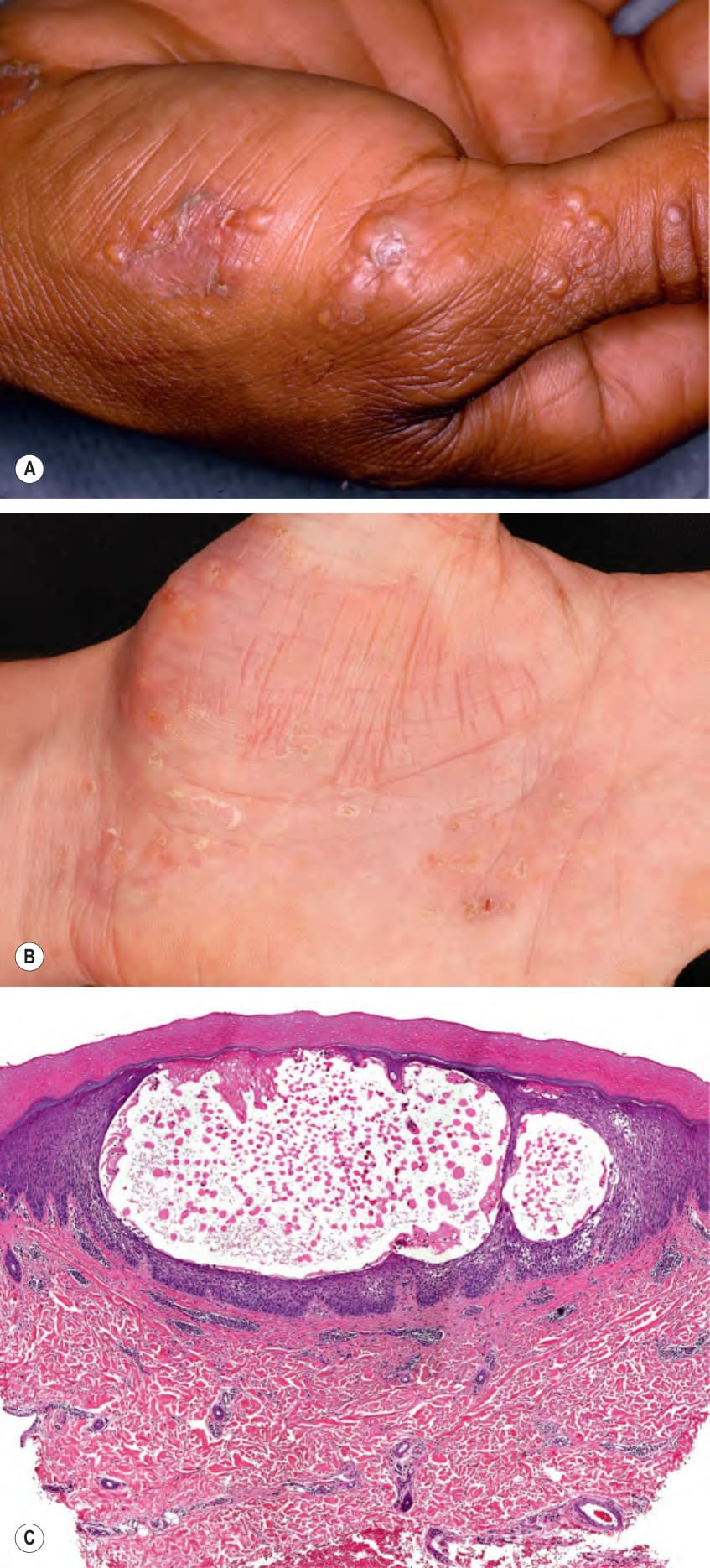
Fig. 13.10 Dyshidrotic eczema – clinical and histopathologic features.A
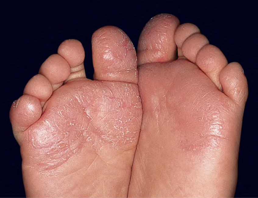
Fig. 13.11 Juvenile plantar dermatosis in a child. Erythema and scaling of the plantar surface of the great toes and fifth toes as well as the ball of the foot bilaterally. Note the glazed appearance of the skin of the left foot. Courtesy Kalman Watsky, MD.
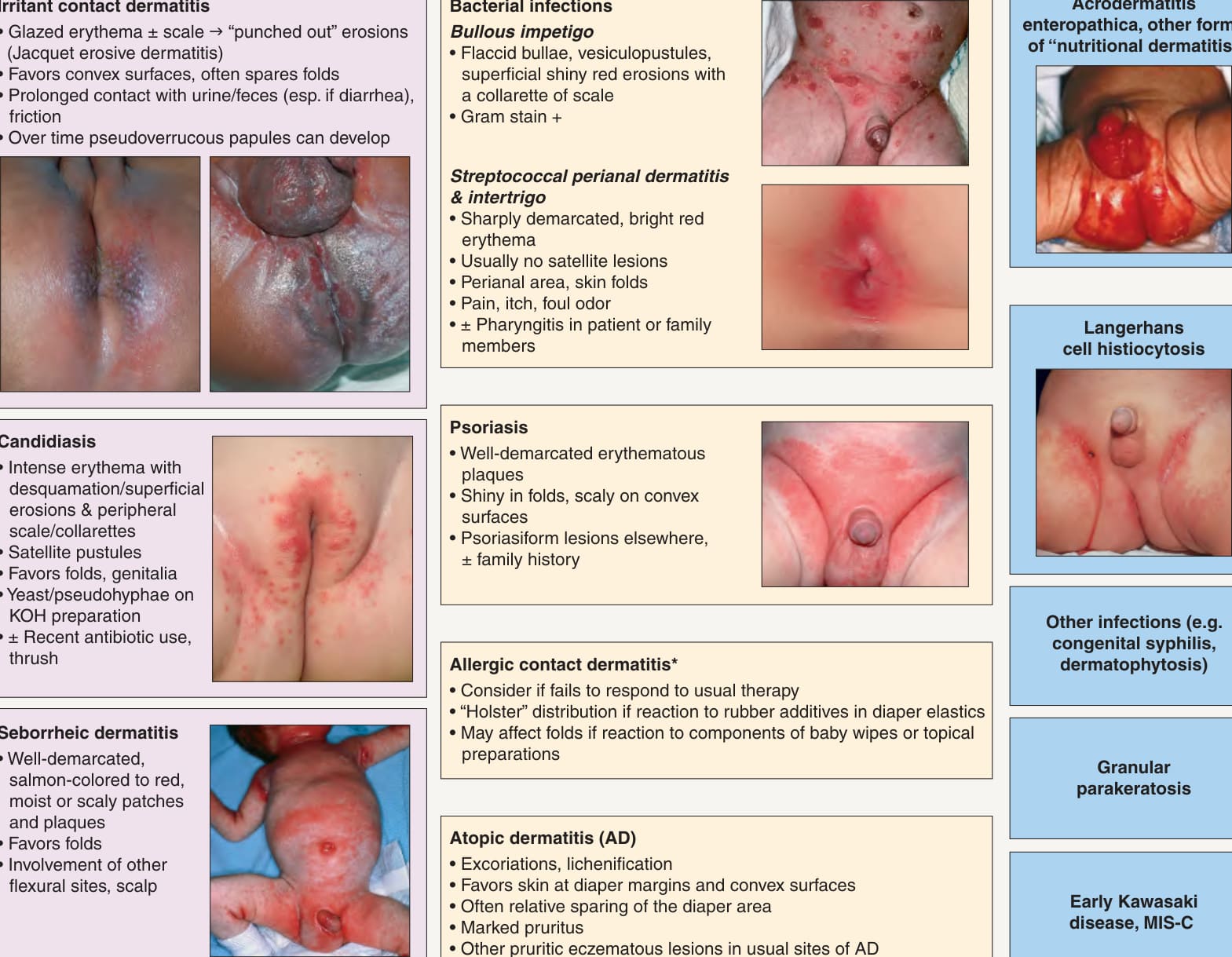
*Fig. 13.12 Differential diagnosis of diaper dermatitis. While the most common etiologies are irritant contact dermatitis, cutaneous candidiasis and seborrheic dermatitis, patients often have a combination with one disorder superimposed on another. The spectrum of “erosive papulonodular dermatosis” due to chronic irritation in the diaper area includes granuloma gluteale infantum, pseudoverrucous papules, and Jacquet erosive dermatitis. Discrete papules or nodules are seen in scabies, whereas congenital syphilis often presents with erosions and even ulcerations, MIS-C, SARS-CoV-2-related multisystem inflammatory syndrome in children. Potential allergens include sorbitan sesquioleate (an emulsifier in diaper balms), fragrances, disperse dyes, rubber additives (e.g. mercaptobenzothiazole), and preservatives in baby wipes (e.g. iodopropynyl butylcarbamate). Insets: Courtesy Robert Hartman MD; Julie V. Schaffer, MD.
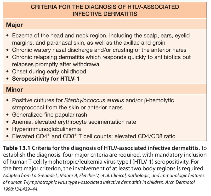
Table 13.1 Criteria for the diagnosis of HTLV-associated infective dermatitis. To establish the diagnosis, four major criteria are required, with mandatory inclusion of human T-cell lymphotropic/leukemia virus type I (HTLV-1) seropositivity. For the first major criterion, the involvement of at least two body regions is required. Adapted from La Grenade L, Manns A, Fletcher V, et al. Clinical, pathologic, and immunologic features of human T-lymphotrophic virus type I-associated infective dermatitis in children. Arch Dermatol 1998;134:439–44.
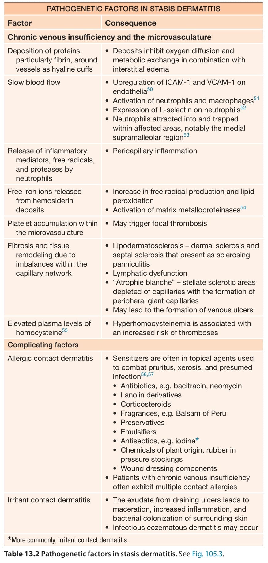
Table 13.2 Pathogenetic factors in stasis dermatitis. See Fig. 105.3.
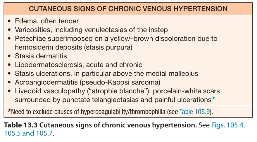
Table 13.3 Cutaneous signs of chronic venous hypertension. See Figs. 105.4, 105.5 and 105.7.
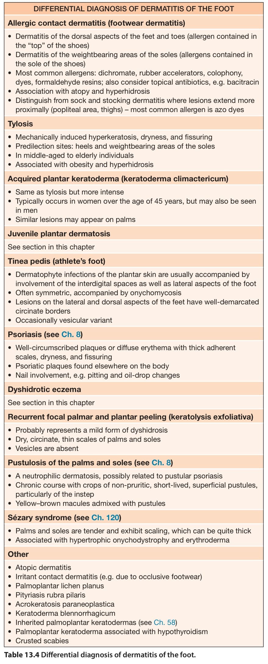
Table 13.4 Differential diagnosis of dermatitis of the foot.
Dermatol. 1897;6:827–846.2. Shuster S. The aetiology of dandruff and the mode of