DESCRIPTION OF TECHNIQUES
Electrodesiccation and Electrofulguration
For very superficial lesions, such as those involving only the epidermis, electrosurgical destruction by electrodesiccation can be achieved with little, if any, scarring. The markedly damped, high-voltage current generated by spark gap electrosurgical units causes superficial tissue damage by dehydration of the treatment site. It is delivered in a monoterminal (concentrative) fashion (see Fig. 140.1). If the electrode is held at a slight distance from the tissue, a spark is formed between the electrode and the tissue. This technique, termed electrofulguration, achieves very superficial destruction because the surface carbonization it produces insulates the underlying tissues from thermal heat spread.
Dermatologists who employ a Hyfrecator® or similar device often utilize this type of waveform. As the surface becomes charred by sparking at the surface, it is common for blood to appear in droplets that penetrate through the surface char. This is because the electrical current does not penetrate deeply enough to seal nearby blood vessels. Therefore, when surgical excision is undertaken, electrocoagulation is a better current to employ in order to provide hemostasis (see below). However, for treatment of small very superficial epidermal lesions, electrofulguration should be considered the waveform of choice. For example, it is very effective for the treatment of dermatosis papulosa nigra.
Electrodesiccation is the method of choice when superficial tissue destruction is desired. For example, it is ideal for treatment of epidermal lesions such as seborrheic or actinic keratoses (Fig. 140.3), acrochordons, verrucae planae, or small epidermal nevi. Hemostasis of mild capillary bleeding can also be achieved by use of electrodesiccation. A standard technique for treating keratoses by this method is to move the electrode slowly across the surface of the lesion (for small lesions) or to insert it directly into the lesion (for larger lesions), while applying current at a low power setting. After a few seconds, the lesion bubbles as the epidermis separates from the underlying dermis. It can then be easily removed with a curette or simply by rubbing a piece of gauze across the treatment site. The clinical endpoint in treating epidermal lesions is punctate bleeding, which is controlled with pressure, by spot electrocoagulation, or by topical hemostatic agents such as aluminum chloride. More profuse bleeding indicates probable damage to the dermis, with a greater likelihood of subsequent scarring. Extremely small superficial lesions can be treated by electrofulguration, which causes the least amount of damage to adjacent tissues.
Electrocoagulation
In electrocoagulation, a moderately damped current is applied in a biterminal manner (both concentrative and dispersive electrodes are used). This current is of higher amperage and lower voltage than that utilized for electrodesiccation. Because this type of current penetrates more deeply, it has the potential for greater tissue destruction.
Electrocoagulation is particularly useful for deep tissue destruction and surgical hemostasis. It is our preferred modality for treating small and uncomplicated primary basal cell and squamous cell carcinomas, as well as other lesions such as trichoepitheliomas, which extend into the dermis. Much of the literature refers to “curettage and electrodesiccation (C&E)” or “electrodesiccation and curettage (ED&C)”, but, in fact, electrocoagulation may be the preferred electrosurgical waveform for all but the most superficial tissue destruction.
When performed properly, electrosurgery plus curettage yields cure rates of 90%–95% for uncomplicated basal cell carcinomas and super-ficial squamous cell carcinomas, making it an efficient, cost-effective treatment. Thorough curettage is likely the key element affecting success. Initially, a large curette (4–6 mm) is used to debulk the tumor, followed by a smaller curette (2 mm). The curette should be scraped across the surface of the treatment site in every direction, from superior to inferior, inferior to superior, left to right and right to left, using fairly firm downward pressure. In this way, all tumor extensions are eradicated.
After thorough curettage, the electrode is brought into direct contact with the tissue to be treated and is moved slowly around the periphery and across the curetted portion of the lesion, which eventually becomes charred. A curette may then be used to gently remove the charred tissue. For treatment of skin cancers, this procedure is repeated two or more times in an attempt to remove any small tumor extensions (Fig. 140.4). During the last curettage, a very small curette (1 mm) may be used to remove the final tiny “roots” of the tumor. Scarring must be expected with this procedure, and wounds following curettage and electrosurgery are allowed to heal by second intention.
Electrocoagulation is also effective for treatment of superficial telangiectasias (Fig. 140.5), unwanted hair (electroepilation), and ingrown toenails (electrosurgical matrixectomy). Hemostasis using electrocoagulation can be achieved by either monopolar or bipolar means. Because the electrosurgical energy may be transmitted for several millimeters along the vessel wall, it is important to use the minimum effective exposure time and power setting in order to prevent delayed bleeding from damaged vessels. We often use monopolar electro coagulation, in which the electrode is touched directly to the bleeding vessel. Coagulation can also be achieved by touching the electrode to a hemostat that has been clamped on the severed vessel. In bipolar electrocoagulation (Fig. 140.6), a bipolar forceps is used to provide more directed pinpoint hemostasis. Electrocoagulation current delivered in this manner causes less adjacent tissue damage but requires a dry operative field to be effective.
Nodular basal cell carcinoma preoperatively after injection of local anesthesia. B The tumor is curetted to debulk all friable tissue. C Further curettage, often with a smaller-diameter curette, should proceed in all directions to remove small extensions of tumor. D Following thorough curettage, the surface and periphery of the wound are treated by electro coagulation current to stop bleeding and potentially remove further tumor. E Final wound – curettage and electrosurgery have been repeated twice more, until all friable tissue has been removed. Healing occurs over 4–6 weeks, depending upon the site.
Electrosection (Cutting)
Electrosection involves the biterminal application of a slightly damped current. The current, of low voltage and high amperage, causes minimal lateral heat spread and tissue damage, and it has the additional advantage of simultaneously achieving hemostasis and cutting. “Pure” cutting can be obtained using a true undamped tube current, which provides the least amount of lateral heat spread and causes vaporization of tissue without hemostasis. Such current has been suggested by Chiarello to be useful in debulking large skin cancers, contouring surrounding skin following shave excisions, scar revision following skin flaps and grafts, and rhinophyma repair. When electrosection is performed using a filtered, fully rectified current, subsequent spot electrocoagulation can be achieved by changing to the electrocoagulation current.
Electrosection can be used to perform rapid and effortless excisions or incisions. Virtually no manual pressure by the operator is required. Electrodes configured as loops, triangles, or diamonds can be used for quick removal of skin tags, papillomas, intradermal nevi, and other exophytic skin lesions. A straight, narrow electrode is most often used to incise the skin and is applied to the tissue in brisk, continuous paintbrush-like strokes. The difference between electrosection and scalpel excision is immediately apparent to the first-time user of electrosection. At the appropriate power setting, the electrode passes smoothly through the tissue like a “hot knife through butter”. If perceptible sparking occurs during incision, the power setting is too high; if the electrode “drags”, the power setting is too low.
Slightly damped currents cause some charring at the margins of the excised tissue. Therefore, when a specimen suitable for histopathologic analysis is required, the filtered current (see Fig. 140.2) should be used because it does not create significant electrosurgical artifact. It is recommended that the new initiate to electrosection develops technical skills and concepts by practicing on beefsteak before utilizing this technique in the clinical arena.
The major advantage of electrosection over scalpel surgery is that when a blended cutting and coagulating current is utilized, hemostasis is achieved immediately as the incision is made. However, larger blood vessels (>2 mm) require additional spot electrocoagulation at the completion of the excision. Another drawback is that vaporization of the tissue generates a smoke plume, which can be unpleasant for both the patient and the operator and may contain potentially infectious viral particles (see below). Consequently, effective smoke evacuation equipment should be available during such procedures.
Electrosection is extremely useful for achieving relatively bloodless excision of large, bulky lesions, such as acne keloidalis nuchae and rhinophyma (Fig. 140.7), in which the surgical defect is allowed to heal by second intention. Electrosurgical excision followed by primary closure can also be undertaken with no impairment of wound healing, as compared with conventional scalpel surgery. This modality has been used, without complication, to create skin flaps and perform cosmetic facial surgery, including blepharoplasty and rhytidectomy, with excellent outcomes.
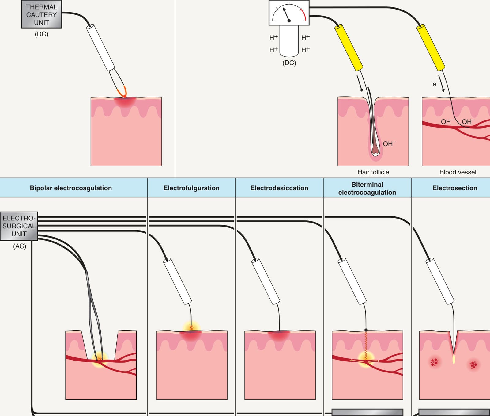
Fig. 140.1 Electrocautery, electrolysis, and different types of electrosurgery. AC, alternating current; DC, direct current; e−, electron; H+, proton; OH−, hydroxide ion.
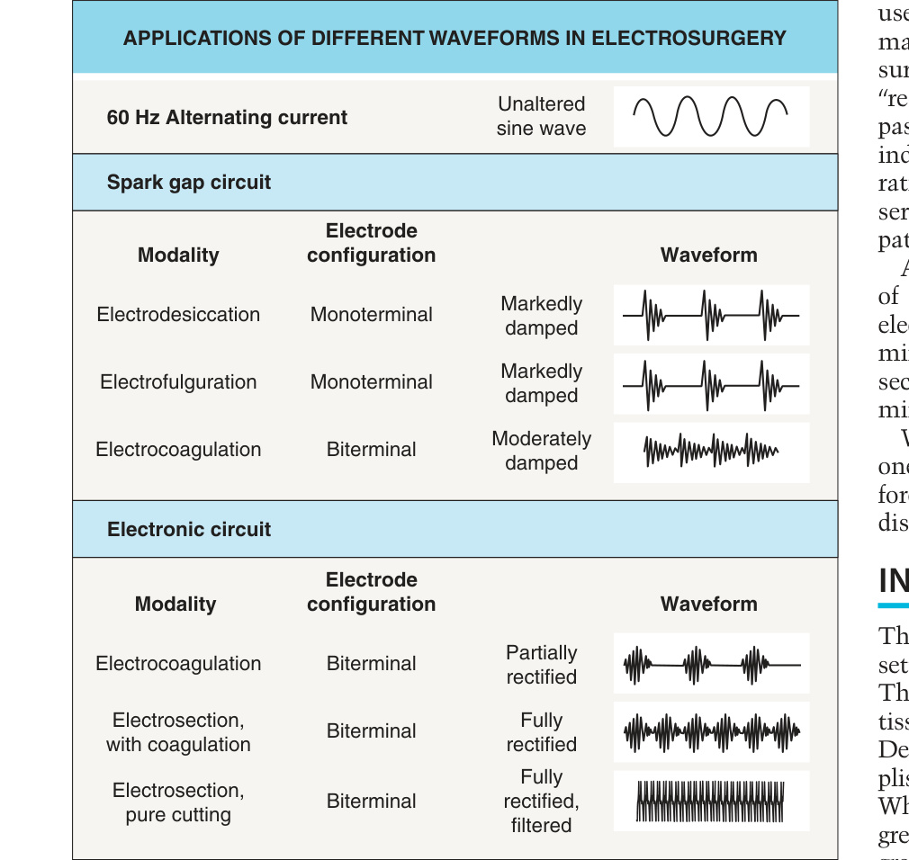
Fig. 140.2 Applications of different waveforms in electrosurgery.
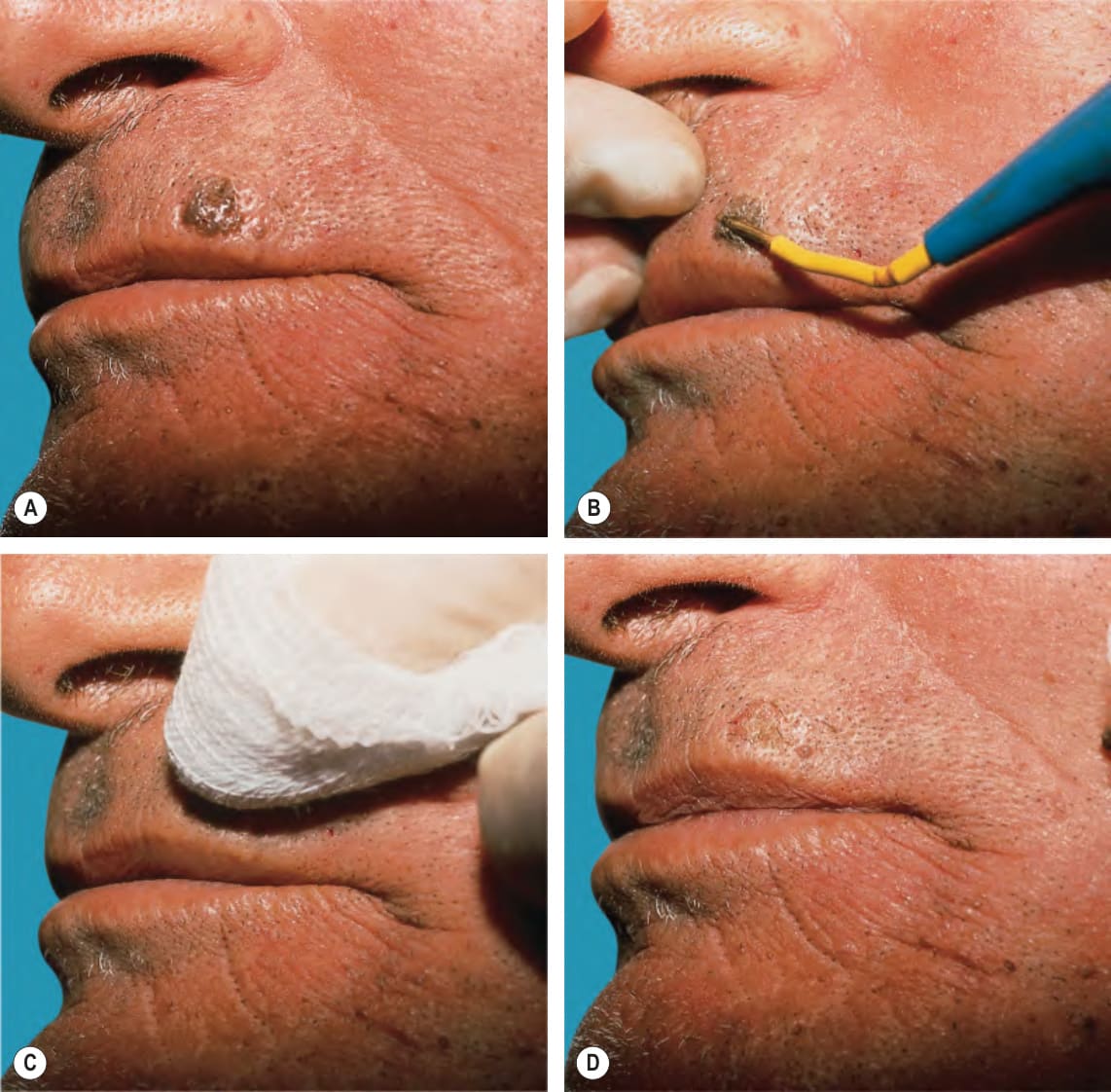
Fig. 140.3 Seborrheic keratosis on the upper lip.A Given its epidermal location, electrosurgical destruction should employ a current that provides the most superficial type of tissue damage. B Under local anesthesia, the lesion is treated by electrodesiccation. Delivery of current is stopped when the lesion begins to “bubble”. C In lieu of a curette, a gauze pad is useful for removing charred tissue after desiccation of super-ficial lesions. D There is minimal bleeding, indicating that damage did not penetrate deeply into the dermis.
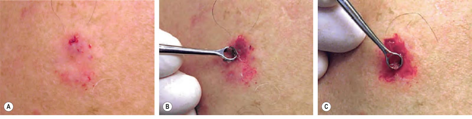
Fig. 140.4 Basal cell carcinoma on the right deltoid area, treated by curettage and electrosurgery.A
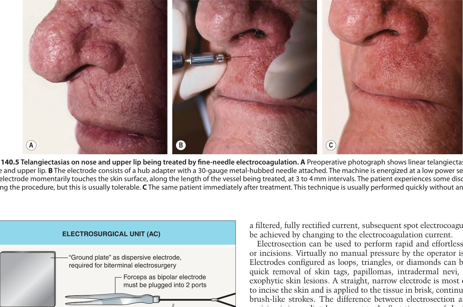
Fig. 140.5 Telangiectasias on nose and upper lip being treated by fine-needle electrocoagulation.A Preoperative photograph shows linear telangiectasias on the nose and upper lip. B The electrode consists of a hub adapter with a 30-gauge metal-hubbed needle attached. The machine is energized at a low power setting and the electrode momentarily touches the skin surface, along the length of the vessel being treated, at 3 to 4 mm intervals. The patient experiences some discomfort during the procedure, but this is usually tolerable. C The same patient immediately after treatment. This technique is usually performed quickly without anesthesia.
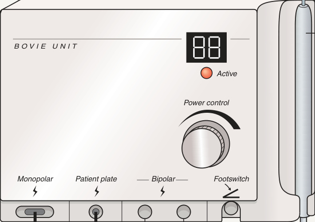
Fig. 140.6 Electrosurgical unit (AC) with attached electrosurgery pencil. The monopolar pencil is used for electrodesiccation and electrofulguration. When used with the grounding plate, it is utilized in a biterminal mode for electro coagulation. Alternatively, the unit can be used with the bipolar forceps to provide pinpoint electrocoagulation for hemostasis. Bipolar electrocoagulation is also recommended for use in patients with cardiac implantable electronic devices. AC, alternating current.
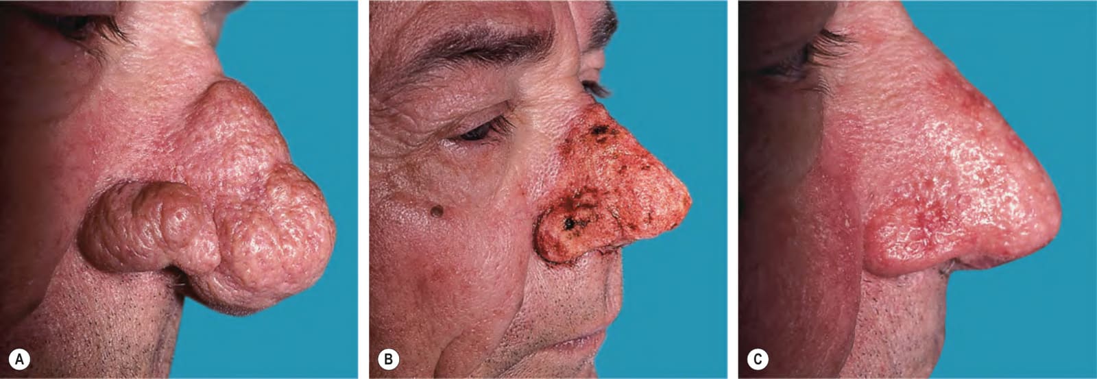
Fig. 140.7 Rhinophyma.A Moderately severe rhinophyma in a middle-aged man. B Immediately after electrosurgical planing of excess sebaceous glands. Care is taken to perform subtotal removal as very aggressive therapy can result in significant scarring and possible deformity caused by scar contracture. C Four weeks later, healing with good cosmetic outcome is noted. During healing, the wound is kept moist with an ointment (e.g. petrolatum) and semi-occlusive dressings.