DISCUSSION
Physiology and Structure
Local anesthetics act by blocking sodium channels in the axon cell membrane, and this prevents sodium from entering the nerve cell. The nerve cell is not depolarized and consequently the action potential is blocked. The cationic form of the anesthetic appears to bind to the inner pore of voltage-gated sodium channels, possibly leading to both narrowing of the pore lumen (steric block) and creation of an electrostatic barrier to permeation. Smaller unmyelinated C-type nerve fibers that conduct pain sensation are blocked more quickly and easily than intermediate fibers that also carry sensations of heat and cold. The myelinated A-type fibers that carry pressure sensation and motor fibers are blocked last. Clinically, this is evident when an area seems fully anesthetized for scalpel surgery, but the patient still feels the pressure of the surgeon’s fingers at the surgical site.
All local anesthetics consist of three parts (Fig. 143.1):
●a secondary or tertiary amine end
●an aromatic end
●an intermediate connecting chain that contains an ester or amide. The aromatic portion is hydrophobic and lipophilic. This is essential to allow the anesthetic to diffuse through nerve cell membranes. The amine portion is hydrophilic and is responsible for the anesthetic’s water solubility, which is important for preparing, storing, and administering the anesthetic.
Local anesthetics are weak organic bases, which, to be water-soluble and injectable, require the addition of a hydrochloride salt. In aqueous
George J. Hruza Anesthesia 143
solution, the salt equilibrates between the ionized and non-ionized form. The ionized form is water-soluble, allowing injection into and diffusion through tissue. However, it is the non-ionized, lipid-soluble base that can diffuse into the nerve cell membrane. Within the sodium channel’s inner pore, it is the ionized cation that is responsible for blocking nerve conduction (see above).
The dissociation constant (pKa) of each anesthetic determines the proportion of the anesthetic base and its cation at a given pH. The pKa of all local anesthetics is higher than physiologic pH. For most local anesthetics at a pH of 7.4, 80% or more is in the cationic ionized form. Alkalinization of the anesthetic solution, as is done with the addition of sodium bicarbonate, will speed its onset of action as more of the anesthetic will be in the non-ionized form. However, if the pH is raised too much, the anesthetic may precipitate out of solution. Anesthetic sensitivity to pH also helps explain why infected tissue is difficult to effectively anesthetize. The inflammatory response surrounding the infection acidifies the site (reduces the pH), which reduces the proportion of the anesthetic in the non-ionized, lipid-soluble form.
Pharmacology
Local anesthetics are classified into two groups, depending on the linkage in the intermediate chain (Table 143.1). Amide anesthetics have an amide linkage and ester anesthetics have an ester linkage (Fig. 143.1). They differ in how they are metabolized and in the risk of sensitization. Ester anesthetics are hydrolyzed by plasma pseudocholinesterase, and the metabolites are excreted by the kidneys. Patients with a deficiency of functional pseudocholinesterase, who are often diagnosed after prolonged paralysis following administration of standard doses of succinylcholine, are at an increased risk of ester anesthetic toxicity. The metabolite para-aminobenzoic acid (PABA) is responsible for the allergic reactions seen with ester anesthetics. Amide anesthetics are metabolized by the hepatic microsomal cytochrome P450 enzyme system, and the metabolites are excreted by the kidneys. Patients with severe liver disease may be at increased risk of amide anesthetic toxicity.
Local anesthetics differ in the speed of onset, duration of action, and potency, depending on each compound’s intrinsic chemical characteristics (Table 143.1). A low pKa leads to rapid onset of anesthesia, as more of the anesthetic will be in the non-ionized form. Greater lipid solubility is associated with higher anesthetic potency, as the compound penetrates the nerve cell membrane more easily. Duration of action is determined by the strength of anesthetic binding to the sodium channel pore.
For pregnant women, the local anesthetic of choice is lidocaine. It is historically classified as FDA pregnancy category B, which means that, in animal studies, no teratogenic effects have been documented. Studies in pregnant women who received lidocaine during the first trimester of pregnancy have shown no increase in anatomic abnormalities in the newborns. However, it is recommended that lidocaine, as with all other pharmacologic agents, be used cautiously during the first 4 months of pregnancy, when maximum organogenesis takes place. Lidocaine crosses the placenta into the fetus. Lidocaine can be safely used in nursing mothers with the realization that some of the anesthetic may be excreted in the mother’s milk.
Lidocaine can be safely used in children, but the maximum recommended dosage should be adjusted downward based on the child’s weight and age. Care must be taken in premature infants due to cardiovascular effects. Parabens, used as preservatives, are bound to albumin. In a jaundiced newborn, they could displace bilirubin from the albumin, worsening the hyperbilirubinemia. For this reason, only paraben-free anesthetics should be used in newborns.
Additions to Local Anesthetics
Epinephrine
All local anesthetics, except for cocaine hydrochloride, relax vascular smooth muscle, which results in vasodilation. This causes increased bleeding at the operative site and reduced duration of anesthetic action as the anesthetic is rapidly removed from the surgical site via the dilated blood vessels. The addition of epinephrine (adrenaline) has the beneficial effect of constricting blood vessels, which prolongs the duration of anesthesia 100% to 200% by slowing removal of the anesthetic from the surgical site. Also, there is reduced intraoperative bleeding due to the vasoconstriction. The addition of epinephrine provides more effective anesthesia by decreasing the volume of anesthetic needed. The reduced absorption rate decreases anesthetic toxicity and allows larger doses to be used safely. The vasoconstrictive effect of epinephrine, manifested by skin blanching, takes about 15 minutes to fully develop. While they usually coincide, blanching does not always denote the anesthetized area.
Epinephrine is premixed with local anesthetics at a concentration of 1 : 100 000 or 1 : 200 000. However, concentrations as low as 1 : 1 000 000 achieve effective vasoconstriction, while concentrations >1 : 100 000 are associated with a greater risk of side effects. The concentration used in a given patient should be individualized. Patients who have relative contraindications to epinephrine should receive lower concentrations, while highly vascular areas such as the scalp should receive higher concentrations. At an epinephrine concentration of 1 : 100 000, the maximum dosage used is determined by the anesthetic with which the epinephrine is premixed (Table 143.1).
Epinephrine is a strong β- and α-agonist and, as such, it must be used cautiously in patients with altered β- and α-receptors. Absolute contraindications to the use of epinephrine include hyperthyroidism and pheochromocytoma. Patients taking β-blockers, monoamine oxidase inhibitors, tricyclic antidepressants, and phenothiazines are more sensitive to epinephrine. Therefore, epinephrine should be used with caution, with the dose and concentration reduced accordingly. In patients taking β-blockers, severe hypertension developing after injection of epinephrine-containing anesthetics has been reported. This is probably due to unopposed α-adrenergic activity with its associated vasoconstriction. Fortunately, this reaction seems to be quite rare, and mainly occurs when higher doses are used. Patients with severe hypertension or with severe cardiovascular disease (especially coronary artery disease) may have their underlying disease exacerbated if large amounts of epinephrine are administered with the local anesthetic. High doses of epinephrine can induce labor. However, the low doses of epinephrine used in cutaneous surgery can be safely used during pregnancy. Epinephrine use in the periorbital area in patients with narrow angle glaucoma should be avoided, as it may aggravate the patient’s glaucoma.
The use of epinephrine on digits has been controversial. Historically, epinephrine was not used on digits for fear of causing vasoconstriction that might result in digital necrosis. More recent studies have not demonstrated any increased risk from epinephrine in digital anesthesia. It appears that most cases of digital necrosis occurred due to vessel compression from too much anesthetic volume being injected (tamponade), constricting circumferential dressings, tourniquets, postoperative hot soaks (possibly due to heat-induced edema), infection, use of vasoconstrictive anesthetics such as cocaine hydrochloride, or non-standard mixing of lidocaine with epinephrine. There is no evidence of digital necrosis due solely to commercially available lidocaine with epinephrine. However, epinephrine is contraindicated for digital anesthesia in patients with peripheral artery disease. We have found that combining lidocaine with dilute epinephrine (1 : 500 000) and small volumes provides safe digital anesthesia. Ring blocks of the digit should be avoided.
Self-limited systemic side effects of epinephrine include palpitations, anxiety, fear, diaphoresis, headache, tremor, weakness, tachycardia, and elevated blood pressure. These signs and symptoms can be seen on occasion even with normal doses used for skin surgery, but they usually resolve within a few minutes (Table 143.2). They are more commonly seen when injecting highly vascular areas, especially the face and scalp. Skin necrosis from vasoconstriction is an extremely rare complication of epinephrine injection and would be an issue only in patients with severe vascular compromise at the injection site.
Serious side effects of epinephrine injection include cardiac arrhythmias (e.g. ventricular tachycardia, ventricular fibrillation), cardiac arrest, and cerebral hemorrhage. None of these should be expected to occur at the doses used for skin surgery. However, it is prudent to limit the total dose injected in patients with severe cardiac disease.
Epinephrine is stable only in an acidic environment. Therefore, when epinephrine is premixed with local anesthetics, the pH is lowered into the 3.5 to 5.5 range with the addition of acidic preservatives such as sodium metabisulfite to stabilize the epinephrine. This acidic solution not only is more painful at the time of injection, but also slows the onset of anesthetic action, as less anesthetic is in the non-ionized form. By preparing the mixture fresh daily and using it by the end of the day, the mixture has a higher pH, which is less painful. Adding 0.5 ml epinephrine (1 : 1000) to 50 ml plain lidocaine will give a final concentration of 1 : 100 000 epinephrine. Alternatively, the lidocaine with epinephrine can be neutralized with sodium bicarbonate (see below).
Sodium bicarbonate
Injecting the standard mixture of lidocaine with epinephrine at a pH of 3.5 to 5.5 is quite painful, with significant stinging due to the acidic pH. The pH can be neutralized with the addition of sodium bicarbonate. Adding one part of 8.4% sodium bicarbonate to ten parts lidocaine with epinephrine will bring the pH into a more physiologic 7 to 8 range. This mixture significantly reduces the pain of anesthetic injection. Both components can be drawn up into the syringe, immediately before injection. Alternatively, 5 ml sodium bicarbonate (8.4%) can be added to a 50 ml bottle of lidocaine with epinephrine. As the epinephrine activity is lost at a rate of 25% per week in an alkaline or neutral environment, the mixture should be labeled with the date prepared, kept refrigerated, and used within about 1 week. There is evolving FDA and United States Pharmacopeia (USP) <https:// www.usp.org/compounding/general-chapter-797> guidance that requires any buffered local anesthetic (in office “compounding”) to be used within 4 hours of preparation. As state boards of pharmacy adopt this guidance, physicians in those states will be required to comply. Several dermatology societies are developing a USP monograph to extend the time that buffered lidocaine can be used after preparation.
Hyaluronidase
Hyaluronidase (derived from bovine testicular hyaluronidase) depolymerizes hyaluronic acid, which breaks up ground substance, allowing anesthetics to diffuse further away from the injection point. In addition, there is less distortion of the injected structures. The greater diffusion of anesthetic may decrease its duration of action. Hyaluronidase is most useful for periorbital surgery and to increase the rate of successful nerve blocks. It is prepared by adding 150 units (U) to 30 ml of local anesthetic. Allergic reactions to hyaluronidase are rare, but they can occur. Some surgeons recommend an intradermal skin test before using hyaluronidase. In addition, the preparation may contain thimerosal preservative, which can cause contact dermatitis.
Anesthetic Mixtures
In an attempt to take advantage of different anesthetic properties, some surgeons combine two local anesthetics in one syringe. For example, a combination of lidocaine, for its rapid onset of action, with bupivacaine hydrochloride, for its longer duration of action, is often used. However, a study looking at such combinations has found that these mixtures do not live up to their promise. The mixture seems to take on the properties of one of the components to the exclusion of the other. Therefore, most surgeons will inject the rapid-onset anesthetic first and the longer-acting anesthetic later, to minimize the pain of injection and maximize the duration of action.
Side Effects
Vasovagal reactions
By far the most common side effect of local anesthetic injection is a vasovagal reaction, in which the vagus nerve discharges due to patient anxiety, resulting in an increase in parasympathetic tone (Table 143.2). Vasovagal reactions are manifested by dizziness, diaphoresis, syncope, bradycardia, and hypotension. Placing the patient in the Trendelenburg position will rapidly relieve the patient’s symptoms. A cold towel on the forehead can also be helpful. Oxygen, fluids, atropine, or epinephrine are usually not necessary. To avoid significant vasovagal reactions, patients should have local anesthetic infiltrated in the recumbent position.
Allergic reactions
The most important side effect of local anesthetics is the development of allergic reactions. The allergy is usually a type I IgE-mediated reaction manifested by urticaria, angioedema, bronchospasm, and, on rare occasions, anaphylaxis with associated hypotension and tachycardia (Table 143.2). Most true local anesthetic allergies have been reported with ester anesthetics; amide anesthetics are implicated only very rarely. Preservatives added to multidose vials, especially methylparaben and sodium metabisulfite, have frequently been shown to be the cause of “local anesthetic” allergy.
There is some allergy cross-reactivity amongst the various ester anesthetics and amongst the amide anesthetics, but there is no cross-reactivity between the ester and amide anesthetic classes. It is PABA
(the metabolite of ester anesthetics) that is thought to be responsible for ester anesthetic allergic reactions. Ester anesthetics can also cause type IV delayed hypersensitivity reactions and cross-react with several contact sensitizers, including PABA, para-amino salicylic acid, and para-phenylenediamine (PPD).
Patients who present with a history of “local anesthetic” allergy need to be questioned in detail about the “allergy” with a review of relevant medical records whenever possible. Often, the “allergy” is a result of a vasovagal reaction or epinephrine sensitivity. If the reaction seems to represent a true allergic reaction and the offending anesthetic is known (usually an ester), using an anesthetic from the other class (usually an amide) in a preservative-free solution is a reasonable option. Skin testing, by an allergist, may be warranted if the offending agent is not certain. The testing should include pinprick followed by intradermal tests of an ester anesthetic, an amide anesthetic, methylparaben, and sodium metabisulfite. Alternatively, for small procedures, adequate anesthesia can be obtained with intradermal injection of 1% diphenhydramine solution. Epinephrine may be added to counteract vasodilation caused by diphenhydramine, to enhance the anesthetic effect, and to reduce systemic antihistamine symptoms. Intradermal normal saline with benzyl alcohol preservative will achieve very brief anesthesia through pressure effects on cutaneous nerve endings and the anesthetic properties of the benzyl alcohol preservative. It can be used for very short procedures such as a shave biopsy.
Limited allergic reactions can be managed with oral antihistamines and prednisone. However, patients developing bronchospasm, angioedema, or hemodynamic compromise require immediate emergency management including subcutaneous epinephrine, bronchodilators, parenteral antihistamines, corticosteroids, intravenous fluids, and oxygen (Table 143.2).
Local side effects
Bruising and edema are frequently seen after local anesthetic infiltration, especially in the periorbital area. Periorbital edema will often develop after surgery on the forehead and frontal scalp. Transient motor nerve paralysis is sometimes seen. This may be delayed for some time after the sensory nerves have become anesthetized, because of the large myelinated nerve fibers involved. The paralysis may persist for several hours after the sensory nerves have returned to normal. Informing the patient when a motor nerve has been affected by the anesthetic will eliminate distressed patient telephone calls. Prolonged sensory nerve paresthesia may develop if a sensory nerve is injured due to intraneural injection. This is most commonly seen after nerve blocks. It can be minimized by avoiding intraneural injections and by using small-gauge needles for injection.
Overdosage
If the local anesthetic dosage administered is kept within recommended ranges, clinical symptoms of local anesthetic overdose are unlikely to be encountered. Maximum recommended dosages of lidocaine are 5 mg/kg plain (i.e. with no epinephrine), 7 mg/kg if with epinephrine and at standard (1%–2%) concentrations, and 35–50 mg/kg for tumescent lidocaine anesthesia consisting of dilute 0.05%–0.1% lidocaine with 1 : 1 000 000 epinephrine (Table 143.1). The symptoms of local anesthetic overdose are directly related to its serum blood level, with worsening CNS and cardiovascular signs and symptoms as the level increases (Table 143.2). CNS symptoms start with circumoral and digital numbness and tingling, followed by lightheadedness, tinnitus, visual disturbances, slurred speech, muscle twitching, and, finally, seizures and coma. Cardiovascular and pulmonary symptoms develop at significantly higher doses than do early CNS symptoms and include hypotension, arrhythmias, respiratory depression, and cardiac arrest.
When lidocaine is administered, there are over 150 potential drug– drug interactions. Most are minor or only relevant when large doses of lidocaine are used. Patients receiving medications that inhibit the liver’s cytochrome P450 system (e.g. erythromycin, ketoconazole, itraconazole; see Ch. 131), and therefore inhibit lidocaine metabolism, can exhibit signs of lidocaine overdose at lower doses. With large doses of lidocaine, patients taking class I and III antiarrhythmic drugs (e.g. amiodarone) are at increased risk of cardiac arrhythmias and those taking CNS depressants (e.g. opiates) are at increased risk of CNS toxicity.
Bupivacaine hydrochloride has a greater risk of cardiac toxicity than lidocaine. Prilocaine hydrochloride metabolizes to ortho-toluidine, which is an oxidizing agent capable of converting hemoglobin to methemoglobin. This can become significant with large doses of more than 500 mg. Benzocaine has also been implicated in causing methemoglobinemia when used on mucosal surfaces. The treatment of methemoglobinemia is methylene blue infusion.
Topical Anesthetics
Skin
Historically, the only anesthetic used for topical anesthesia of keratinized skin was benzocaine, which is an ester anesthetic. Anesthesia was generally effective only when applied to traumatized skin. Little, if any, anesthesia was achieved in intact normal skin. Of greater concern was the high rate of allergic contact dermatitis seen with benzocaine.
There are now several topical anesthetics that achieve moderate super-ficial anesthesia of intact skin (Table 143.3). EMLA® is available in a cream formulation or as a disc. It is a eutectic mixture of 2.5% lidocaine and 2.5% prilocaine hydrochloride. EMLA® is able to achieve super-ficial anesthesia, with the degree of anesthesia related to the amount and duration of application before surgery. The cream is applied as a thick layer under occlusion at least 1 hour before the procedure while the disc itself provides occlusion. It is especially useful for reducing the pain of non-ablative laser procedures and to reduce the pain of local anesthetic or other injections. Some dermatologists who perform CO laser ablative resurfacing solely under EMLA® anesthesia prepare the skin with vigorous degreasing followed by two applications of EMLA® 1 hour apart under occlusion. Additional EMLA® is applied to the skin after the first pass with the laser has removed the epidermis.
EMLA® appears to be safe, but some caution is advised when using large amounts on skin with a damaged skin barrier and in infants who might be susceptible to methemoglobinemia from too much prilocaine hydrochloride absorption. For recommended applications in children, see Table 143.4. It should not be used in patients with G6PD deficiency. EMLA® often blanches the skin after application, followed by vasodilation 30–60 minutes after its removal. The vasodilation is usually of no clinical significance, but there are reports of petechiae and purpura developing at the site of EMLA® application. Additional reported side effects include occasional burning and irritation, chemical injury to the eye, contact urticaria, and allergic contact dermatitis, with the latter two reactions most often due to the prilocaine component.
Other preparations that have been optimized for epidermal penetration include: LMX®, containing 4% or 5% lidocaine (Table 143.5); Topicaine®, containing 4% lidocaine; 4% tetracaine gel; 30%–40% lidocaine in acid mantle cream; and various other compounded mixtures of lidocaine, benzocaine, and tetracaine (Table 143.3). Due to safety issues reported with compounded medications (see below), the FDA has severely restricted access to compounded topical anesthetics.
In a comparison study, EMLA® and LMX® 4% were found to achieve superior anesthesia to tetracaine 4% gel, which was superior to Betacaine LA®, which was superior to control. According to the manufacturer, LMX® does not require occlusion to be effective. A comparison of EMLA® under occlusion and LMX® unoccluded found that LMX® achieved equivalent anesthesia to EMLA® after less than one-third of application time. None of the topical anesthetics have any significant effectiveness for palmar or plantar surfaces.
Pliaglis® is a 7% lidocaine/7% tetracaine preparation that, when cold, is a cream, but when placed on the skin and exposed to air it becomes a flexible membrane. Once anesthesia has been achieved (after about 30 minutes), the membrane can be peeled off prior to the procedure. This product is useful for laser procedures. The anesthetic level is equivalent to that of the other topical anesthetics.
Iontophoresis of 1%–4% lidocaine with epinephrine can enhance the depth and effectiveness of topical anesthesia. It is practical only for rather small areas, but anesthesia can be achieved within a few minutes with a 1 mA current. The main drawback is the additional equipment and supplies needed. Ultrasound or warm steaming of the skin also enhances the effect of topical anesthetics, probably by enhancing anesthetic penetration. In addition, fractionated ablative lasers and skin microneedling devices have been shown to enhance penetration of topical anesthetics, leading to improved local anesthesia. When performing multi-pass laser treatments of large areas, these techniques may reduce the need for injectable anesthetics.
2.5%/prilocaine 2.5% (EMLA®) in children. Because of the prilocaine, the product should not be used in: (1) premature neonates with a gestational age <37 weeks because of increased risk of methemoglobinemia due to immature reductase pathways; and (2) young children (especially those <1 year of age) receiving medications that may increase the risk of methemoglobinemia (e.g. acetaminophen, antimalarials, dapsone, nitrofurantoin, nitroglycerin, phenobarbital, phenytoin, sulfonamides).
Topical skin anesthetics are useful for primarily non-invasive laser, intense pulsed light, and other energy-based therapies (see Ch. 137). They are very helpful in reducing the pain of needle insertion as well as some of the pain of injection when injecting local anesthetics, botulinum toxin, or filler materials such as the various hyaluronic acid-based filler materials that, due to their acidity, sting on injection. Of note, filler materials are now frequently premixed with lidocaine to reduce the pain of their injection (see Table 158.4). However, anesthesia is too unpredictable to use topical skin anesthetics even for minor surgical procedures such as skin biopsies, curettage, or electrodesiccation.
The various preparations have different recommendations regarding times of application and need for occlusion. However, for maximal effectiveness, the anesthetic should be applied as a thick layer under occlusion to the proposed treatment area for at least 1 hour. Caution should be exercised when large areas are to be anesthetized, especially when using compounded mixtures with high lidocaine concentrations, as several deaths have been reported in patients applying compounded topical anesthetics under occlusion to their entire lower extremities.
Mucous membranes
Topical anesthetics for mucous membranes are far more effective than skin anesthetics, as the stratum corneum barrier is absent (Table 143.3).
application in children. Cream should be applied to intact skin, with or without occlusion. Currently not FDA approved for children <2 years of age, so its application in that age group represents an off-label use. Courtesy Julie V. Schaffer, MD.
One to two drops of 0.5% tetracaine into each eye achieves complete conjunctival anesthesia after a few seconds of stinging. This allows for insertion of eye shields or painless injection through the conjunctiva. Proparacaine (proxymetacaine) hydrochloride is an equally effective alternative conjunctival anesthetic that has less associated stinging sensations.
A 2%–10% solution of cocaine hydrochloride is the topical anesthetic of choice for intranasal anesthesia because of its excellent vasoconstrictive and hemostatic properties. However, onerous record-keeping requirements make it relatively impractical for in-office use. Due to its significant cardiac stimulatory effects, cocaine hydrochloride should be used with caution in patients with significant heart disease.
Oral and anal mucosa can be effectively anesthetized within a couple of minutes with 2%–4% lidocaine jelly or viscous lidocaine as well as various benzocaine-containing preparations. Topical intraoral anesthesia is especially helpful in reducing the pain of nerve block injections done via the intraoral route.
Cryoanesthesia
Rapid cooling of the skin surface is used routinely for many non-ablative laser procedures. The anesthesia is achieved with one of the following: a short burst of cryogen sprayed onto the skin surface (Dynamic Cooling
Device™); a cold glass window placed directly onto the site being treated (contact cooling); an iced gel placed on the surface being treated (passive cooling); or −5°C refrigerated air blown onto the skin surface with a coupling gel in place (Cryo 5™). All of these methods cool the skin to reduce pain of laser treatment and to protect the epidermis from laserinduced heat injury.
Cryoanesthesia can also be helpful in skin surgery. Dermabrasion can be done entirely under cryoanesthesia by freezing the skin with a cryogen spray before dermabrading. Unfortunately, the most effective and safe cryogen sprays (Frigiderm®, Fluro-Ethyl®) contain chlorofluorocarbons that are harmful to the ozone layer, and their manufacture has been discontinued. Ethyl chloride spray can, through evaporative cooling, achieve brief anesthesia to reduce the pain of needle insertion. An ice cube or −5°C forced air cooling (see above) will likewise numb the skin just long enough to reduce the pain of needle insertion. We have found forced air cooling helpful in reducing the pain of palmar and finger injections of botulinum toxin A for hyperhidrosis. However, if the cold air is aimed directly onto the needle, the liquid within the needle may freeze momentarily, making it seem that the needle is clogged. Moving the cold air blower away from the needle for 5 to 10 seconds will permit the contents of the needle to thaw out, allowing injection to continue.
Anesthetic Injection Techniques
Local infiltration
The great majority of dermatologic surgical procedures are done under anesthesia achieved with local infiltration. There are several maneuvers that can significantly reduce the pain of injection. The pain of needle insertion is reduced by reassurance, verbal distraction, and mechanical distraction such as a pinch at the site of injection. Mechanical distraction works via the gate theory of pain. The pinching stimulates cutaneous nerves, making them somewhat refractory to the immediately following pinprick sensation from needle insertion. A very useful adjunct, which also takes advantage of the gate theory of pain, is the use of vibration at or immediately proximal to the injection site. This is very useful when injecting anesthetic in very anxious or young patients and has made it feasible for us to eliminate the need for nerve block anesthesia when injecting botulinum toxin A into the palms and soles in many patients. Care must be taken when applying a vibrator to the injection site, as the needle may move unpredictably across the skin surface due to the vibration before being inserted into the skin. Small 30-gauge needles that are inserted quickly further reduce pain. Slow, timid needle insertion is felt more by the patient than a quick needle insertion. Topical anesthesia, as noted above, can almost completely eliminate the pain of needle insertion.
The actual injection of the anesthetic causes a significant stinging sensation that is usually far more painful than the needle insertion. Buffering the anesthetic to a physiologic pH and using an anesthetic that has been warmed to body temperature will reduce the stinging sensation. Injecting the anesthetic as slowly as practical will reduce the pain from tissue distention by the anesthetic fluid, and this also allows the injection of additional anesthetic to be done through an already numb area. Additional sticks should be made through an already numb area, and the injection should start on the side that the sensory innervation is coming from and proceed distally. Using the smallest practical syringe size, usually 1 or 3 ml, will allow for low pressures of injection, which are less painful. Subcutaneous anesthetic injection is less painful than intradermal injection, but the onset of anesthesia is slower and of shorter duration. We usually start by creating a small intradermal wheal, followed by subcutaneous injection, and finishing with intradermal injection at the incision line to enhance anesthesia and hemostasis.
There are also compressed air delivery systems for administering lidocaine into the dermis and they may be useful in children (Table 143.6).
Field block anesthesia
Field block anesthesia involves injecting a ring of anesthetic around the proposed surgical site. It is useful for anesthetizing large areas while conserving the amount of anesthetic used and minimizing distortion of the surgical site. Injecting a ring of anesthesia around a cyst will avoid puncturing the cyst. Ring block anesthesia is practical only in areas where innervation arrives horizontally through the skin, such as is seen on the scalp, rather than vertically from deeper tissues, as is seen on the eyelid. A ring block may achieve anesthesia, but there will be no hemostasis at the incision site. Therefore, the addition of intradermal anesthetic with epinephrine at the proposed incision site is recommended. The most frequent use of ring block anesthesia is on the scalp, nose (for rhinophyma repair), ear pinna, and on the trunk and extremities (where the injection is started as a ring block but often finished with infiltration inside the ring block).
Tumescent anesthesia
Tumescent anesthesia involves the subcutaneous infiltration of large amounts of dilute 0.05%–0.1% lidocaine with 1 : 1 000 000 epinephrine (Table 143.7). It has been used most extensively for liposuction under local anesthesia (see Ch. 156), ambulatory phlebectomy, hair transplantation, and dermabrasion. The anesthetic is infiltrated with the help of a pump, with the injection carried out through long 18- to 20-gauge 3.5 inch (8.9 cm) spinal needles or specially designed multiport cannulas. The infiltration is started slowly with small needles and then gradually speeded up utilizing spinal needles and finally infiltration cannulas. Infiltration of the deep subcutaneous plane is done first, followed by the superficial fat compartment. The solution is injected until firm tumescence of the tissue has been achieved. Anesthesia and epinephrine-induced hemostasis develop within about 20 minutes and last for several hours, which is significantly longer than the duration of anesthesia achieved with conventional concentrations of lidocaine with epinephrine. In the subcutaneous fat, dilute lidocaine is absorbed at a much slower rate than standard lidocaine concentrations. Doses as high as 35–50 mg/kg lidocaine have been found to be safe when used in tumescent anesthesia.
Nerve Block Techniques
Nerve blocks are an effective and efficient way to anesthetize large areas using the least amount of anesthetic, minimizing both patient discomfort and distortion of the operative site. When injecting in the
region of a nerve, care must be taken not to inject the accompanying blood vessels. Using larger, 25- to 27-gauge needles allows one to draw back to make sure that the needle is not intravascular. To reduce pain of injection, an intradermal wheal using a 30-gauge needle should be placed first. The aim of nerve block injections is to deposit the anesthetic near, but not into the nerve, as intraneural injection may cause nerve injury and subsequent dysesthesia. Insertion of the needle into a nerve is usually felt by the patient as a sharp pain radiating along the nerve. If this happens, the needle should be pulled back and repositioned slightly. Intra-foramina injections should also be avoided to minimize risk of nerve injury. With proper placement, most nerves can be blocked with a 1–2 ml injection. The addition of hyaluronidase will enhance diffusion of the anesthetic and increase the rate of nerve block “take”.
For most nerve blocks, 1% lidocaine with epinephrine and sodium bicarbonate is used. If prolonged anesthesia is desired, 0.25% bupivacaine hydrochloride with epinephrine can be added. As the sensory nerves are large and myelinated, anesthetic effect may take 10 to 20 minutes to develop. Using 2% lidocaine may increase success of the nerve block. Articaine 4% with or without epinephrine supplied in dental carpules seems to be especially effective for relatively painless facial nerve blocks, with very small volumes (often <0.2 ml/injection site) of anesthetic needed for effective anesthesia. This seems to be due to its more effective diffusion from the site of injection compared to lidocaine. Some surgeons omit epinephrine from the nerve block anesthetic as it is not needed for vasoconstriction at the site of the nerve. We use it as part of our anesthetic mix to prolong the duration of the nerve block. Once a nerve has been effectively blocked, local infiltration of lidocaine with epinephrine along the proposed incision line to achieve local vasoconstriction may be helpful in reducing bleeding.
Facial nerve blocks
The forehead and frontal scalp above the eyebrows are innervated by the supraorbital nerve (Fig. 143.2), which exits the skull through the supraorbital foramen at or immediately above the orbital rim in the midpupillary line (Fig. 143.3). The notch can often be palpated on the patient. Approximately 1 ml of anesthetic is infiltrated through a 0.5 inch (1.3 cm) needle inserted perpendicularly into the skin immediately superficial to the periosteum at the orbital rim in the midpupillary line (Fig. 143.4). The supratrochlear nerve innervates the midforehead, glabella, and frontal scalp (Fig. 143.2). It exits the skull at the upper
medial portion of the orbit (Fig. 143.3). Injecting 1 ml of anesthetic immediately superficial to the periosteum at the superomedial orbital rim at the junction of the glabella and eyebrow will block the supratrochlear nerve (Fig. 143.4). To achieve complete forehead and frontal scalp anesthesia requires extending a line of anesthetic injected into the subcutaneous plane laterally from the supraorbital nerve to the region immediately superior and posterior to the attachment of the ear pinna. This will block the auriculotemporal nerve and greater auricular nerve branches that innervate the forehead, temple, and frontal scalp (see Ch. 142).
The infraorbital nerve exits the skull through the infraorbital foramen (Fig. 143.3) and innervates the medial cheek, upper lip, and nasal ala (Fig. 143.2). With a 1 inch (2.5 cm) needle inserted perpendicularly to the skin, 2 ml of anesthetic is injected approximately 1 cm inferior to the orbital rim in the midpupillary line, immediately superficial to the maxillary bone periosteum (Fig. 143.4). A less painful and more reliable alternative is to block the infraorbital nerve through the intraoral approach. The needle is inserted between the first and second premolars (bicuspids), moving cephalad parallel to the periosteum until the horizontal portion of the maxillary bone is reached (Fig. 143.5). The needle is pulled back slightly and the anesthetic is injected. The pain of needle insertion can be lessened by applying a topical anesthetic to the mucosa a couple of minutes prior to injection. The infratrochlear nerve, which innervates the lateral and dorsal nose (Fig. 143.2), can be blocked by injecting anesthetic superficial to the periosteum at the inferomedial orbital rim at the superior end of the nose–cheek concavity (Fig. 143.4). The nasal tip receives innervation from the external nasal branch of the anterior ethmoidal nerve (Fig. 143.2), which emerges at the nasal cartilage and bone junction slightly lateral from the midline (Fig. 143.3). Perichondrial infiltration of anesthetic at this spot will anesthetize the nasal tip (Fig. 143.4). To complete nasal anesthesia, a few drops of anesthetic at the base of the columella may be needed as well.
The lower lip and portions of the chin are innervated by the mental nerve (Fig. 143.2), which exits the mandible through the mental foramen in the midpupillary line (Fig. 143.3). A 0.5 inch (1.3 cm) needle is inserted perpendicular to the skin to the level of the periosteum in the midpupillary line halfway cephalad along the mandible, and 1 ml of anesthetic is injected to block the mental nerve (Fig. 143.4). The intraoral route is less painful and more reliable. The needle is inserted between the first and second premolars (bicuspids) caudally along the periosteum to a halfway point down the mandible (Fig. 143.6).
Digital nerve blocks
Nerve blocks for the fingers and toes are technically very similar. There are two options to anesthetize a digit. The technique most commonly
used by dermatologists blocks the sensory nerves coursing laterally along the digit. A 0.5 inch (1.3 cm) needle is inserted perpendicular to the digit near its base in the horizontal plane until the bone is touched. At this point, the needle is withdrawn slightly, positioned dorsally and anesthetic is injected. Next, the needle is positioned ventrally and additional anesthetic is injected. This process is repeated on the other side of the digit. This block can be done without epinephrine or with dilute 1 : 500 000 epinephrine, and small volumes (usually <1.5 ml) should be injected to minimize the risk of ischemic injury from mechanical circumferential compression of the blood supply to the digit. However, as noted previously, there is no documented risk of ischemic injury from standard 1 : 100 000 epinephrine concentrations when used for digital anesthesia in patients without significant peripheral vascular compromise (as long as the injected anesthetic volume is limited).
Alternatively, the digit can be blocked at the level of the metacarpal or metatarsal bone. A 1.5 inch (3.8 cm) needle is inserted dorsally between the metacarpal/metatarsal bones, and anesthetic is infiltrated as the needle is advanced ventrally, with most of the anesthetic injected just dorsal to the palmar/plantar skin. This injection will block the metacarpal/metatarsal nerve, which travels immediately superficial to the flexor retinaculum. The process is repeated on the other side of the metacarpal/metatarsal bone of the digit being anesthetized. The advantage of this block is that there is less risk of compression injury than can occur with the more commonly performed digital block.
Penile nerve block
The dorsal nerve of the penis is a branch of the pudendal nerve. It divides into major anterior (dorsal) and minor posterior (ventral) branches at the base of the penis. By injecting a ring of anesthesia without epinephrine or with 1 : 500 000 epinephrine in the subcutaneous plane around the base of the penis, most of the penis will be anesthetized. The only areas that may require additional anesthetic infiltration will be the periurethral region of the glans and, sometimes, the ventral glans and frenulum.
Hand nerve block
With the popularity of botulinum toxin A for palmar and plantar hyperhidrosis, a method to anesthetize the palmar surface is necessary. Topical anesthetics are ineffective on palmoplantar skin, and local anesthetic injections into the palms and soles are very painful. Nerve block of the median nerve will anesthetize the palmar surface of the first three and a half fingers plus two-thirds of the palmar surface (Fig. 143.7). Block of the ulnar nerve will anesthetize the rest of the fingers and palm (Fig. 143.7). The median nerve lies in the carpal tunnel, deep to the palmaris longus tendon and between the flexor digitorum super-ficialis and flexor carpi radialis tendons. By apposing the thumb and fifth finger with the wrist slightly flexed, the palmaris longus tendon will become apparent. A 0.5–1 inch (1.3–2.5 cm) needle is inserted at the first (proximal) wrist crease, immediately medial (ulnar) to the palmaris longus tendon (Fig. 143.8). It is then advanced deep into the
carpal tunnel and a few milliliters of anesthetic are injected. A slight “popping” or reduction in resistance can be felt as the needle enters the carpal tunnel.
The ulnar nerve is most easily blocked at the elbow where it travels between the olecranon process and the epicondyle of the humerus. The ulnar nerve is the “funny bone”. The patient’s arm is flexed, and the needle is inserted between the bones with several milliliters of anesthetic injected.
An alternative is to have an anesthesiologist perform a Bier block, in which the blood is pushed out of the arm with a compressive bandage, a tourniquet is applied, and dilute lidocaine is injected intravenously. Anesthesia of the hand will develop in 5 to 10 minutes and last for about 1 hour. Once the procedure is complete, the tourniquet is gradually released and the anesthetic is released into the general circulation. Therefore, the dose administered should be monitored to keep it at a safe level.
Foot nerve block
Anesthesia of the plantar surface requires nerve blocks of the posterior tibial nerve, which innervates the heel and middle of the sole of the foot; the sural nerve, which innervates the fifth toe and the lateral side of the sole of the foot; the superficial peroneal nerve, which innervates the skin of the toes; the saphenous nerve, which innervates the instep; and the deep peroneal nerve, which innervates the skin between the first and second toes (Fig. 143.9). Before starting injections around the ankle, the area should be thoroughly prepped with antiseptic to minimize the risk of infection.
The patient is placed in the prone position. The posterior tibial artery is palpated or located using a Doppler ultrasound probe, and a 1.5 inch (3.8 cm) needle is directed anteriorly and laterally, immediately lateral to the arterial pulse, until the bone is touched. The needle is pulled back slightly, and several milliliters of anesthetic are injected in the groove between the medial malleolus and the Achilles tendon (Fig. 143.10). The sural nerve is blocked by injecting anesthetic in the groove between the lateral malleolus and Achilles tendon in an identical fashion to the tibial nerve injection (Fig. 143.10).
With the patient in the supine position, the saphenous and super-ficial peroneal nerves are blocked by infiltrating anesthetic subcutaneously from malleolus to malleolus along the dorsum of the foot (Fig. 143.11). The deep peroneal nerve is blocked by inserting a 1.5 inch (3.8 cm) needle lateral to the extensor hallucis longus tendon (toward the middle of the foot) down to the bone (Fig. 143.11). Next, the needle is pulled back slightly, and several milliliters of anesthetic are injected. The tendon is identified by having the patient dorsiflex his or her great toe against resistance. Alternatively, local anesthetic can be infiltrated between the first and second toes for easier and more expeditious anesthesia.
Higher Levels of Anesthesia
In anxious patients, or when relatively large areas are to be anesthetized, oral or sublingual anxiolytics such as triazolam (Halcion®) 0.25–0.5 mg
or zolpidem tartrate (Ambien®) 5–10 mg will reduce anxiety and may even generate a certain degree of amnesia for the procedure. Diazepam (Valium®) 5–10 mg is useful for longer procedures due to its longer duration of action, but it does not produce amnesia for the procedure. The addition of a narcotic analgesic such as oxycodone (10 mg)/ acetaminophen (325 mg) orally (Percocet®) or meperidine hydrochloride (Demerol®) 50 mg intramuscularly will act synergistically with the anxiolytics to achieve a greater degree of sedation as well as reducing the pain of local anesthetic infiltration. We find these useful for longer procedures done under tumescent anesthesia. Of course, consent for the procedure has to be obtained before taking any of these medications and patients must have someone else take them home after the procedure.
Great toe dorsiflexion aids in visualizing the extensor hallucis longus tendon.
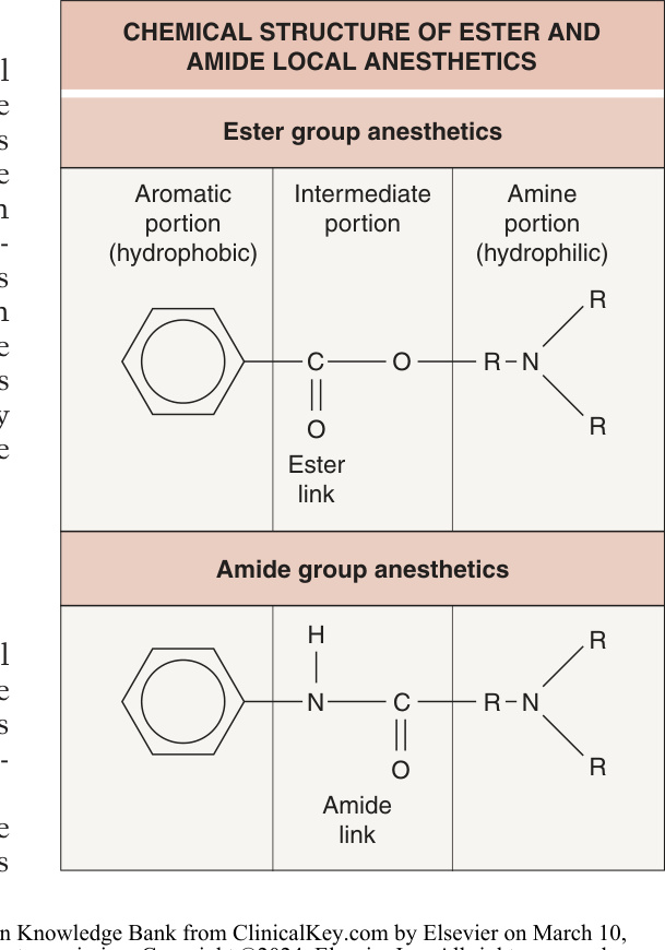
Fig. 143.1 Chemical structure of the ester and amide group local anesthetics. The aromatic (hydrophobic and lipophilic) end is joined to the amine (hydrophilic) end with an ester or amide linkage.
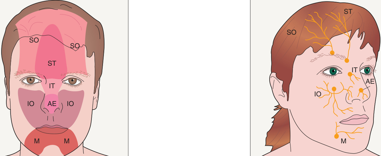
Fig. 143.2 Sensory innervation of the central face.
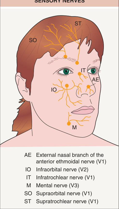
Fig. 143.3 Skull exit points of central face sensory nerves.
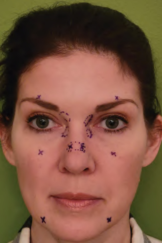
Fig. 143.4 Locations of needle insertions for central face nerve blocks. From superior to inferior, the supraorbital, infraorbital, and mental nerves are marked with an “x” and the supratrochlear, infratrochlear, and external nasal nerves are marked with a rectangle.
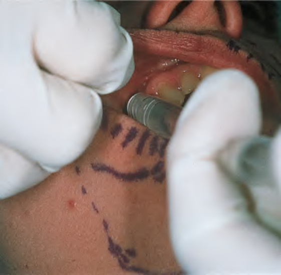
Fig. 143.5 Intraoral route for infraorbital nerve block. The needle is inserted cephalad between the premolar (bicuspid) teeth in the midpupillary line.
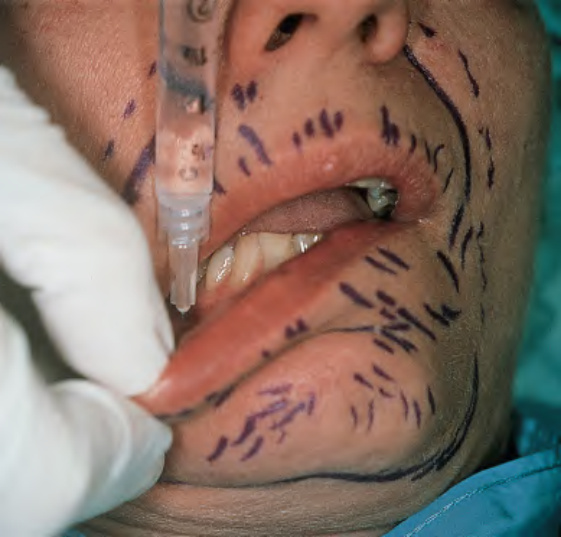
Fig. 143.6 Intraoral route for mental nerve block. The needle is inserted caudally between the premolar (bicuspid) teeth in the midpupillary line.
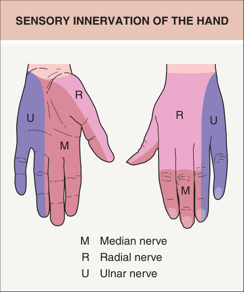
Fig. 143.7 Sensory innervation of the palmar surface and dorsal surface of the right hand.
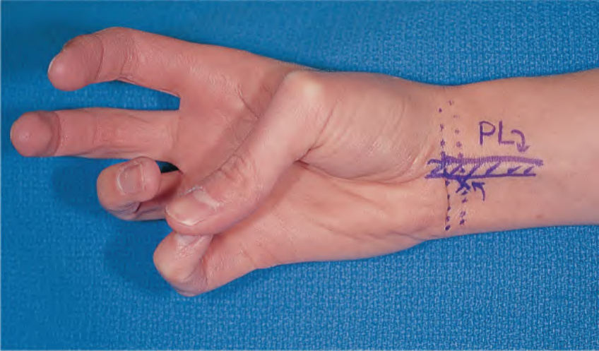
Fig. 143.8 Cutaneous markers for the median nerve location at the wrist. Note thumb and little finger apposition and wrist flexion to visualize the palmaris longus (PL) tendon. X, needle entry point.
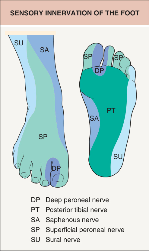
Fig. 143.9 Sensory innervation of the dorsal surface and plantar surface of the right foot.
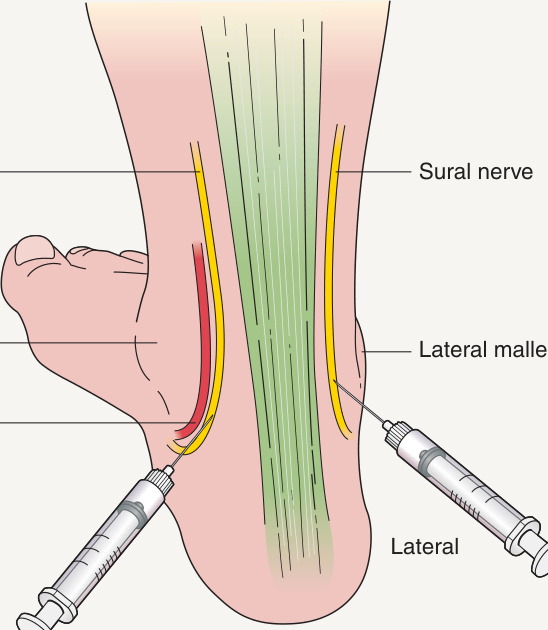
Fig. 143.10 Posterior tibial and sural nerve blocks.
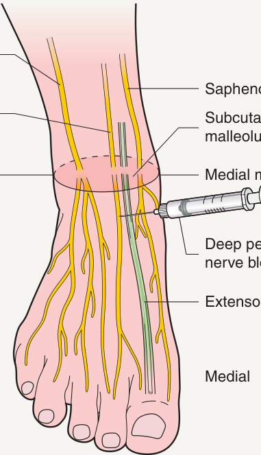
Fig. 143.11 Superficial peroneal, saphenous, and deep peroneal nerve blocks.
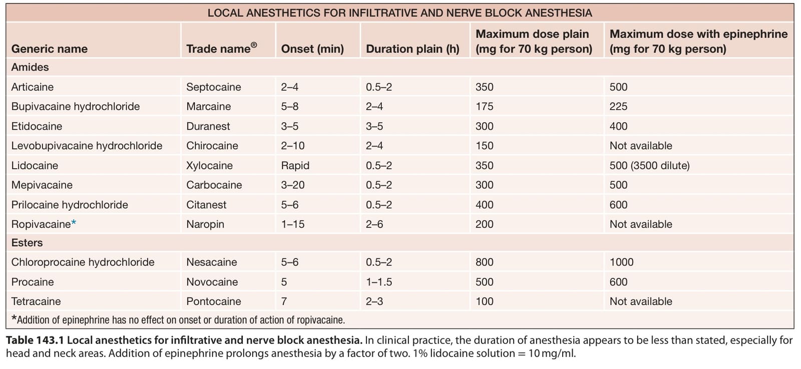
Table 143.1 Local anesthetics for infiltrative and nerve block anesthesia. In clinical practice, the duration of anesthesia appears to be less than stated, especially for head and neck areas. Addition of epinephrine prolongs anesthesia by a factor of two. 1% lidocaine solution = 10 mg/ml.
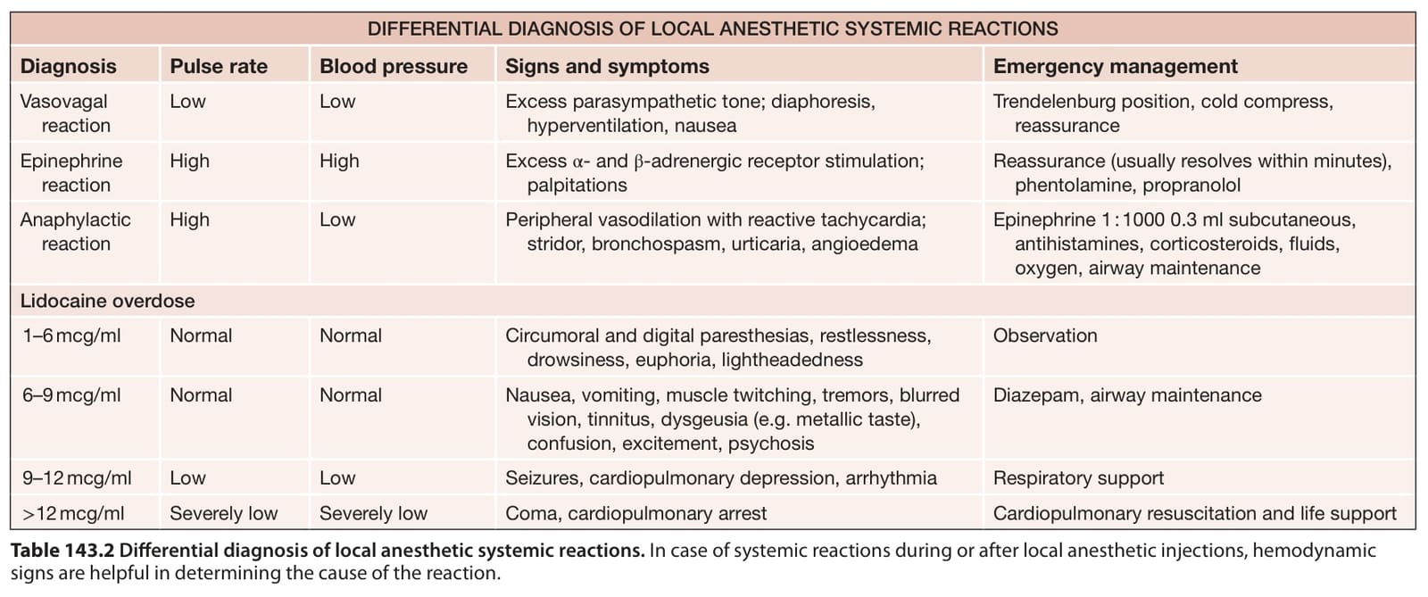
Table 143.2 Differential diagnosis of local anesthetic systemic reactions. In case of systemic reactions during or after local anesthetic injections, hemodynamic signs are helpful in determining the cause of the reaction.
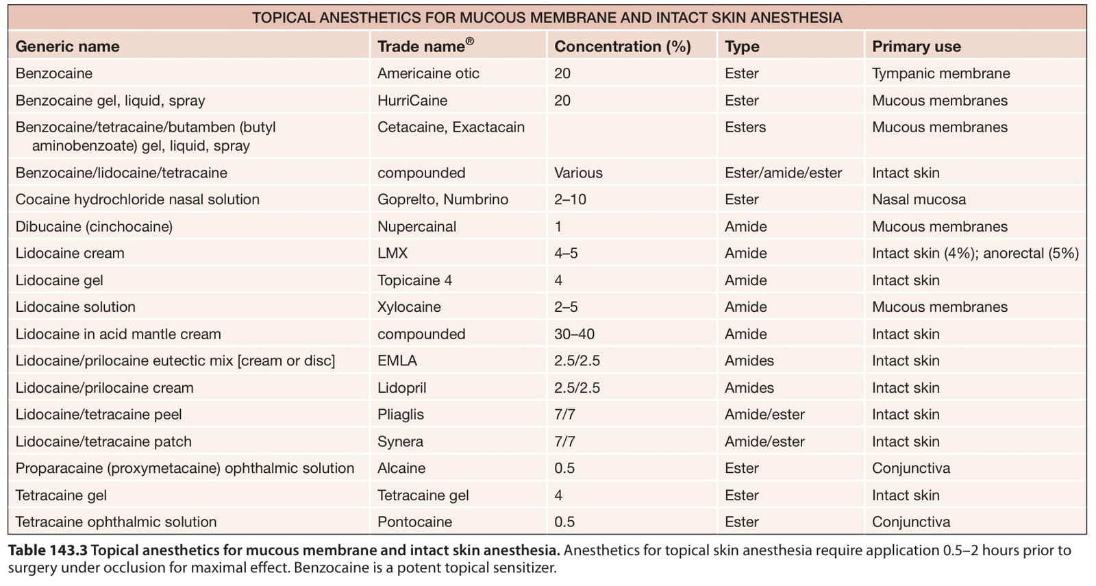
Table 143.3 Topical anesthetics for mucous membrane and intact skin anesthesia. Anesthetics for topical skin anesthesia require application 0.5–2 hours prior to surgery under occlusion for maximal effect. Benzocaine is a potent topical sensitizer.
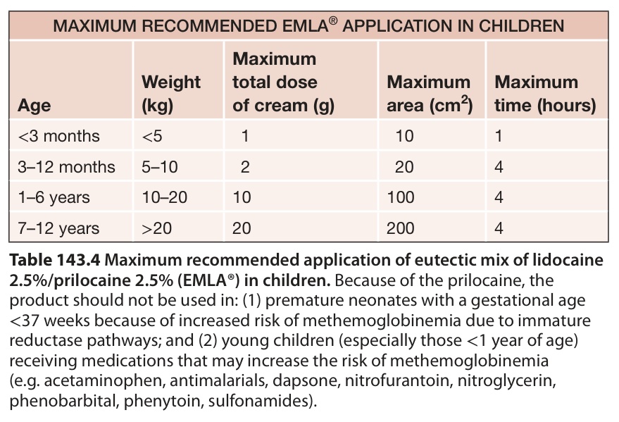
Table 143.4 Maximum recommended application of eutectic mix of lidocaine
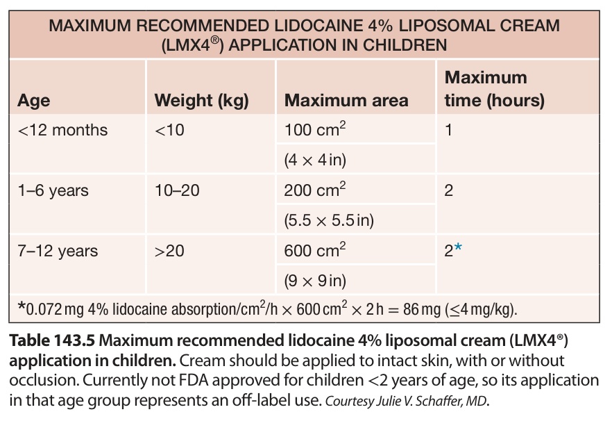
Table 143.5 Maximum recommended lidocaine 4% liposomal cream (LMX4®)

Table 143.6 Compressed air delivery systems for lidocaine. Lidocaine is delivered into the dermis.
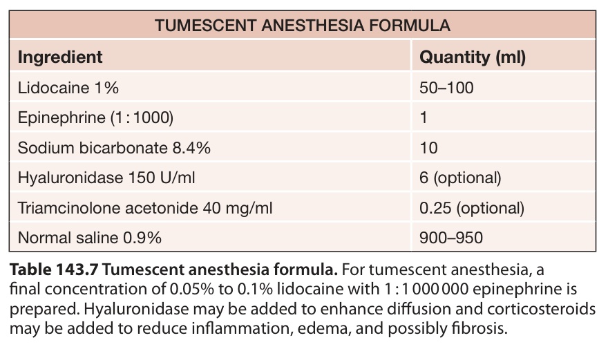
Table 143.7 Tumescent anesthesia formula. For tumescent anesthesia, a final concentration of 0.05% to 0.1% lidocaine with 1 : 1 000 000 epinephrine is prepared. Hyaluronidase may be added to enhance diffusion and corticosteroids may be added to reduce inflammation, edema, and possibly fibrosis.
Credentialing for higher levels of anesthesia may be regulated by state or national (outside the US) medical licensure authorities and hospitals, and the physician must be in full compliance with the rules in his or her particular state or country. Most of the regulations require procedures done under general anesthesia or with intravenous sedation to be done in an ambulatory surgery center or hospital with a supervising anesthesiologist. Higher levels of anesthesia involving the use of intravenous medications such as midazolam hydrochloride (Versed®) and fentanyl should be done in a monitored environment with appropriate resuscitative equipment readily available. Nitrous oxide administration is a useful form of moderate analgesia, with the degree depending on the concentration administered. At a 20% concentration, the analgesia is equivalent to giving a narcotic analgesic. At an 80% concentration, most patients will become unconscious. The advantage of nitrous oxide is the rapid reversal of anesthetic effect as soon as the gas is turned off. With proper training, nitrous oxide can be safely administered in the office, as it has been done for many years in many dental offices. An easier version of nitrous oxide analgesia, Pro-Nox™ (Clarion Medical Technologies), delivers a fixed ratio of 50% nitrous oxide and 50% oxygen. The patient holds the mouthpiece and controls how much they inhale, remaining conscious throughout the procedure. General inhalation anesthesia or deep intravenous sedation with propofol should generally be performed by anesthesiologists.