DISCUSSION
Sutures
Since prehistoric times, humans have been using various materials to close skin wounds. The first known reference appears in the Edwin Smith Papyrus (c. 1600 BCE). Although the precise suture material was not described, examination of artifacts from that time period suggests that it was most likely linen. Hippocrates has been credited with being the first to use the term “suture” in reference to sewing human tissue in 400 BCE.
Over the centuries, suture materials that have been employed include cotton, leather, flax, insect mandibles, horse and even human hair. Ancient Roman physicians would braid the hair of gladiators tightly to close their scalp wounds and by doing this, these innovative surgeons demonstrated the understanding that sutures can produce immediate hemostasis in addition to wound edge approximation.
The ideal suture, were it to exist, would handle easily, hold a secure knot, and have high tensile strength. It would neither cause inflammation nor promote infection, and it would gradually dissolve, thereby obviating the need for suture removal. Unfortunately, the perfect suture material does not exist. However, the ever-widening array of choices available to the dermatologic surgeon and a knowledge of each suture’s individual characteristics increases the likelihood of choosing the best possible materials for each surgical situation.
Suture materials are divided into two categories: (1) absorbable; and (2) non-absorbable. Studies of wound-healing by Levenson et al. have shown that during the first 1–2 weeks after surgery, the intrinsic tensile strength of the wound is ~7%–10% of native bursting pressure. At 5 weeks, the intrinsic tensile strength is ~60%, and, in general, after 1 month, dehiscence is unlikely with normal activity. During this time period, absorbable buried dermal sutures are utilized to alleviate tension and maintain wound edge approximation. Transepidermal, non-absorbable sutures are non-tension-bearing, but allow for fine adjustments of the epidermal edges.
The characteristic attributes ascribed to a suture are defined and standardized by the United States Pharmacopeia (USP) as follows:
●Two physical configurations exist: monofilament and multifilament. Multifilament suture can be spun, twisted, or braided. Braiding serves to increase pliability, strength, knot security and handling, but may abrade or shear tissue. Braiding also potentially increases the risk of infection by allowing bacteria to become entrapped within filament strands. Monofilament sutures have a low coefficient of friction and slide easily through tissue (see below). Their drawback is that they are traditionally stiffer than braided suture and may have decreased knot security, requiring additional throws to ensure that the knot is secure.
●Capillarity is defined as the ability of a suture to absorb and transfer fluid. Multifilament sutures have greater capillarity than monofilament.
●The USP size of a suture is determined by the diameter needed to achieve a given tensile strength, and this is expressed in multiples of zeros; the smaller the cross-sectional diameter of the suture, the more zeros. For example, 7–0 polyglactin 910 (Vicryl®, Polysorb®) is a much finer suture than 3–0 polyglactin 910. The actual diameter of a given USP size varies depending upon the composition of the suture, with a 4–0 surgical gut being a larger-diameter suture than 4–0 polypropylene because polypropylene is innately stronger. In general, the smallest suture that will provide adequate tensile strength for the indicated repair should be utilized.
●Elasticity refers to the ability of a suture to regain its original length after being stretched. An elastic suture, such as polybutester, allows for tissue swelling and then maintains tension on the wound edges after the edema has resolved.
●The coefficient of friction determines the ease with which a suture will pull through tissue. A suture with a low friction coefficient, such as polypropylene, slides easily through tissue. For this reason, polypropylene is commonly used for the running subcuticular suture (see Ch. 146). Knot strength is directly proportional to the friction coefficient of the suture material. The more slippery the suture material, the more likely it is that the resulting knot will unravel. Hence, when suturing with polypropylene, it is prudent to place several additional throws for each knot.
●Memory is defined as a suture’s tendency to retain its natural configuration and is determined by the elasticity and plasticity of the suture material. Memory is a useful property for maintaining closure during the postoperative period. High-memory sutures, such as nylon, are widely used for surface approximation. Drawbacks of high-memory sutures are that they do not handle very easily and have a relatively low knot strength. As with sutures that have a low coefficient of friction, a few extra throws may be helpful to ensure a secure knot. Suture material with low memory, such as silk, is easy to handle and rarely becomes untied.
●Plasticity is the ability of the suture to be molded or retain its new length and form as well as tensile strength after it has been stretched. This is an important characteristic for a suture material that is to be used in situations with tissue edema.
Sutures with plasticity, such as polypropylene, will stretch to accommodate edema and not cut into tissue.
●Pliability refers to how easily the suture can be bent. Braided suture materials, such as silk, are the most pliable, and they are capable of being tied readily into a knot.
●The tensile strength of a suture is determined by the force required to snap it divided by the cross-sectional diameter of the suture. In general, synthetic materials are stronger than natural materials. A suture that has been knotted has approximately one-third the tensile strength of the same material unknotted.
●Tissue reactivity is the degree of foreign body inflammatory response evoked by a suture when placed into a wound. In general, natural materials (such as surgical gut) are degraded by proteolytic enzymes and cause a far greater inflammatory response than do synthetic materials (such as poliglecaprone 25) which are absorbed by hydrolysis.
Types of Suture Material
Sutures are characterized as absorbable or non-absorbable based upon their ability to be enzymatically digested or hydrolyzed. Most absorbable sutures lose the majority of their tensile strength long before they are fully absorbed. Sutures that are absorbable do not require removal. In contrast, non-absorbable sutures are resistant to hydrolysis and enzymatic degradation. Non-absorbable sutures such as nylon and polypropylene are usually used as surface or cutaneous sutures, and they are routinely removed 5–14 days after surgery (earlier on the face and later on the trunk and extremities).
Absorbable sutures
The most commonly used absorbable sutures are described below and summarized in Table 144.1.
Surgical gut (catgut), one of the first sutures ever mass-produced, is composed of 97%–98% purified collagen fibers derived from bovine or ovine intestine. It is a natural tan fiber that is packaged wet in alcohol and dries quickly when exposed to air. Plain surgical gut is not commonly used nowadays because of high tissue reactivity and slow digestion but rapid loss of tensile strength. Specifically, when used as a buried suture, plain gut loses its tensile strength within 7 to 10 days but requires 60 to 70 days for complete digestion.
Chromic gut is plain gut that has been processed with chromium salts to increase its resistance to enzymatic degradation and decrease its tissue reactivity. The tensile strength of chromic gut typically lasts for 10 to 14 days. Chromic gut is still widely used for suturing of mucosal surfaces.
Fast-absorbing gut is plain gut that has been heated to begin breakdown of the collagenous material within the suture prior to use. It maintains its tensile strength for 5 to 7 days. Fast-absorbing gut has become popular for suturing skin grafts and for placing surface sutures in wounds that are well approximated by buried sutures. No increase in wound infections has been observed in comparative studies, and this suture is convenient for the surgeon and the patient because it does not require removal. However, its relatively high coefficient of friction leads to noticeable tissue drag when suturing skin. This disadvantage
can be ameliorated by the addition of a thin layer of petrolatum ointment (known as “greasing the gut”).
Polyglycolic acid (Dexon®), introduced in 1970, was the first synthetic absorbable suture and its reduced tissue reactivity and predictable absorption profile represented a vast improvement over surgical gut. It is a braided glycolic acid polymer with easy handling qualities, and it maintains 20% of its tensile strength at 3 weeks. Polyglycolic acid suture is available plain or coated with poloxamer 188. This lubricant coating allows the suture to pass more easily through tissue and makes it more pliable, which facilitates knot tying.
Polyglactin 910 (Vicryl®, Polysorb®), a synthetic, braided copolymer of glycolide and L-lactide, was first introduced in 1974 and supplanted polyglycolic acid as the most popular buried absorbable suture in cutaneous surgery. The water-repelling properties of lactide delay penetration of water and delay the loss of tensile strength. Polyglactin 910 maintains 75% of its tensile strength at 2 weeks and 50% at 3 weeks. Although stronger than polyglycolic acid, polyglactin 910 absorption is generally complete by 90 days, whereas polyglycolic acid is usually still being absorbed at 120 days.
Polyglactin 910 is available either undyed or dyed violet. Although the violet color usually dissipates, some surgeons avoid the dyed form because occasionally it may be visible when embedded in the skin. Polyglactin 910 comes coated with a mixture of polyglactin 370 and calcium stearate that serves as a lubricant, facilitating the passage of the suture through tissue. Polyglactin 910 is immensely popular due to its easy handling characteristics and because it holds knots well and does not tear tissue.
Although polyglactin 910 is traditionally used as an absorbable dermal suture, it may be used safely for wound edge approximation as a running cutaneous suture and removed in 5–14 days. This method still requires suture removal, but the use of a single suture for both the dermal and surface wound closure results in a 50% savings in suture cost per reconstruction. In the authors’ experience, polyglactin 910 is also an excellent choice for the vermilion lip as it does not irritate the opposing mucosal surfaces and leads to much less inflammation than silk.
Polyglactin 910 is also available in a partially hydrolyzed form (Vicryl Rapide®), which is useful as a percutaneous suture that does not need to be removed as it spontaneously dislodges in 7–14 days. This suture is indicated for wound edge approximation where only 1–2 weeks of support will be needed and suture removal may be painful or is inconvenient.
Polydioxanone (PDS II®) is a monofilament polymer made from the polyester poly (p-dioxanone). The primary advantage of polydioxanone over polyglycolic acid and polyglactin 910 is prolonged tensile strength. It maintains ~70% of its tensile strength at 2 weeks and 50% at 4 weeks. Traces of buried polydioxanone are present in 6-month histologic preparations. Polydioxanone is employed in high-tension areas, such as the proximal extremities and trunk. When compared with polyglycolic acid in closure of skin wounds under tension, polydioxanone was associated with less scar spread and a lower incidence of hypertrophic scar formation.
Similar to polyglycolic acid and polyglactin 910, polydioxanone has minimal tissue reactivity although it is slightly higher than poliglecaprone 25 in a murine model. As a monofilament suture, it retains packaging memory and can be relatively stiff and difficult to tie. However, polydioxanone glides through tissue easily.
Polyglyconate (Maxon®), a copolymer of glycolic acid and trimethylene carbonate, is also an absorbable monofilament suture. Its overall properties and indications are similar to polydioxanone. It maintains 80% of its tensile strength at 2 weeks and 60% at 4 weeks, with complete absorption occurring via hydrolysis by ~180 days. Despite this prolonged absorption, there is minimal tissue reactivity. Polyglyconate may be most useful for large surgical procedures on the trunk or extremities that are under substantial tension and require prolonged, suture-based approximation during healing.
Poliglecaprone 25 (Monocryl®) is an absorbable monofilament suture consisting of a copolymer of glycolide and ε-caprolactone. It exhibits very high initial tensile strength, but this declines rapidly in the early postoperative period with ~50%–60% of its tensile strength at 1 week, only 20%–30% at 2 weeks (undyed), and essentially 0% at 21 days. This absorption profile limits its use in areas where prolonged dermal support is desired. Complete absorption occurs at ~90 days.
Poliglecaprone 25 offers supple handling and minimal tissue reactivity, which led to a lower incidence of hypertrophic scarring when compared to partially hydrolyzed polyglactin 910 (Vicryl Rapide®) for skin approximation in a breast reduction scar model. Furthermore, poliglecaprone 25 demonstrates better knot tying and security than other commonly used absorbable monofilament suture materials. Because poliglecaprone 25 induces less inflammation than polyglactin 910 and glides easily through tissue, for many dermatologic surgeons it has become the absorbable suture of choice for low tension closures in the head and neck region. In a randomized study of deep dermal sutures for repair of Mohs defects, a statistically higher rate of suture extrusion occurred with polyglactin 910 (11%) versus poliglecaprone 25 (3%), but there was no difference in ultimate cosmesis.
Its minimal tissue reactivity permits the use of poliglecaprone 25 for epidermal approximation (with removal at 5–14 days), thereby eliminating the need for separate dermal and surface sutures. A randomized, blinded, split-scar study comparing running 5–0 poliglecaprone 25 versus 6–0 polypropylene for facial surface closures found no significant differences in cosmetic outcome at 1 week or 4 months. Poliglecaprone 25 can also achieve epidermal approximation as a running subcuticular suture that does not require removal.
Glycomer 631 (Biosyn®) and polyglytone 6211 (Caprosyn®) are absorbable monofilament sutures with comparable physical characteristics to poliglecaprone 25. Complete absorption of glycomer 631 occurs between 90 and 110 days, with low tissue reactivity. Polyglytone 6211, introduced in 2002, was designed to offer the most rapid absorption profile within this class of suture (20% tensile strength at 10 days; complete absorption at 56 days). This property was postulated to underpin its reported low rate of suture extrusion.
Non-absorbable sutures
The most commonly used non-absorbable sutures are described below and summarized in Table 144.2.
Silk is a braided, natural suture. It represents the gold standard for ease of handling, knot formation and knot stability, by which newer synthetic sutures are measured. Because braided silk is very soft and pliable, it is commonly used on mucosal surfaces and in intertriginous regions. Despite these advantages, there are major drawbacks to its use in cutaneous surgery. Silk has a low tensile strength, its braided configuration produces a high coefficient of friction, and the high capillarity increases the risk of infection. In fact, the presence of one braided silk suture in a contaminated wound was shown to increase the virulence of staphylococci by 10 000-fold. As an organic, foreign protein (fibroin) made by the silkworm, silk has very high tissue reactivity.
Nylon (Ethilon®, Dermalon®), a monofilament polymerized polyamide, was the first synthetic non-absorbable suture to become commercially available. Because of its low cost, high tensile strength and low tissue reactivity, nylon is the most commonly used non-absorbable suture in dermatologic surgery. It is also available as a braided suture (Surgilon®, Nurolon®) which handles more easily but is more expensive.
Nylon’s elasticity allows it to expand and contract with transient wound edema but its stiffness and memory lead to decreased knot security. In practice, this can be easily counteracted by firmly setting knots and increasing the number of knot throws. This monofilament is available as clear or dyed in a black or green color. The green suture can be helpful when working in dark hair-bearing areas.
Traditionally, nylon is placed as a cutaneous suture but clear nylon may be used as a tension-bearing deep suture that is not removed.
Adapted from Garrett AB. Wound closure materials. In: Wheeland R (ed). Cutaneous Surgery. Philadelphia: WB Saunders, 1994:199–205.
It can be very useful when approximating the orbicularis oris muscle in a lip wedge repair or tacking tissue to the periosteum in situations where prolonged wound strength is desired.
Polypropylene (Prolene®, Surgilene®, Surgipro®) is a monofilament polymer of propylene. It is an inert plastic that has minimal tissue reactivity. The major advantage of this suture is its low coefficient of friction, which allows for a gentle, smooth pull through tissue. Even after extended periods of time, it can be withdrawn easily without disrupting the wound (Fig. 144.1). This characteristic makes it a good choice for the running subcuticular suture. Polypropylene also has high plasticity and stretches with tissue swelling, thus reducing the likelihood of post-operative track marks. Disadvantages of this suture are its memory (which compromises knot security) and increased cost compared to nylon. It is available in clear and dyed blue forms. Similar to nylon, polypropylene may be used as a subcutaneous suture in areas of high tension to provide prolonged support.
Polyester sutures (Dacron®, Mersilene®, Ethibond®) are braided, multi filament sutures with high tensile strength, very good handling, and low tissue reactivity. Ethibond® comes coated with polybutilate, which decreases its drag through tissue (decreased friction coefficient). Similar to polyglactin 910, polyester sutures are of great assistance for approximation of mucosal tissues, particularly the vermilion lip. They combine the soft feel of silk with a lack of tissue reactivity.
Polybutester (Novafil®) is a monofilament suture of polyglycol terephthalate and polybutylene terephthalates. Its major advantages are good handling, low coefficient of friction, high elasticity, and low tissue reactivity.
Suture Selection
In general, a surgeon should select the smallest suture that will provide adequate strength for a given closure (Table 144.3). For facial repairs under low tension, 4–0 or 5–0 polyglactin 910 or poliglecaprone 25 are common absorbable sutures, and 5–0 or 6–0 nylon, polypropylene or fast-absorbing gut are typical cutaneous sutures. Poliglecaprone 25 is popular as a dermal suture due to its relatively fast absorption and
reduced rate of suture extrusion. Polypropylene (removed at 7–14 days) or poliglecaprone 25 (buried and left in place) are the most commonly used sutures for running subcuticular closures. Polyglactin 910 (buried and left in place) may also be employed as a subcuticular suture despite concerns over tissue reactivity with such superficial placement. Indeed, in a randomized controlled trial (RCT), polyglactin 910 resulted in better scar appearance than polypropylene removed at 14 days.
For larger truncal defects, 3–0 or 4–0 polyglactin 910 or poliglecaprone 25 for buried sutures and 3–0 or 4–0 nylon for superficial closure are good choices. Alternatively, running subcuticular closures are particularly valuable on the trunk and extremity to eliminate the possibility of suture track marks, which are common in these locations. Polydioxanone and polyglyconate offer the most prolonged dermal support and are a consideration for wounds under significant tension, while poliglecaprone 25’s rapid loss of tensile strength makes it a poor choice for such closures. An alternative to traditional sutures are skin staples. They offer a quick, strong method for surface closure of large scalp and truncal defects.
Fast-absorbing gut (5–0 or 6–0) is a convenient suture for skin grafts and for any head and neck reconstructions that are well approximated with buried sutures. While some surgeons are concerned that fastabsorbing gut’s relatively higher tissue reactivity might increase early peri-incisional erythema and suture track marks, this has not been the
authors’ experience. An RCT comparing 5–0 fast-absorbing gut and 5–0 polypropylene did find a small, but statistically significant, difference in cosmetic outcome that favored polypropylene; no difference in erythema was observed. Another properly powered RCT focusing on facial skin closures found no difference in cosmetic outcomes between 5–0 and 6–0 fast-absorbing gut. However, this study did not include repairs on the nose, lip, or eyelid.
Cyanoacrylate tissue adhesives (e.g. 2-octyl-cyanoacrylate [Dermabond®], a blend of 2-octyl-cyanoacrylate and n-butyl-cyano acrylate [GluSeal®, LiquiBand®]) offer a practical and rapid alternative method of epidermal closure for wounds that are well-approximated with deep sutures. Wound closure tapes offer a non-traumatic alternative to epidermal sutures, and they are useful when applied to healing wounds following suture or staple removal. Additionally, they can be used over a running subcuticular suture at the time of surgery, relieving tension on the wound edges and providing a cosmetic, convenient alternative to a daily dressing change. Maloney and colleagues demonstrated that the optimal application of wound closure tapes is in a parallel non-overlapping pattern, following complete coating of the skin surface with liquid gum mastic adhesive. MicroMend® is a wound closure device that mimics a butterfly closure and can be removed by the patient. It consists of two arrays of microstaples anchored onto the skin and connected by a bridge. Although this device is an efficient alternative to sutures, studies are needed to determine its value compared to other wound closure options.
Complications
Suture-related complications are generally associated with placement technique, e.g. tied too tightly with strangulation of the tissue, tied too loosely and not approximating the wound edges well, or placed too superficially and extruding through the suture line. However, suture materials can also create complications due to their inherent properties. Some suture material, such as surgical gut and silk, are quite inflammatory. Synthetic absorbable sutures, such as polyglactin 910 and poliglecaprone 25, generally create little tissue reaction, but occasionally they may cause a granulomatous foreign body reaction (see Fig. 151.18).
Patients can develop a sterile pustule or suture abscess with associated inflammation ranging from mild to marked with focal ulceration (Fig. 144.2). These reactions can be distressing, especially if the patient has not been warned of the possibility that they may occur. In patients receiving anticoagulants, the reaction may be hemorrhagic rather than purulent (see Fig. 144.2C,D). Rarely, they may become secondarily infected, and those reactions that are particularly purulent or exudative should be cultured for bacteria. Deep sutures may also extrude or “spit,” sometimes creating a sinus track within the skin.
Removal of the offending suture is the basic treatment for both suture abscess and suture extrusion. If it is readily accessible, the suture should be grasped with a smooth forceps and snipped. A comedone extractor may help to express a suture that is slightly embedded; otherwise, the intact skin overlying the suture may need to be nicked with a #11 blade and then gently probed with a tissue forceps. Antibiotics are rarely necessary. Removing the foreign suture material is usually sufficient to resolve the reaction.
Needles
The ideal needle is sharp and strong, and closely matches the suture diameter. The needle should be strong enough to hold its shape and retain its sharpness, pass after pass. The best needles are made of high-quality stainless steel, and the size and shape of the needle should be selected to correspond to the thickness and toughness of the tissue to be sutured.
There are several major brands of suture materials and each manufacturer uses a different needle nomenclature (Fig. 144.3). Most manufacturers, however, provide needle comparison reference guides. In general, the needle is the most expensive component of the needle/suture and the latter is the most expensive disposable used in dermatologic surgery.
A needle is composed of three parts: the shank, the body and the point (Fig. 144.4). In dermatologic surgery, most needles have a swaged shank with a hollow proximal end into which the suture is inserted and then crimped. The suture track is determined by the size of the needle shank, not the suture size. The body of the needle can be straight, 1/4 circle, 3/8 circle, 1/2 circle, or 5/8 circle (Fig. 144.5). The most common shape used in skin surgery is the 3/8 circle. Alternatively, 1/2 circle needles are valuable for suturing in tight spaces such as placing deep dermal sutures within small (<1 cm) nasal and eyelid defects or fitting multiple buried sutures into high tension wounds on the back or scalp. For the former, the authors use the P-2 needle and for the latter, find the RB-1 taper point needle an excellent choice.
The most common needle used in cutaneous surgery is the triangular, reverse-cutting type (see Fig. 144.4). Triangular “cutting” needles pass through dense tissues more easily than round taper point needles. The conventional cutting needle has its sharp edge on the inside arc of the needle while the sharp edge of the reverse cutting needle faces outward. Reverse cutting needles are preferred as they minimize the risk of tearing through the wound edge during suture placement. Taper point needles do not pierce dense tissue easily, but they are less likely to tear delicate tissues such as nasal mucosa.
For the majority of wound closures on the face, the Ethicon P (“plastic”) and PS (“plastic skin”) or an equivalent series are adequate and economical (see Fig. 144.3). The PS-2 and P-3 needles are particularly suited to dermatologic surgery. They are reverse cutting and their tips are electropolished for added sharpness. The PC (“precision cosmetic”) series represents Ethicon’s superior skin surgery needles, and they can be used for fine, delicate work. These needles have the sharpest tips and have flattened bodies for better grasping and needle strength. If wound edges are fragile, one must exercise some caution when using PC needles with conventional cutting points. The PC-1 needle is a very fine, sharp, 13 mm needle ideal for delicate facial repairs and is the only PC needle attached to fast-absorbing gut.
The FS (“for skin”) and equivalent series of needles are not finely honed, and they are less sharp and less expensive. These needles are adequate for skin surgery on the trunk or extremities. SC-1 is a 13 mm straight needle that the authors believe facilitates full-thickness through and through quilting sutures placed to conform flaps and grafts to the topography of the ear.
The number after the series designation denotes the needle size. Most needles are drawn to size on the package (Fig. 144.6). The smallest needle/suture unit that will provide adequate tensile strength for a
given repair should be utilized. In areas with thick dermis such as the back, larger needles (e.g. PS-1 or PS-2) are much easier to pass through the tissue without bending.
Instruments
Instruments that provide the greatest precision and speed for a given procedure should be selected. There is a wide variation in the cost and quality of surgical instruments. Low-quality, low-cost instruments should be avoided as they are poorly finished and, therefore, are more difficult to use and lack durability. A high-quality instrument will more than make up for its cost by performing well day after day in countless procedures. Modern instruments are a blend of surgical stainless steel with carbon alloy, chromium, nickel and tungsten carbide. Tungsten carbide is a very hard alloy that enhances the function and durability of the blades of scissors and the jaws of needle holders. Quality instruments are usually covered by a warranty which will range from 1 to 5 years or even the life of the instrument, depending on the instrument type and cost.
It is important to keep instruments in good condition by properly caring for them and keeping them sharp. For example, using delicate scissors to cut suture will dull them quickly. Rough handling of delicate forceps and needle holders during surgery or the sterilization process may cause them to become misaligned. Any instrument that is not fully functional should be removed from the surgical tray and sent to the manufacturer for repair.
Some of the authors’ instruments of choice for given procedures are listed and shown in Figures 144.7 through 144.9.
Curettes
Developed in the late 1800s, the dermal curette has been used to treat a wide variety of cutaneous neoplasms, both benign and malignant. Common curettes are straight with a round head (Fox) or possess an oval head set at an angle from the stem (Cannon). They are labeled by size, depending upon the aperture of the cutting surface of the curette.
Mancuso–Fox and Mancuso–Cannon curettes are also available, which have a ruler on the handle. For standard curettage and electrodesiccation, a set of curettes ranging from 6 mm down to 2 mm may be employed. Generally the larger curette is used first to debulk the tumor. This is followed by the smaller curette, which is used to remove small pockets of residual tumor. Smaller curettes (1–2 mm) are excellent instruments for obtaining subungual keratotic debris for fungal culture and KOH examination or detection of scabies mites. Curettes can also be used to debulk tumors, thereby better delineating their margins prior to excision. With repeated use, curettes will become dull and must be periodically sharpened. There are disposable, single-use curettes; however, they tend to be very sharp and may cut into healthy tissue.
Scalpels
The choice of scalpel handle and blade is based on the surgical site and personal preference of the surgeon (Fig. 144.10). The flat, standard #3 handle is the one used most frequently in dermatologic surgery as it is durable, inexpensive, and adequate for the majority of cutaneous procedures. This instrument is available with or without a ruler on the handle. Ergonomic refinements have led to the introduction of several additional handles that offer more precise control and better grip, e.g. the thinner, longer, grooved #7 handle. The Siegel handle, which is a thin, round, knurled and well-balanced handle, is well suited for Mohs micrographic surgery. It is easily rolled with the fingers to facilitate very subtle adjustments in the cutting angle. For small procedures, such as those on the eyelid and/or ear, some surgeons prefer short, round, knurled blade handles that hold slender specialized mini-blades (see below).
The most commonly used blades in dermatologic surgery are #15, #10, and #11. The #15 blade is the most popular blade. It is gently curved and is appropriate for the majority of skin surgery. The #10 blade is a wide blade, similar in shape, but larger than the #15 blade; it is favored when cutting through sites with thick dermis such as the
back. The #11 blade is tapered to a sharp point; it is used to create stab incisions for drainage procedures, the removal of milia, precise standing cone removal, and “through and through” excisions. It is often used with the cutting surface up, which is opposite to the positioning of the #10 and #15 blades. Scalpel blades are made of either stainless steel or sharper, more expensive, Teflon®-coated carbon steel. Disposable scalpels attached to plastic handles are available, but they are not weighted and are usually less sharp.
Needle holders
Needle holders for facial and hand surgery are small and light with narrow, fine jaws, while larger needle holders with wide sturdy jaws are designed for work on the trunk and proximal extremities. The jaws of needle holders are either smooth or serrated (Fig. 144.11). Serrations prevent twisting of larger needles (e.g. Ethicon PS-2 and FS-2) during suturing. Smooth jaws are less damaging to fine-caliber needles (e.g. Ethicon P-3) and will not tear finer sutures (6–0), but suture needles may slip if not grasped carefully. Alloy inserts of tungsten carbide serve to increase the strength and hardness of the jaws and these inserts improve the grip on needles in smooth-jawed needle holders
From left to right: #3 handle with #10 blade, Siegel handle (authors’ preference) with #15 blade, and Beaver handle with #64 blade.
(see Fig. 144.11). Instruments with tungsten carbide inserts are readily identified by their gold finger handles; they are more expensive but are typically guaranteed for five years. There are also small needle holders that have delicate serrated jaws, which allow for a good grip on fine needles without shredding fine sutures. Large needles should not be used with fine needle holders as this may lead to damage to the inserts of these delicate instruments.
Popular models such as the 4.5- to 5-inch long Webster and Halsey needle holders, which have smooth or delicately serrated jaws and tapered tips, are ideal for the small needles and fine suture materials that are commonly used for cutaneous facial surgery (see Figs. 144.7 and 144.11).
However, use of these instruments for procedures on the trunk will quickly lead to their misalignment. The Crile-Wood needle holder has a gently tapered blunt tip; it is designed to hold larger needles and is more appropriate for skin surgery on the trunk or extremities. Baumgartner and Mayo–Hegar holders are durable, strong drivers with short tips and serrated jaws, providing a mechanical advantage with larger needles in thicker skin (see Figs. 144.8 and 144.11). The Olsen–Hegar needle holder has suture-cutting scissors behind the jaws and is convenient to use when working alone although it is easy to inadvertently cut the suture when attempting to regrasp the needle.
Scissors
Surgical scissors are required for cutting skin, undermining the subcutis and deeper fascial layers, cutting sutures, and removing wound dressings (Fig. 144.12). Scissors may have long or short handles, and the blades are straight or curved and serrated or smooth. The tips may be sharp or blunt. Scissors used in cutaneous surgery can be either completely stainless steel (most popular, least expensive) or have tungsten carbide inserts to strengthen the blades.
Gradle scissors are small, delicate, sharp-tipped, and tapered to a very fine point with a gentle curve. Due to their sharpness and precision, Supercut® Gradle scissors are ideal for removing thin stages during Mohs micrographic surgery and for removing skin tags. Gradle scissors must be used with care, and they should never be used to cut sutures. With improper use, they are quickly dulled, the cutting surfaces are nicked, and the tips are easily malpositioned.
Tissue scissors have relatively short handles and sharp tips. They are available in straight or curved models, with or without the serrations that prevent tissue motion during cutting. Of the various models available, the authors prefer curved Supercut® iris scissors to cut tissue and for sharp dissection. Supercut® iris scissors have the sharpest edge and they are easily recognized by their black handles. These scissors have a fine beveled angle at the cutting edge, and they are available with smooth edges or one serrated edge. The “razor-like” edge of these scissors enables the surgeon to cut tissue in a smooth, easy motion.
Westcott and Castroviejo scissors are delicate, spring-loaded tissue scissors with very sharp tips (see Fig. 144.9). The configuration of their handle and spring-loaded action make them ideal for manipulation in small delicate sites. For this reason, they are popular with oculoplastic surgeons. They should only be used for cutting thin tissue, such as that encountered in eyelid surgery, or they will dull quickly.
Large, less expensive scissors are sufficient for cutting sutures. Tissue scissors should never be used to cut sutures. Specially designed sutureremoval scissors with a half-moon hook on the lower blade are available
This Webster needle holder (right) features tungsten carbide jaw inserts which add strength and improve the grip on fine needles in smooth-jawed needle holders. The serrated jaws of the larger Mayo–Hegar needle holder (left) provide a firm grip on large needles, but may shred fine suture and optimally should only be used with 2–0 to 4–0 suture.
and the small hooked tip easily grasps the loop and prevents accidental sticks.
Undermining scissors are usually blunt-tipped (for safety) and have longer handles (for comfort). They are available in different sizes to accommodate the various anatomic regions in which skin surgery is performed. Baby Metzenbaum scissors have a high handle-to-blade length ratio, and, with the resultant small blade arc, they have become the most widely used scissor for sharp or blunt undermining (see Fig. 144.12). Larger Metzenbaum scissors are appropriate for extensive undermining in fascial planes on the scalp, trunk, and extremities (see Fig. 144.8). Stevens tenotomy scissors and Supercut® Shea undermining scissors are utilized for more superficial, delicate undermining (see Fig. 144.7). The tips of these scissors allow for sharp, less traumatic undermining.
For bandage-cutting, 5.5-inch Lister scissors are the most popular, with their angled blades and large blunt tips that easily slide under a dressing without damaging the underlying skin. The Universal scissor is also a popular choice. The serrated edges and larger rings provide greater cutting power.
Forceps
Proper forceps are essential for delicate, safe handling of tissue and suture needles during skin surgery. The most useful forceps in skin surgery are lightweight and have fine tips. The tips may be toothed, serrated, or smooth. Serrated forceps can exert excessive pressure on tissues, resulting in crush injury. Toothed forceps have opposing fine teeth and thus exert less pressure overall. Only toothed forceps should be used to grasp the epidermis and superficial dermis. For cutaneous surgery, the most popular tissue forceps (Adson) has 1×2 teeth. Some forceps have both distal teeth for handling of tissue and a more proximal raised platform for firmly grasping suture needles, allowing the surgeon to avoid manual handling of sharp needles.
Forceps are available with delicate (≤0.9 mm), regular (1–1.5 mm), or heavy (≥1.6 mm) tips. Adson forceps are the standard large forceps used for excisional surgery on the trunk and proximal extremities. Bishop– Harmon forceps and Foerester forceps are very lightweight, fine-tipped, and ideal for delicate work on the face and hand. Because these forceps are easily bent and misaligned, they must be handled with care, and they should not be used for manipulating the thicker skin of the trunk or proximal extremities. Castroviejo forceps usually feature a suturing platform and offer the delicate tip of a Bishop–Harmon forceps, but with a sturdier build and wider grip (see Fig. 144.7).
Both epilating and jewelers’ forceps have fine, sharp points and are useful for suture removal. Splinter forceps have fine, extra-fine or super-fine delicate pointed tips, and they are used in hair transplants, for splinter removal, and for removing embedded sutures.
Hemostats
Hemostats are used to grasp bleeding vessels prior to ligation. The most popular hemostat is the Halsted Mosquito model which is available in 3.5-inch and 5-inch lengths, either curved or straight, and either delicate- or regular-tipped. A modified instrument known as an Allis clamp is particularly helpful during cyst or lipoma removal (Fig. 144.13). It has finger grips and a locking mechanism similar to a hemostat, however, the jaws have opposing sharp teeth that grasp tissue firmly. Since these teeth can devitalize tissue, the Allis clamp is only used to provide traction on tissue that is being excised, such as a cyst wall.
Skin hooks
Skin hooks enable the surgeon to handle tissue with minimal trauma, and they are particularly useful for elevating flaps and reflecting skin edges during undermining. Many surgeons also use skin hooks to visualize bleeding vessels for hemostasis and for placement of dermal sutures. Skin hooks possess a sharp tip that has the potential to inflict a perioperative sharps injury. Thus, these instruments should be handled with caution and the tips shielded on the tray when not in use as is done with scalpels and needles.
Skin hooks are available in single-, double- or multiple-pronged patterns. The multiple-pronged instruments are used mainly for rhytidectomies and larger truncal procedures in which large flaps are being elevated. Single-pronged hooks are used most frequently for undermining delicate skin or for placing sutures. The single shepherd hook – which has a more circular shape – holds tissue better than does the single standard hook, but it does not release as easily. Some surgeons prefer the double-pronged skin hook due to its lower risk of puncture injury to the operator or assistant.
Special Instruments
For delicate work near the eye, the dermatologic surgeon needs fine instruments that allow for precise cutting and carefully controlled tissue manipulation. The Beaver handle, which comes in a variety of sizes, can be rolled with one’s fingertips, providing greater precision and control (see Fig. 144.10). Beaver handles are fitted with specialized smaller, sharper blades. The #64 blade has a rounded tip that one may prefer for work around the eye. The #67 and #65 blades are smaller versions of the #15 and #11 blades, respectively. However, these mini-blades dull more rapidly than do the standard #15 blades. Angled Beaver blades (Beaver® 6600) are useful for taking Mohs stages or biopsies in the ear canal.
The Castroviejo needle holder is ideal for suturing the delicate skin near the eye. This needle holder has spring handles and an optional
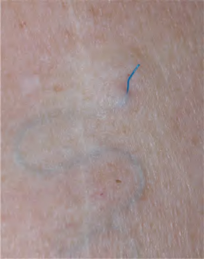
Fig. 144.1 Polypropylene suture. The blue color of the polypropylene suture is visible under the skin one year after its placement as a running subcuticular closure; the patient had neglected to return for suture removal. The suture was easily removed even after one year given its very low coefficient of friction and minimal tissue adherence.
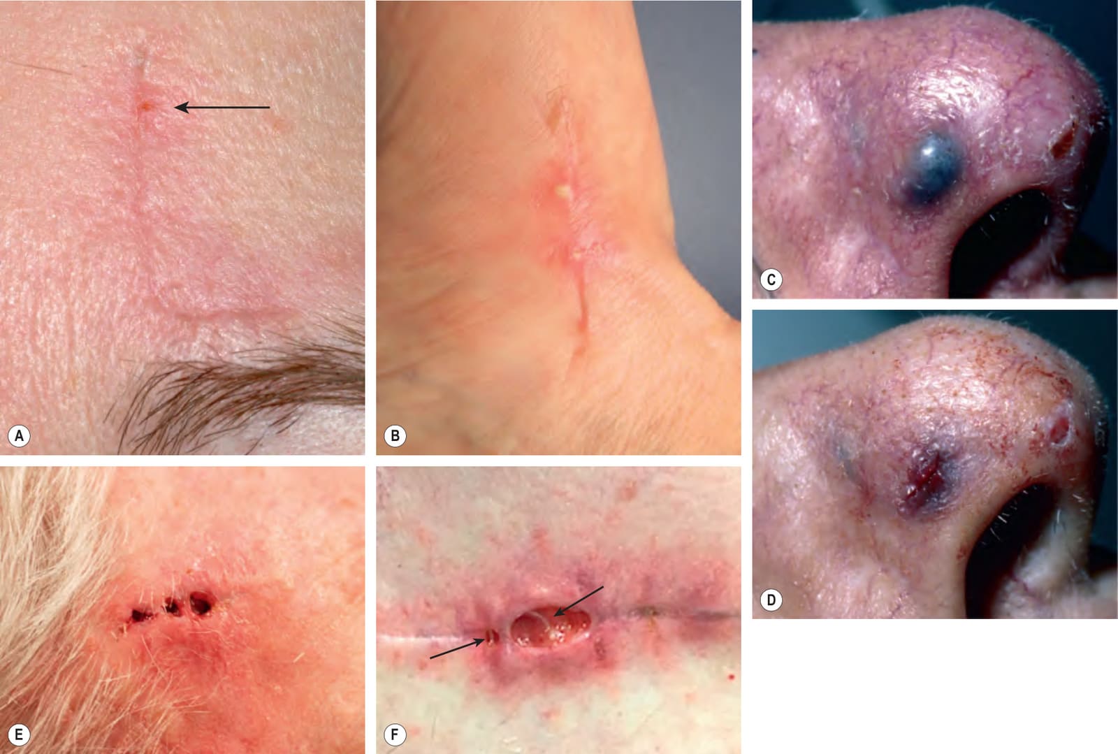
Fig. 144.2 Suture reactions.A Mild reaction at the superior edge of the wound (arrow). B Moderate reaction with two pustules and an inflammatory rim. C, D Hemorrhagic suture abscess in a patient on warfarin and appearance following incision (#11 blade) and drainage of small coagulum. E Severe inflammatory reaction leading to several focal ulcerations. F Severe ulcerative and granulomatous suture reaction, with visible polyglactin 910 suture material (arrows).
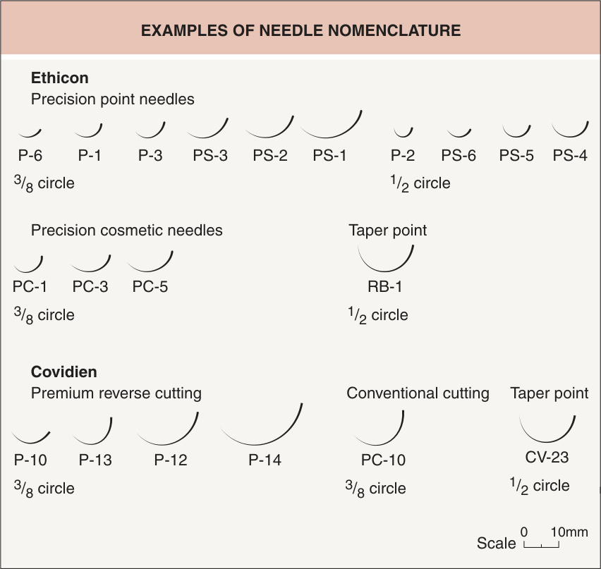
Fig. 144.3 Needle nomenclature by two representative manufacturers. P, plastic; PC, precision cosmetic; PS, plastic skin.
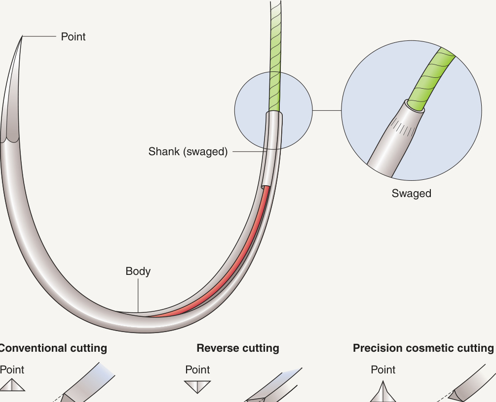
Fig. 144.4 Needle anatomy. The shank body and point are shown as are specific needle points for different types of cutting. Needles should be grasped at approximately one-third of the distance from the swaged area to the point. Avoid placement on or near the swaged area or near the tip.
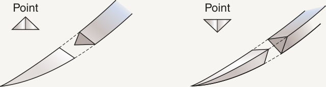
Fig. 144.5 Needle shapes. The most common curvature used in skin surgery is the 3/8 circle. The 1/2 circle curvature is useful when working in confined spaces.
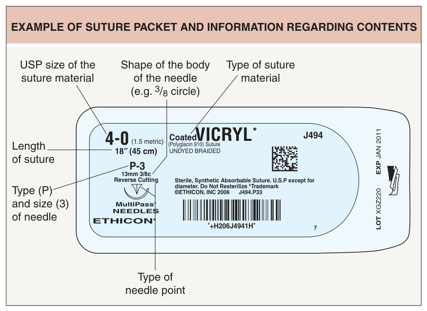
Fig. 144.6 Example of suture packet and information regarding contents.
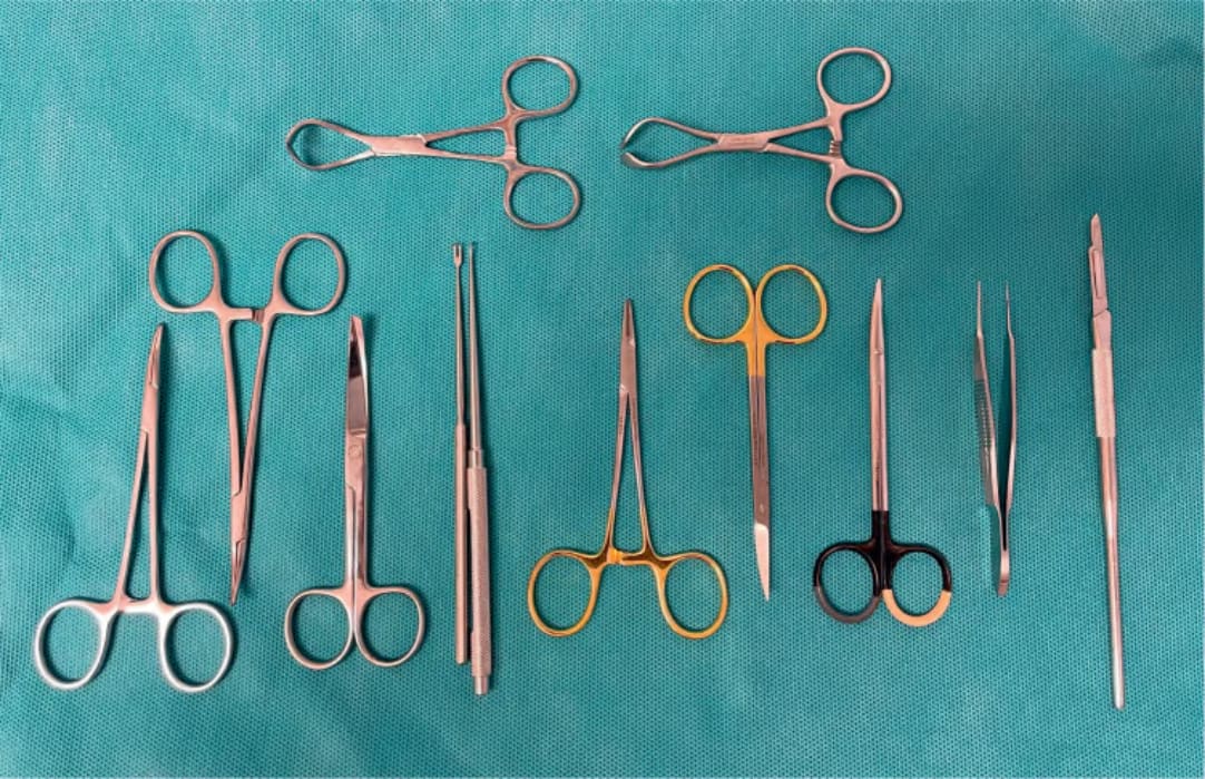
Fig. 144.7 Facial surgical instruments. Authors’ tray for facial surgery/repair includes (from left to right) two curved Mosquito hemostats, suture scissors, a double-pronged and a single-pronged skin hook, delicate Webster needle holder, curved iris scissors, delicate Supercut® Shea undermining scissors, 0.9 mm Castroviejo forceps, and Siegel blade handle with #15 blade. Two towel clamps are at the top.
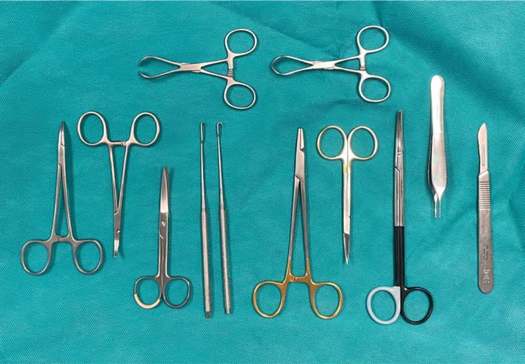
Fig. 144.8 Trunk or extremity surgical instruments. Authors’ tray for larger procedures in thicker skin includes (from left to right) curved Mosquito hemostats, suture scissors, skin hooks, Mayo–Hegar needle holder, curved iris scissors, Supercut® Metzenbaum undermining scissors (excellent when extensive undermining is anticipated; otherwise, baby Metzenbaum or Shea scissors are sufficient), Adson toothed forceps, and #3 handle with #10 blade. Two towel clamps are at the top.
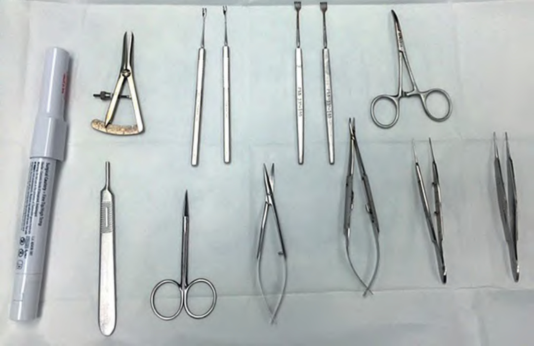
Fig. 144.9 Eyelid tray. Tray setup for eyelid surgery includes (bottom row, left to right) hand-held electrocautery, #3 blade handle, iris scissors, Westcott (or Castroviejo) scissors, Castroviejo needle holder, and two 0.5 mm Castroviejo forceps, in addition to (top row, left to right) Castroviejo calipers (used for measuring dimensions of eyelid skin grafts and planning tarsoconjunctival flaps), 2-pronged delicate skin hooks, 4-pronged skin hooks (rakes), and curved Mosquito clamp.
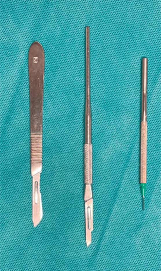
Fig. 144.10 Scalpel handles and blades.
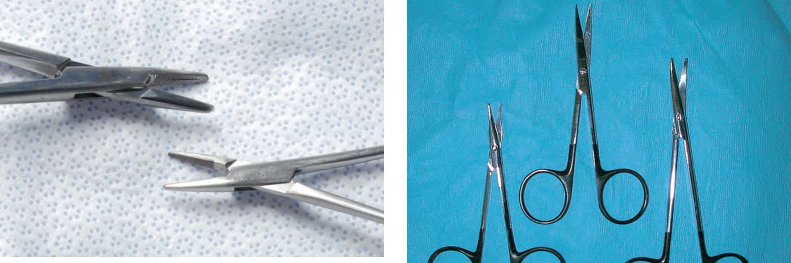
Fig. 144.11 Comparison of delicate Webster and Mayo–Hegar needle holders.
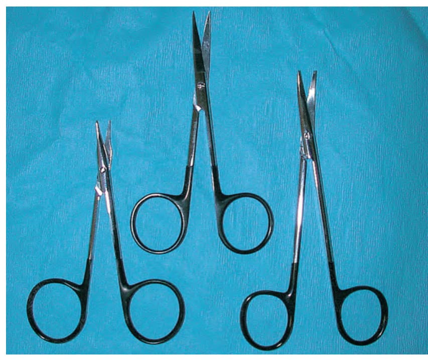
Fig. 144.12 Standard scissors. From left to right: Supercut® Gradle scissors, Supercut® iris scissors, and Supercut® baby Metzenbaum scissors. Courtesy Glenn Goldman, MD.
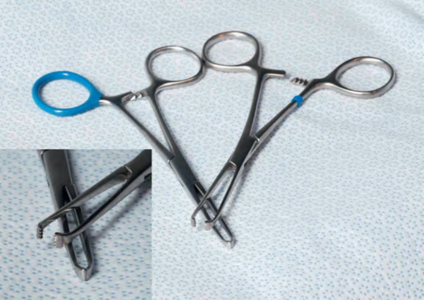
Fig. 144.13 Allis clamps. The inset highlights the sharp teeth which can firmly grasp a cyst or lipoma to facilitate removal.
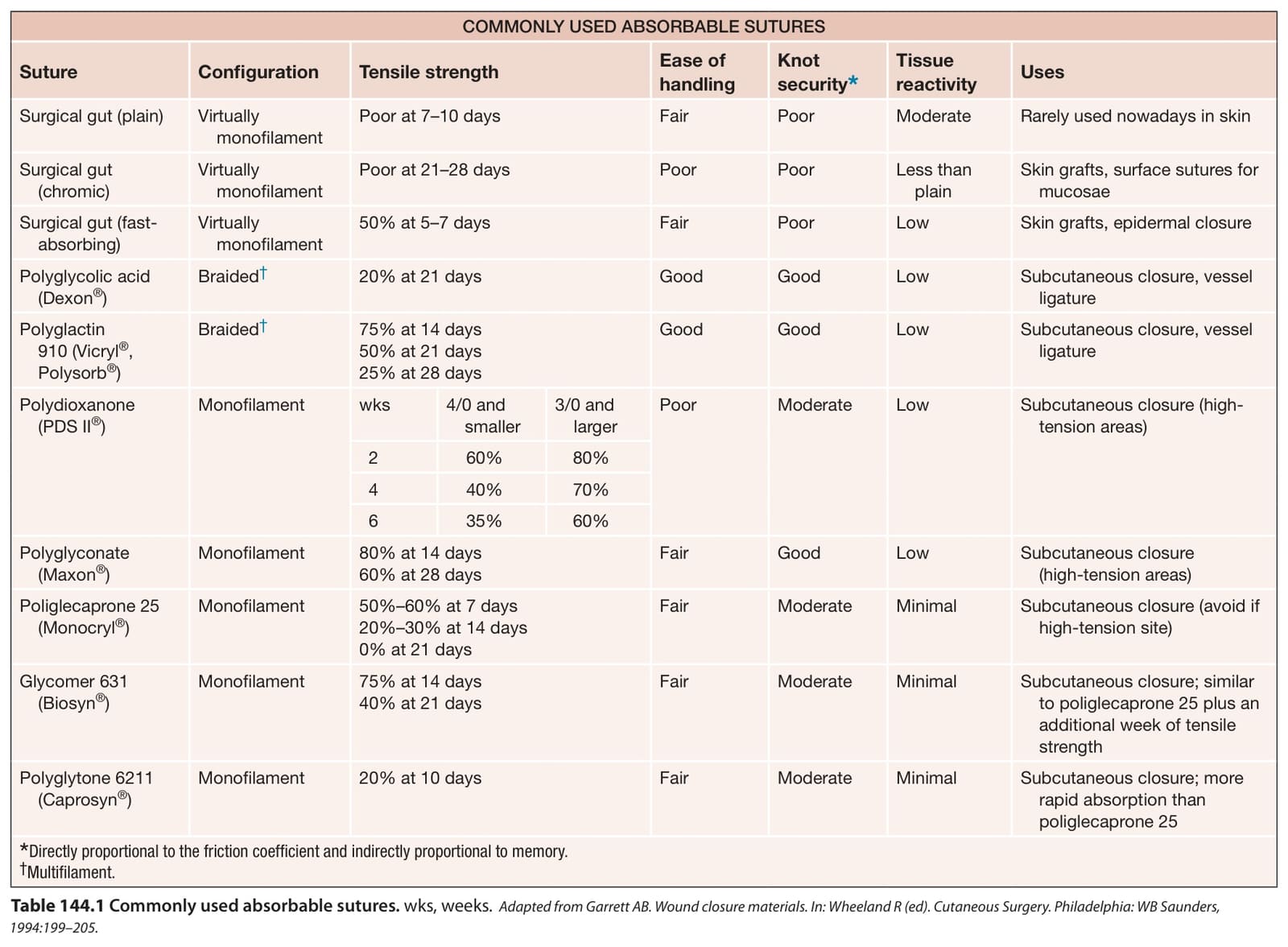
Table 144.1 Commonly used absorbable sutures. wks, weeks.Adapted from Garrett AB. Wound closure materials. In: Wheeland R (ed). Cutaneous Surgery. Philadelphia: WB Saunders, 1994:199–205.
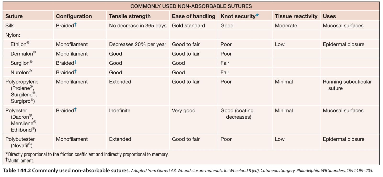
Table 144.2 Commonly used non-absorbable sutures.
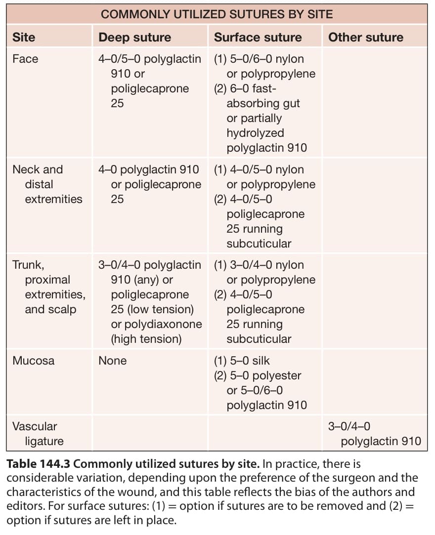
Table 144.3 Commonly utilized sutures by site. In practice, there is considerable variation, depending upon the preference of the surgeon and the characteristics of the wound, and this table reflects the bias of the authors and editors. For surface sutures: (1) = option if sutures are to be removed and (2) = option if sutures are left in place.
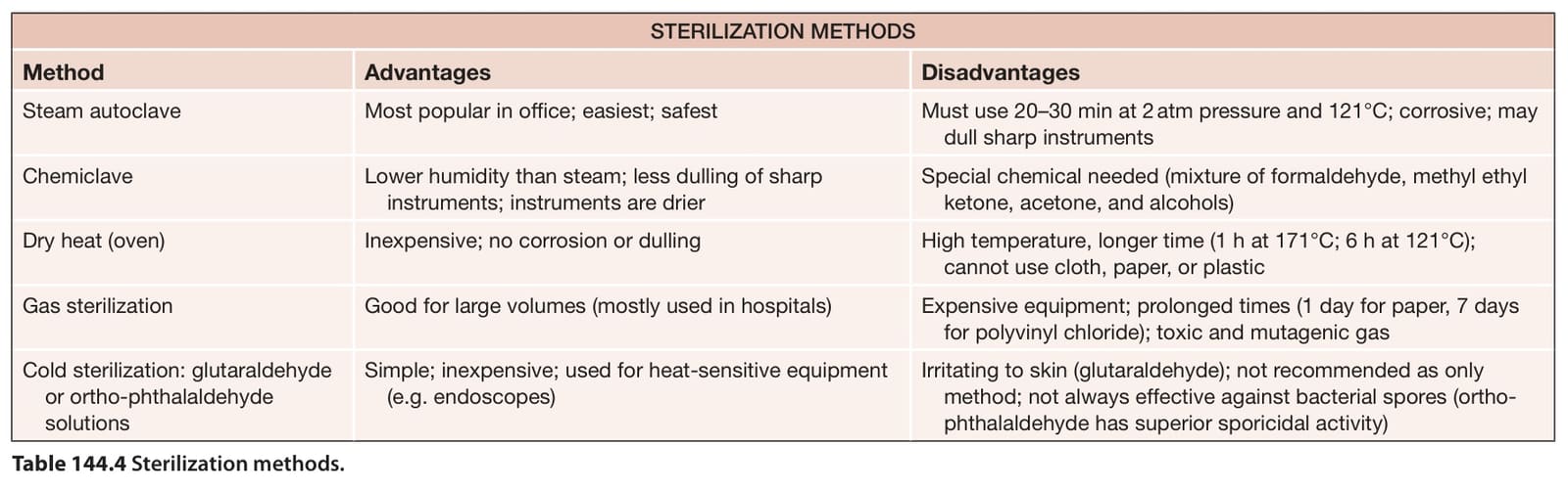
Table 144.4 Sterilization methods.
self-locking device. Castroviejo and Westcott scissors (see above) provide meticulous control when cutting eyelid skin. Lightweight forceps, such as Bishop–Harmon, Foerester or Castroviejo, aid in achieving the delicate handling of tissues necessary in this region. Plastic, Teflon®, and metal eye shields may be used to protect the globe when the lid margin will be breached. Plastic shields are popular as they do not conduct heat or electricity from electrosurgical devices. However, they must never be used with lasers; instead a non-reflective stainless steel shield should be employed.
Chalazion clamps are commonly used for immobilizing earlobes, lips, and tongues during procedures on these areas and provide excellent hemostasis in these highly vascularized tissues. Nail nippers, splitters, and elevators are necessary for nail surgery and are reviewed in Ch. 149.
A summary of sterilization methods is presented in Table 144.4.
Philadelphia: W.B. Saunders; 1994.17. Jenkins H, Hrdina LS, Owens Jr FM, Swisher FM. Absorp-
Table 144.4 Sterilization methods.